Table of Contents
Clinical features | Diagrams / tables | Microscopic (histologic) description | Microscopic (histologic) images | Cytology imagesCite this page: Weisenberg E. Strongyloides. PathologyOutlines.com website. https://www.pathologyoutlines.com/topic/lungnontumorstrongyloides.html. Accessed December 4th, 2024.
Clinical features
- Endemic in Southeastern United States, South America, Southeast Asia and Sub-Saharan Africa
- Infection occurs when larvae in soil penetrate the skin and travel to the lung via the venous circulation; the worms then travel up the trachea to the oropharynx and are swallowed setting up infection in the small intestine
- Female worms in the small intestinal mucosa produce eggs asexually (parthenogenesis) that are passed to the soil where they hatch to continue cycle of infection
- Larvae hatched in the intestines may penetrate the colonic mucosa to travel to the lung to reinitiate infection (autoinfection)
- Disease usually affects the gastrointestinal tract
- In immunocompromised patients, severe disease with large burden of parasites may occur; patients may present with alveolar hemorrhage (Prim Care Respir J 2009;18:337)
- May predispose to invasive infections caused by enteric organisms (Am J Clin Pathol 2007;128:622)
Microscopic (histologic) description
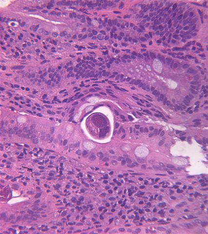
- In the lung, eosinphilic pneumonia with hemorrhage may occur
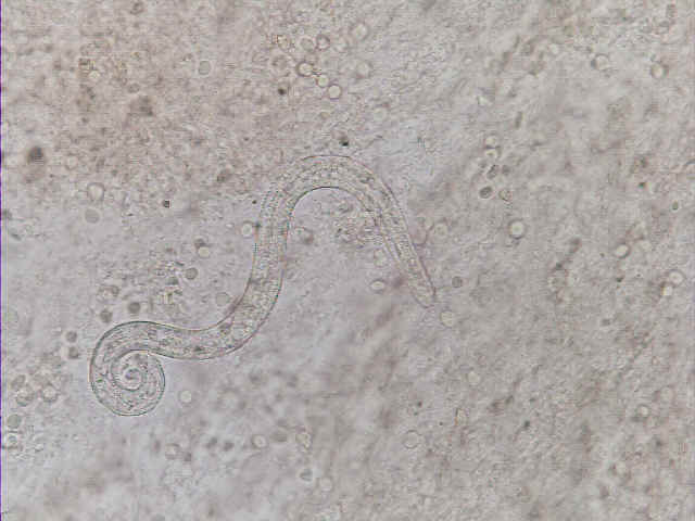
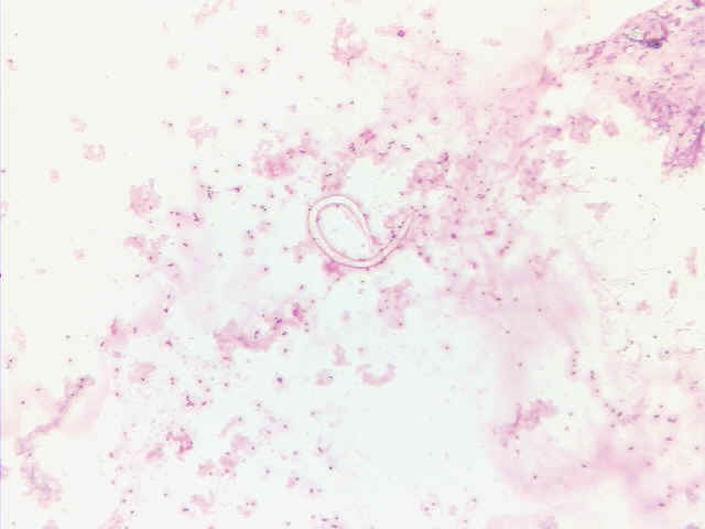
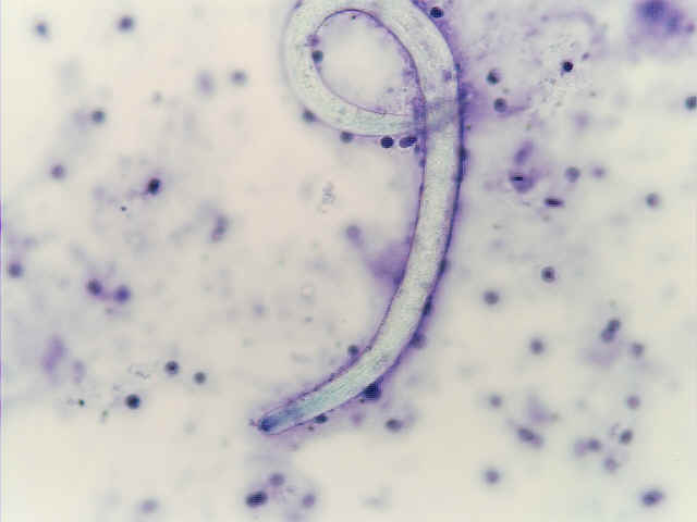
- Worms may be found in airways, alveoli and blood vessels
Microscopic (histologic) images


