Table of Contents
Definition / general | Case reports | Gross images | Microscopic (histologic) description | Microscopic (histologic) images | Cytology images | Positive stains | Electron microscopy description | Differential diagnosisCite this page: Weisenberg E. Nodular amyloid (amyloidoma). PathologyOutlines.com website. https://www.pathologyoutlines.com/topic/lungnontumoramyloidnodular.html. Accessed December 25th, 2024.
Definition / general
- Rare, presents as asymptomatic nodule
- Usually in asymptomatic elderly with nodule on chest Xray, but no evidence of systemic disease
- Good prognosis; typically doesn't progress to lymphoproliferative disorders
- Nodular involvement may be associated with systemic involvement
Case reports
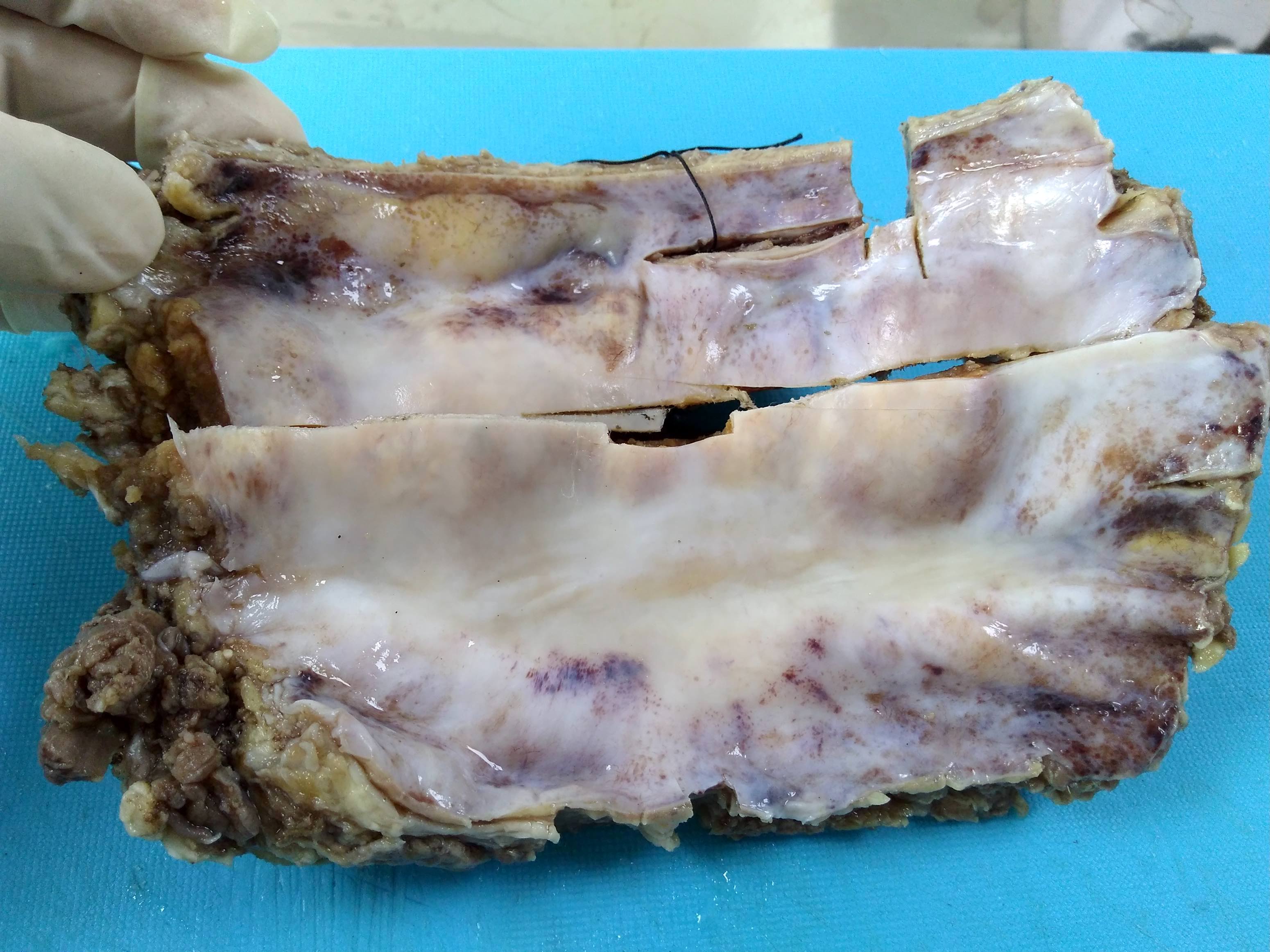
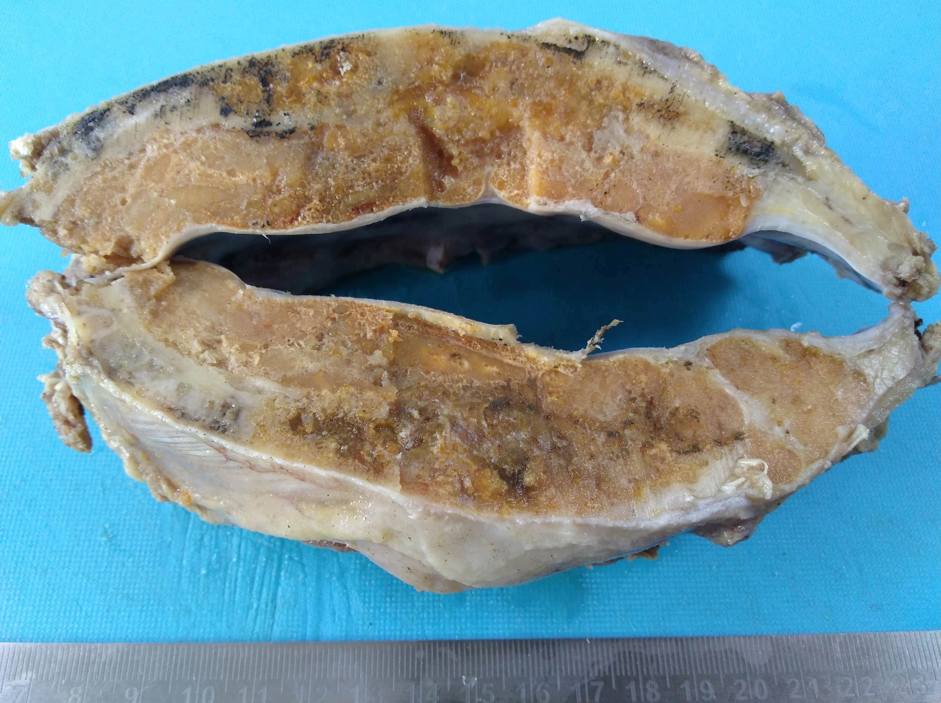
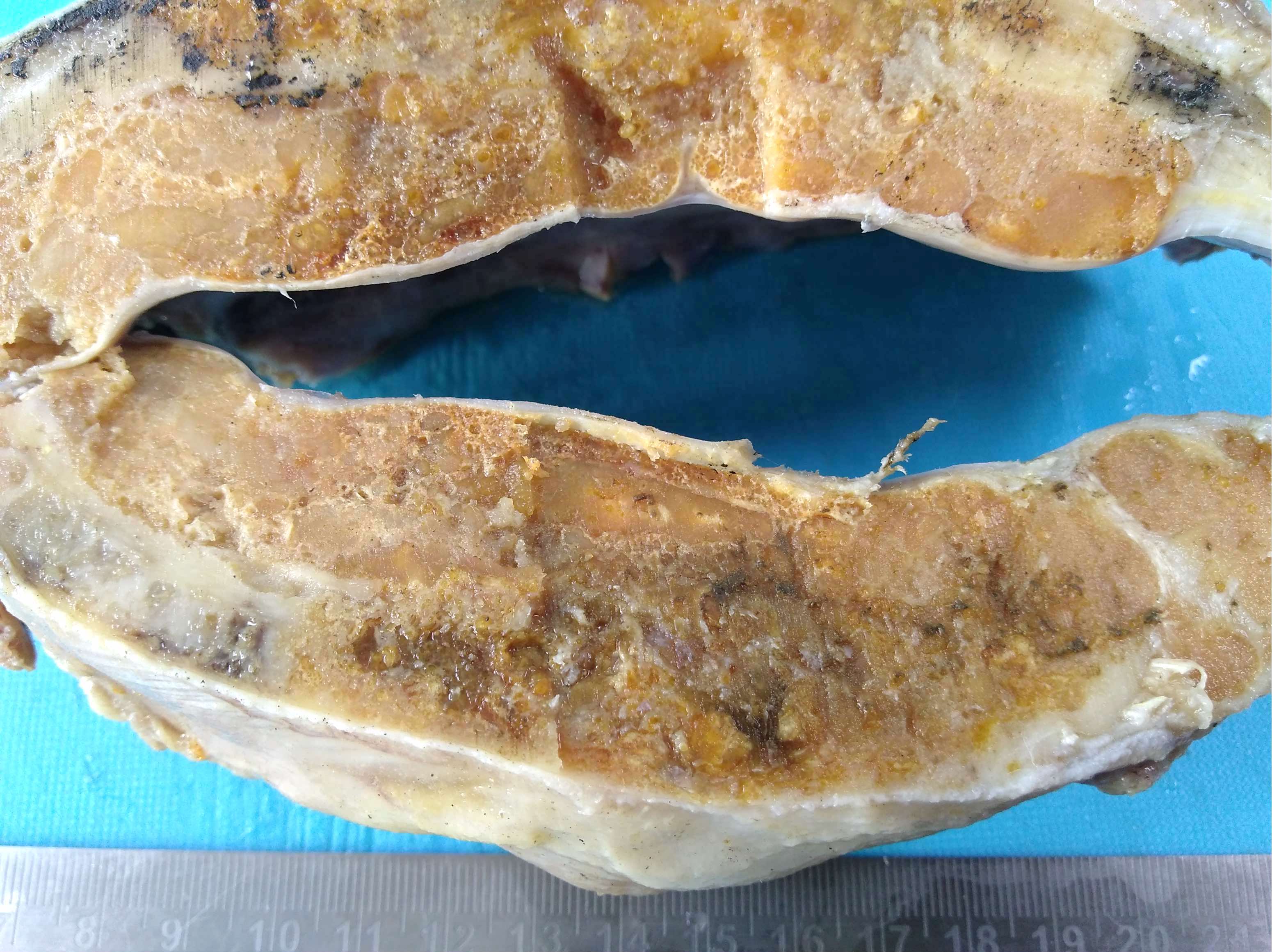
- 85 year old woman with asymptomatic pulmonary nodules (Case #197)
Gross images
Microscopic (histologic) description
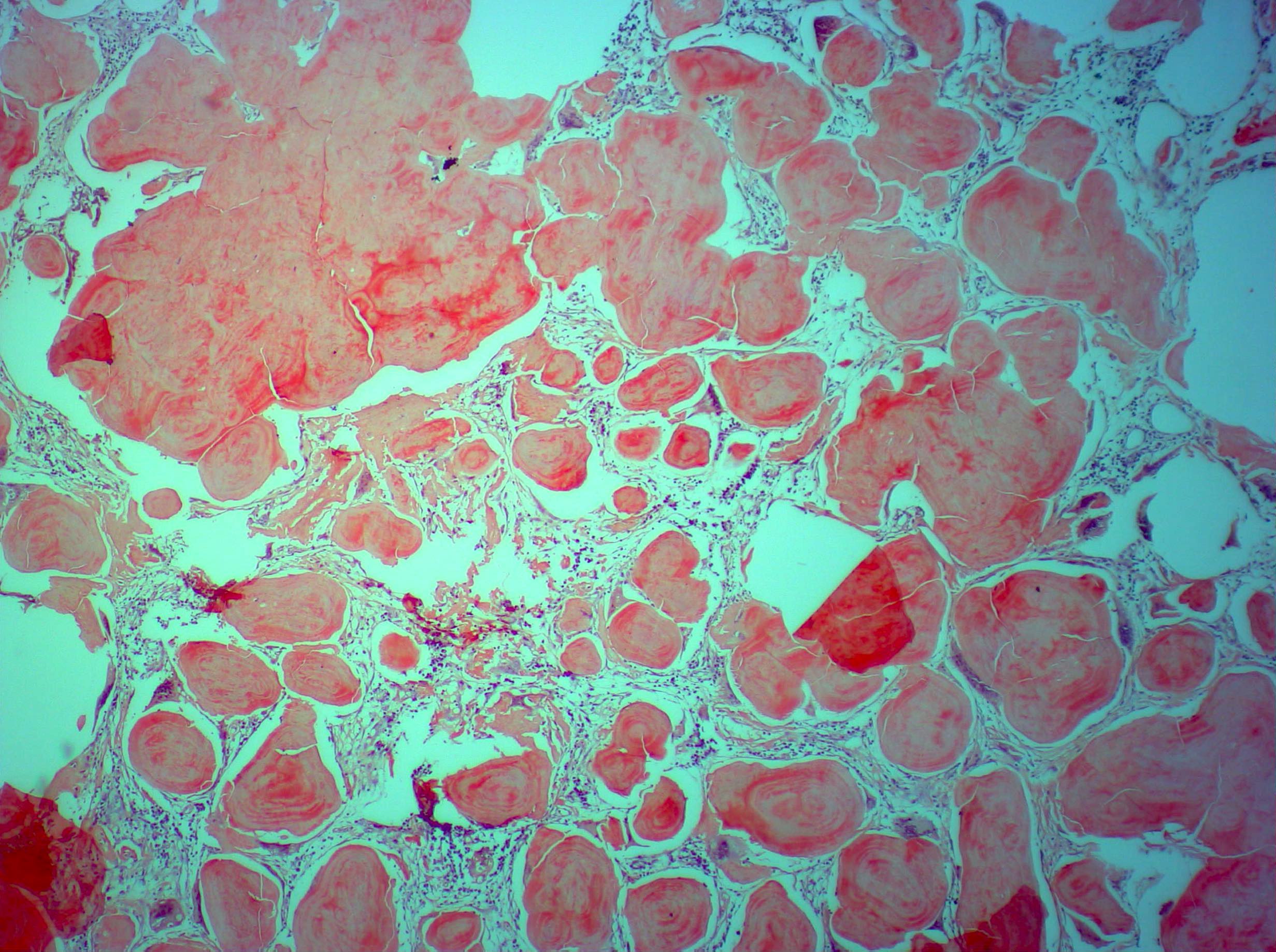
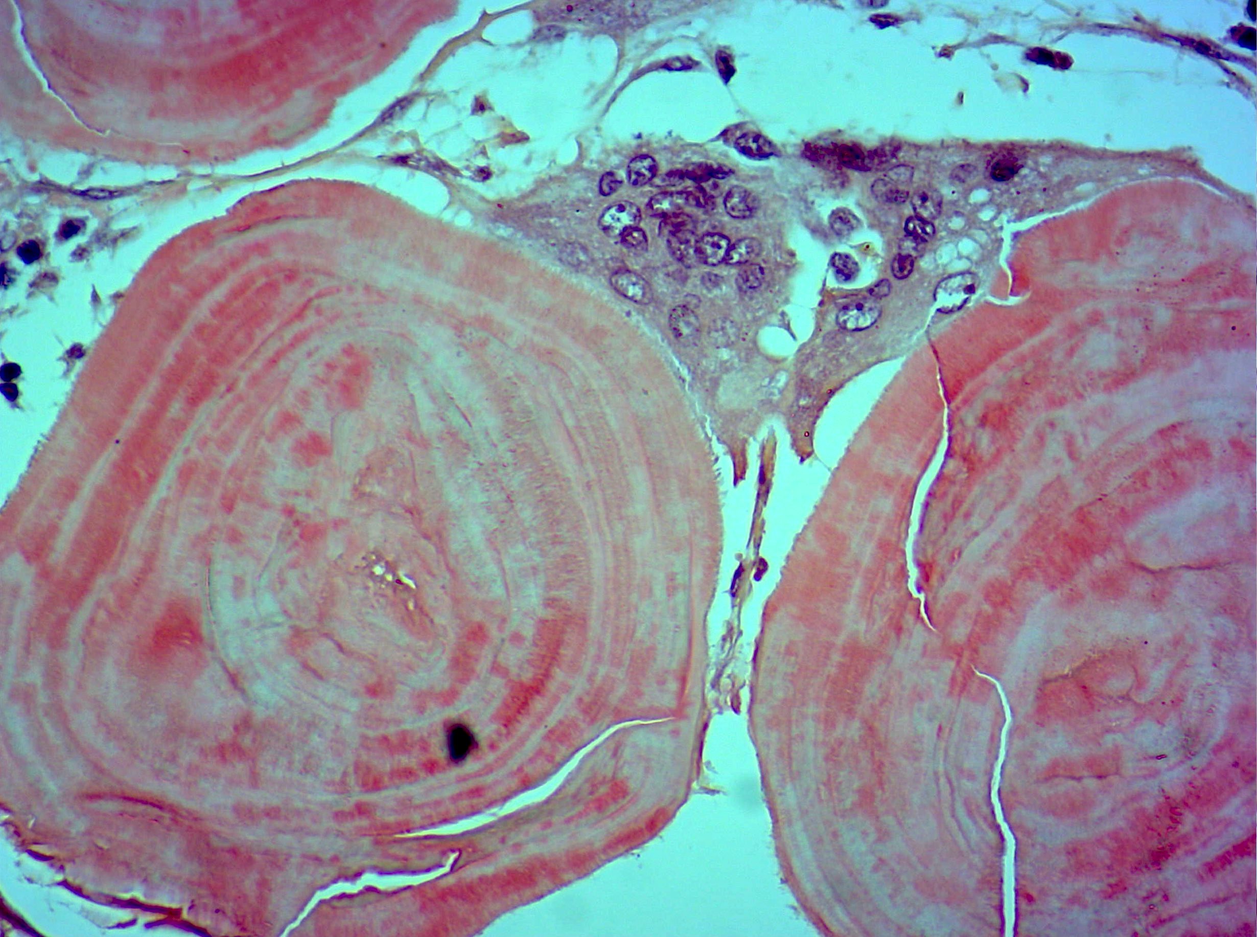
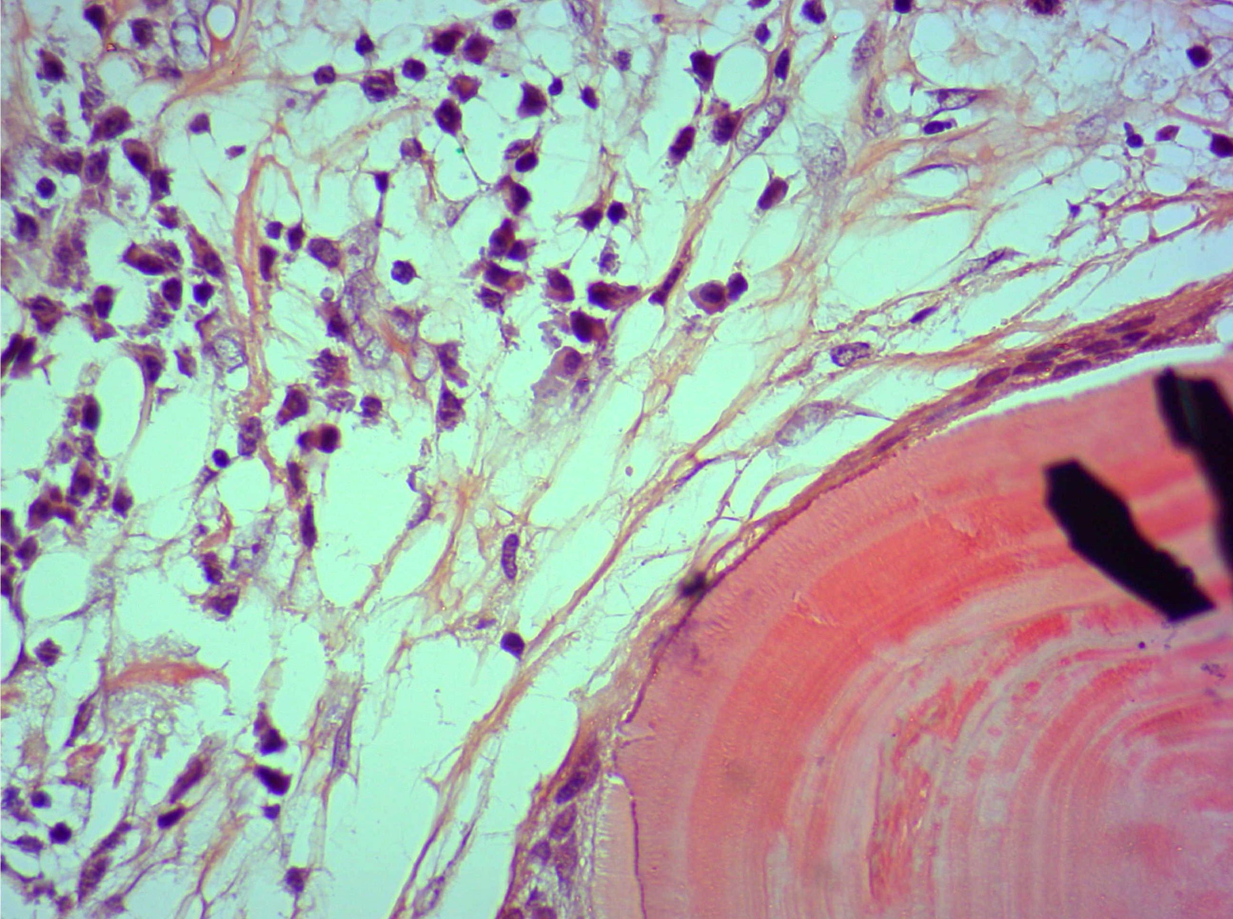
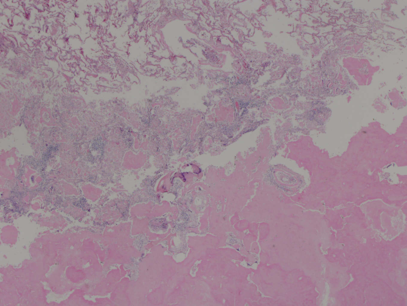
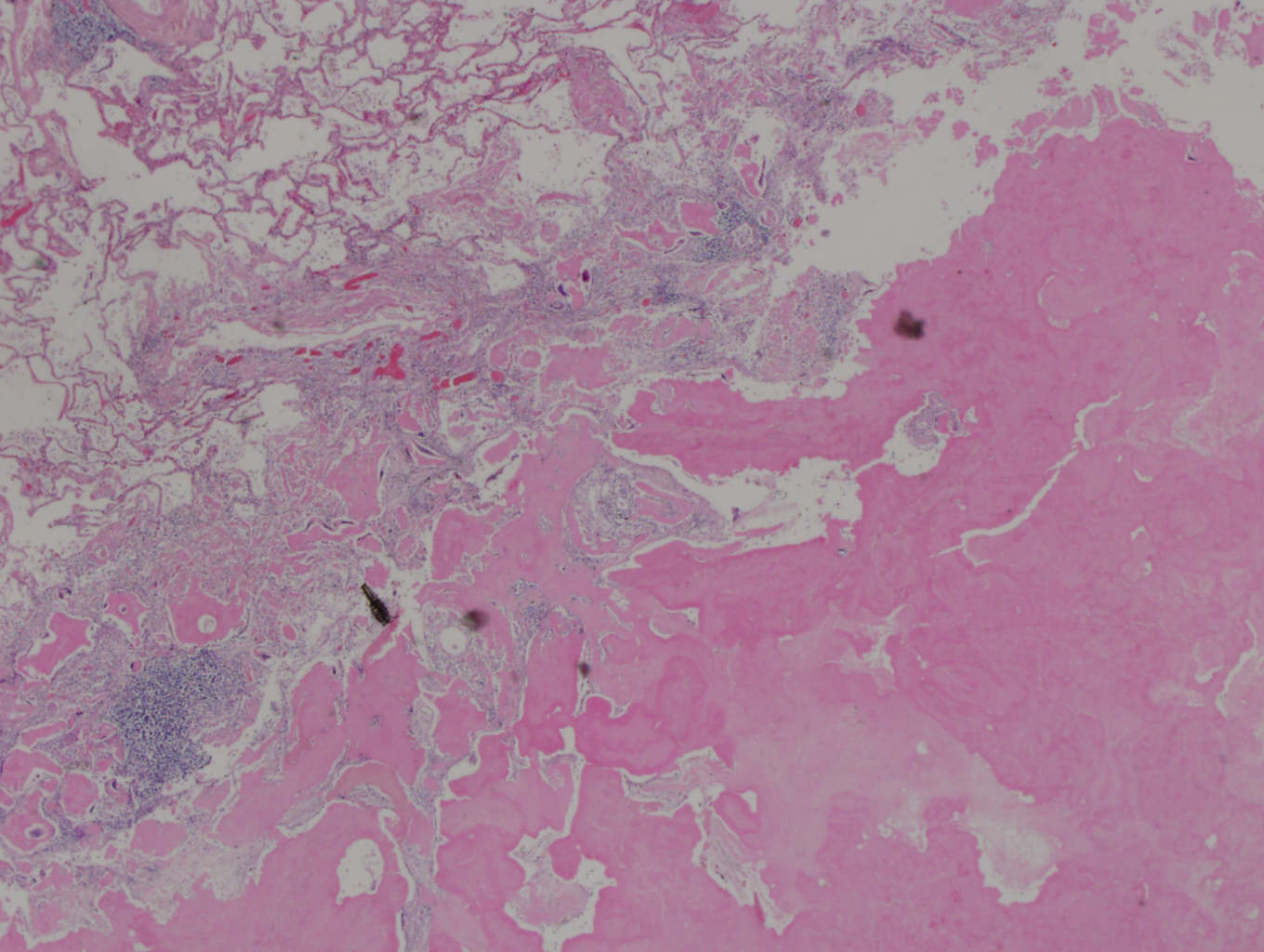
- Well circumscribed amyloid, often with lymphocytes (T cells) and plasma cells
- Granulomatous reaction to amyloid common, often calcification and ossification
Microscopic (histologic) images
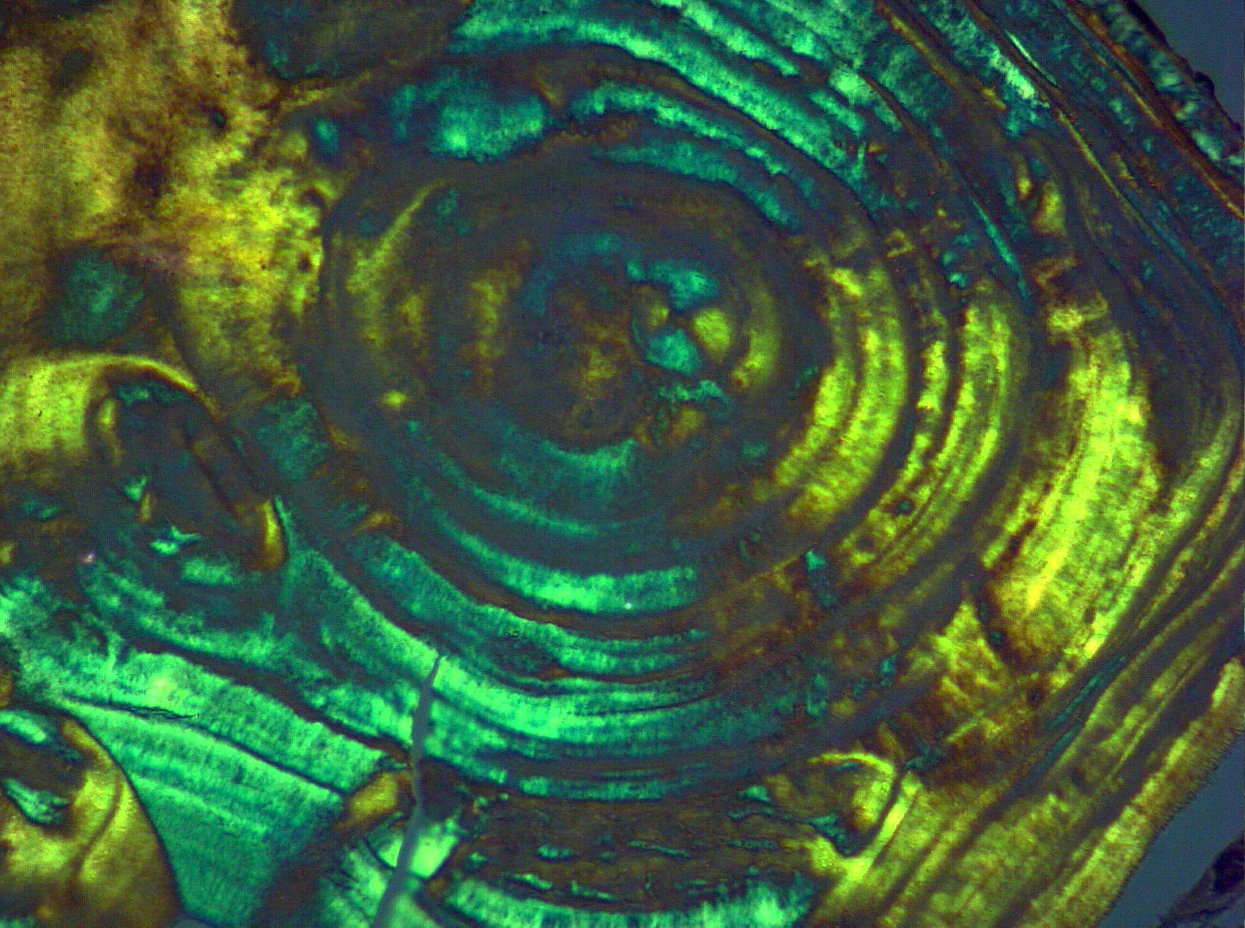
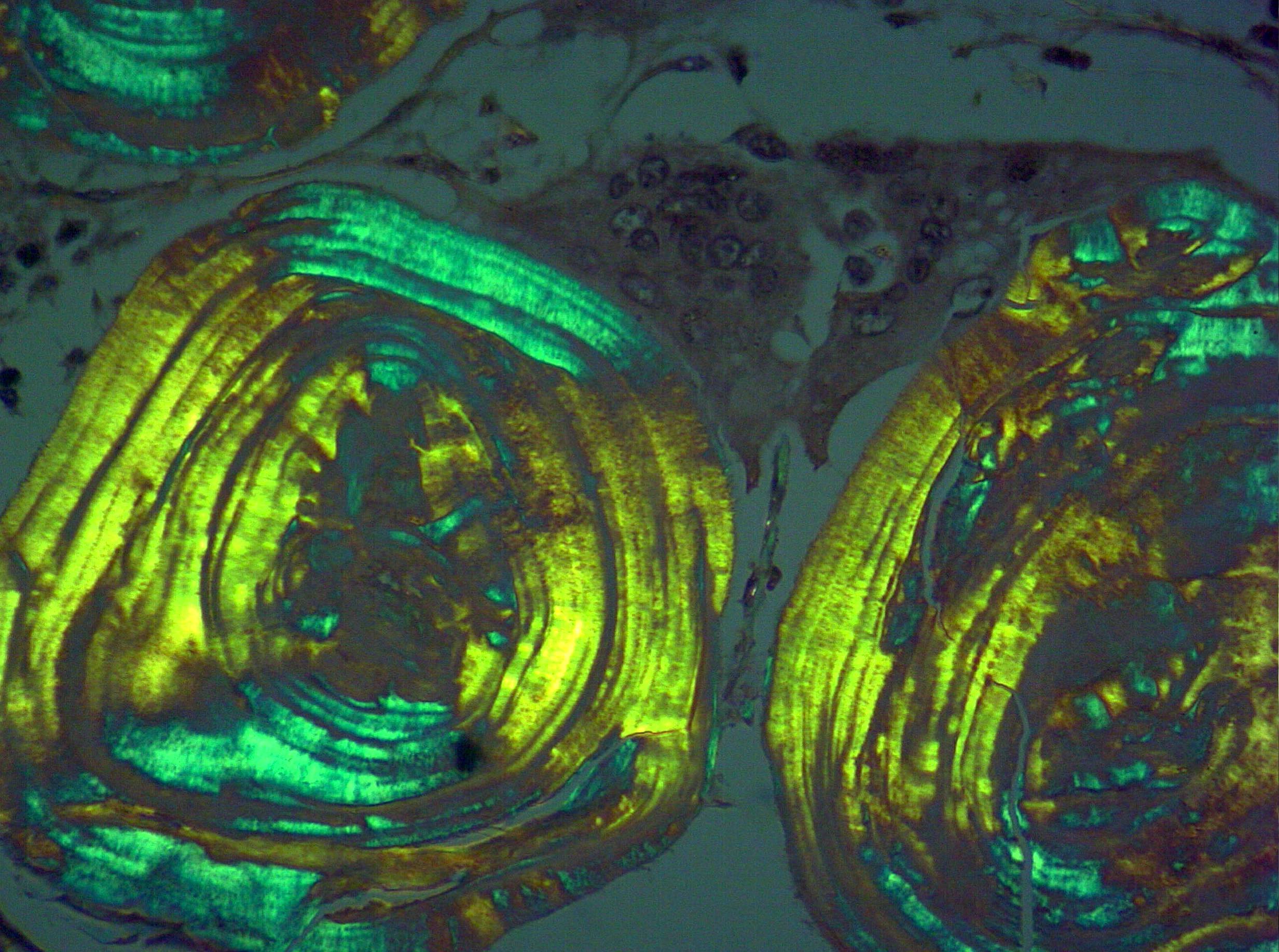
Contributed by Dr. Sajna V.M. Kutty, MVR Cancer Centre and Research Institute, Kozhikode, Kerala, India
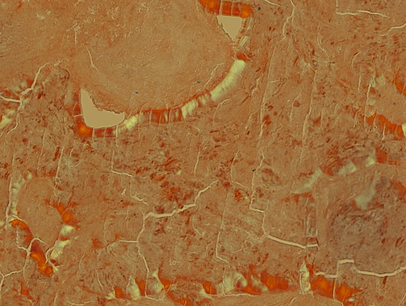
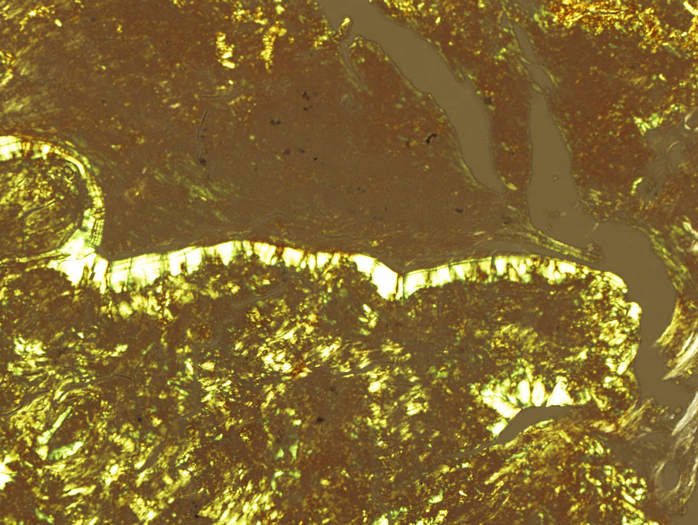
Case #197
Cytology images
Positive stains
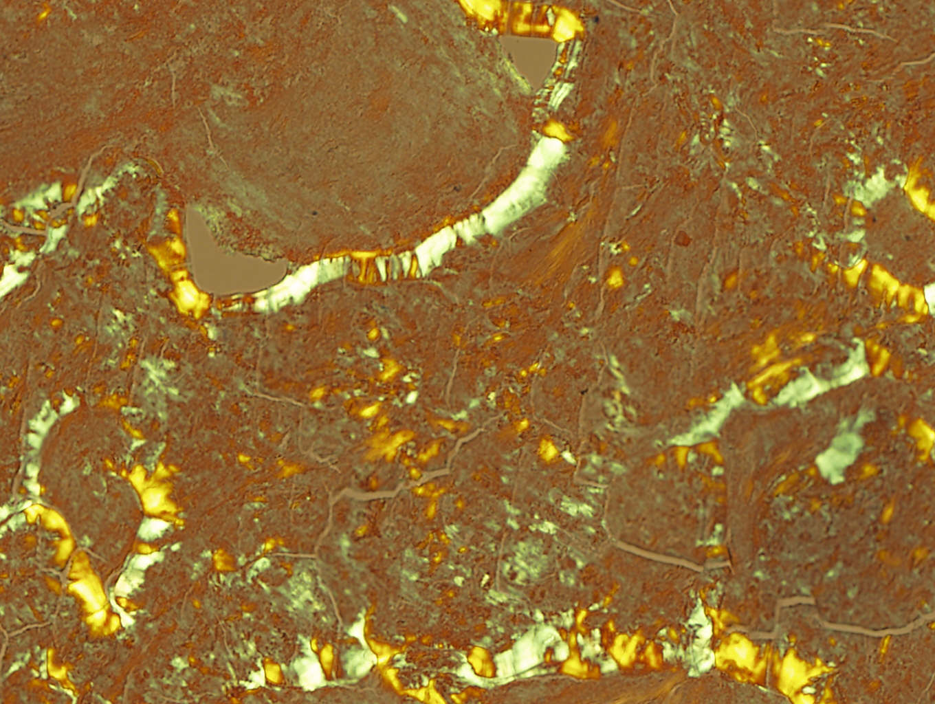
- Congo red staining: glassy, salmon pink amorphous material with apple green birefringence under polarized microscopy
- Clonal restriction of plasma cells (kappa or lambda staining, not both)
Electron microscopy description
- Amyloid fibrils
Differential diagnosis
- Primary pulmonary lymphomas with amyloid production: < 1% of pulmonary lymphomas have amyloid deposits, usually age 70+ years with marginal zone or SLL / CLL subtypes, often with lymphatic tracking and reactive lymphoid follicles; the presence of evenly distributed lymphocytes in the nodule and invasion of the pleura is specific for lymphoma
- Hyalinizing granuloma: history of exposure to Histoplasma or TB, collagen is Congo red negative


