Table of Contents
Definition / general | Case reports | Treatment | Clinical images | Gross description | Gross images | Microscopic (histologic) description | Microscopic (histologic) images | Positive stains | Negative stains | Electron microscopy description | Differential diagnosisCite this page: Jain D. Undifferentiated embryonal sarcoma. PathologyOutlines.com website. https://www.pathologyoutlines.com/topic/livertumorundiffsarcoma.html. Accessed March 30th, 2025.
Definition / general
- Also called malignant mesenchymoma, mesenchymal sarcoma
- 9 - 15% of pediatric hepatic tumors (#3 after hepatoblastoma and hepatocellular carcinoma) - usually ages 6 - 10 years
- In children, often associated with mesenchymal hamartoma (Pediatr Dev Pathol 2011;14:111), rarely with vaginal embryonal rhabdomyosarcoma (Ann Diagn Pathol 2011;15:250)
- Rare in adults (Cancer 2008;112:2274)
- Appears to be a primitive mesenchymal neoplasm with possible foci of differentiated sarcoma, such as angiosarcoma
- Typically presents with pain, fever, abdominal mass and normal serum AFP (J Pediatr Surg 2008;43:1912)
- Good prognosis (Cancer 2002;94:252) but large tumors may rupture and cause death (J Pediatr Hematol Oncol 2007;29:63)
Case reports
- 10 year old girl with abdominal pain and anorexia (Case of the Week #134)
- 16 year old girl with palpable liver mass, fever and vomiting (Arch Pathol Lab Med 2003;127:e163)
- 74 year old woman with tumor arising from mesenchymal hamartoma (Int J Surg Pathol 2012;20:297)
Treatment
- Complete resection and chemotherapy (J Gastrointest Surg 2007;11:73), possibly liver transplant (J Pediatr Hematol Oncol 2013;35:451)
Gross description
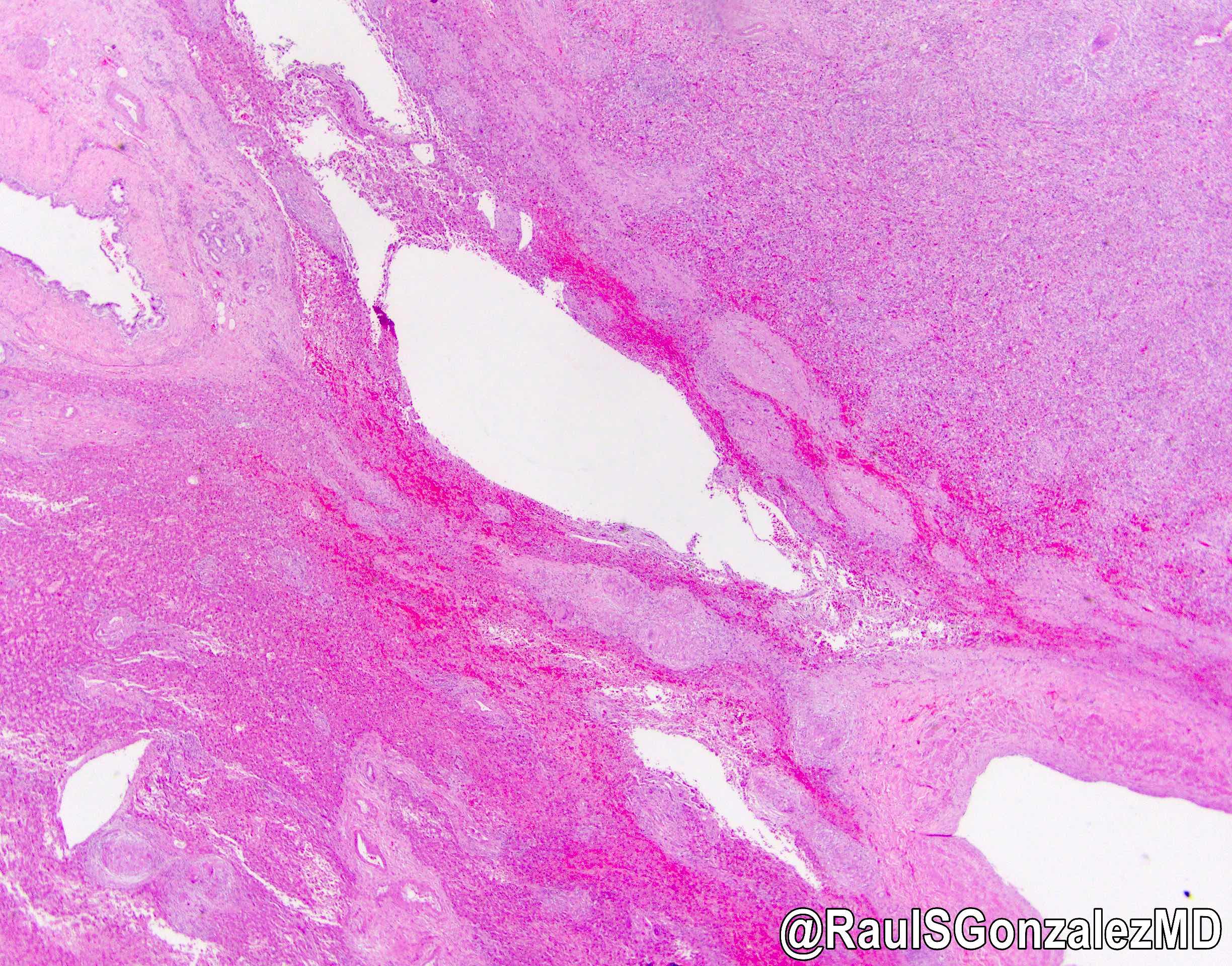
- 10 - 30 cm, solitary, well demarcated, soft tumor with cystic, gelatinous, hemorrhagic and necrotic foci
Gross images
Microscopic (histologic) description
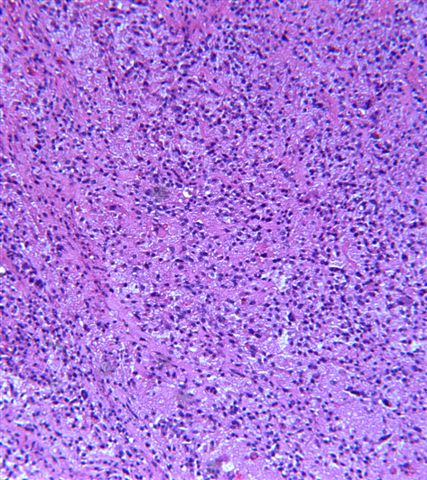
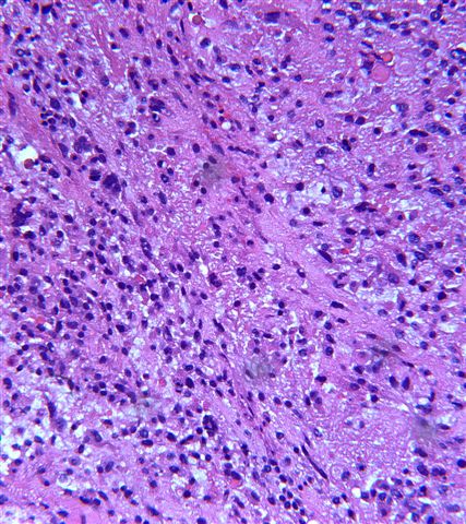
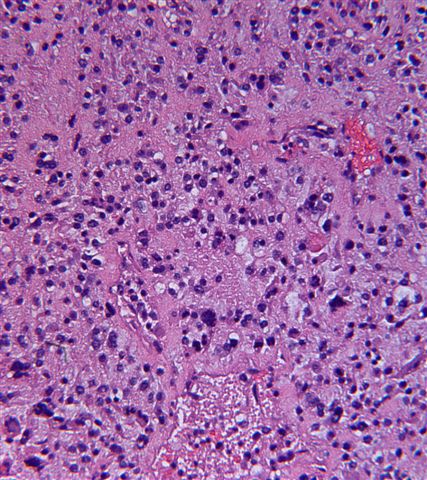
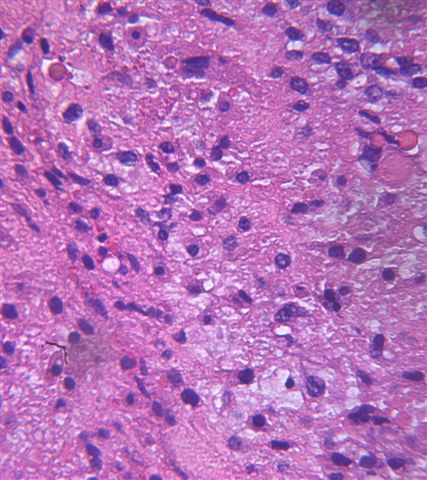
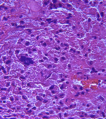
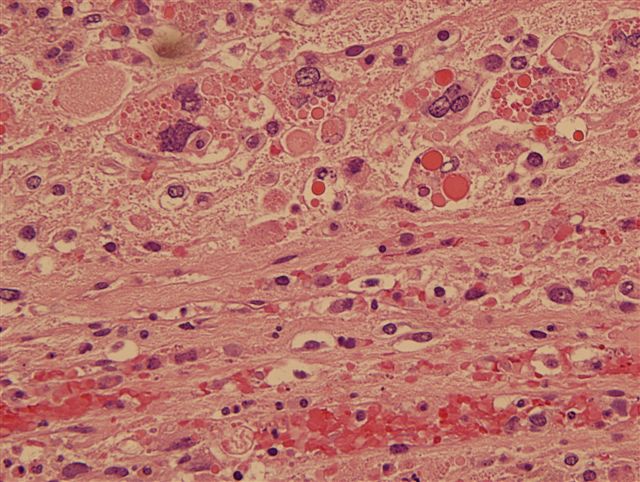
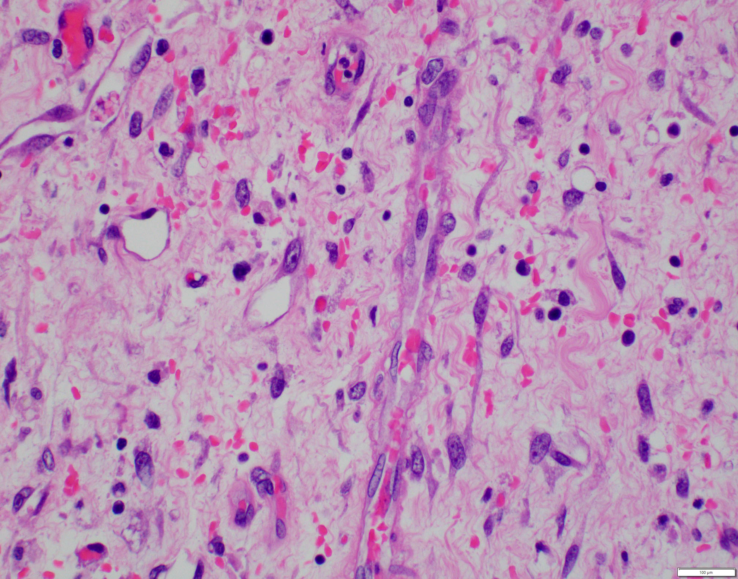
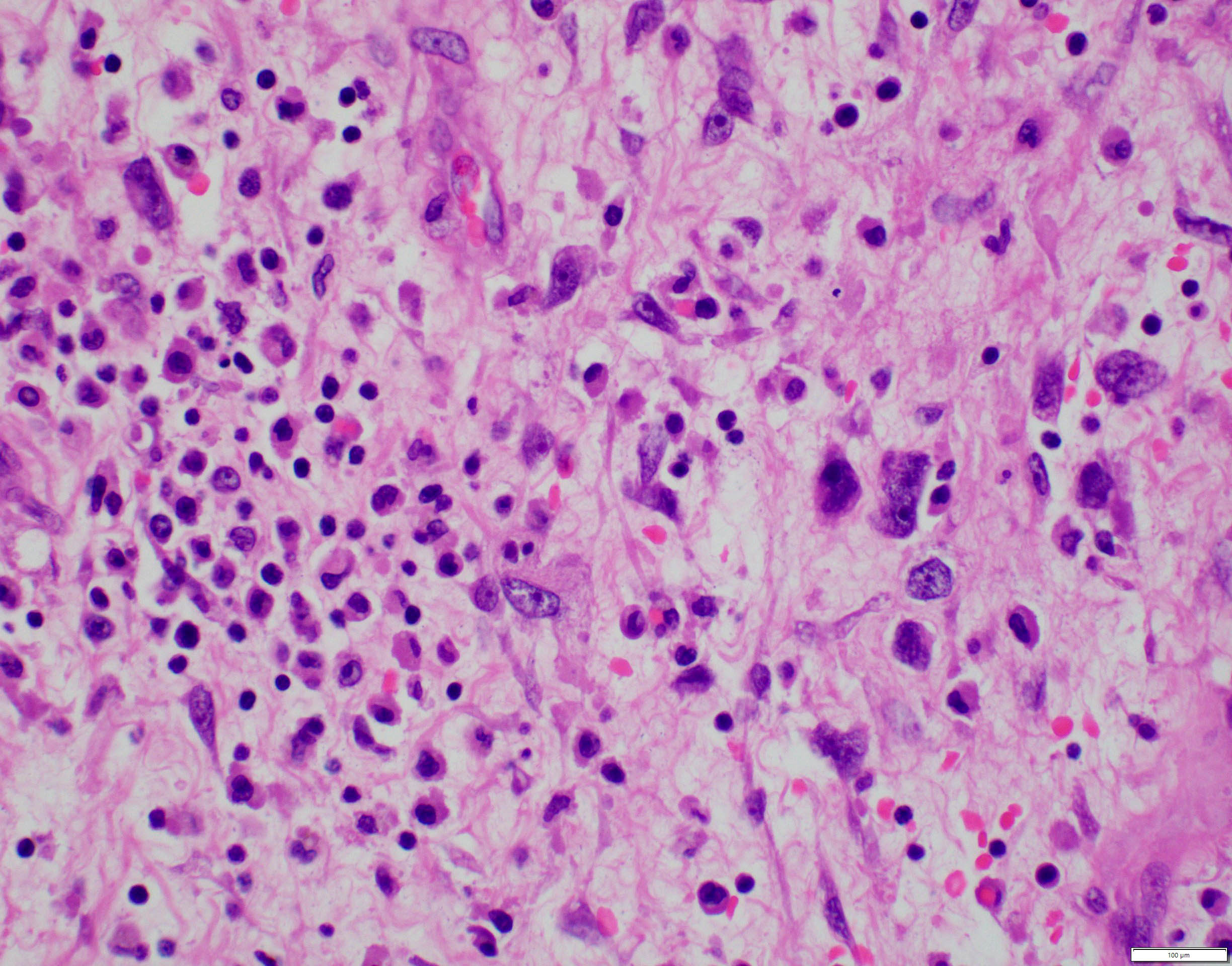
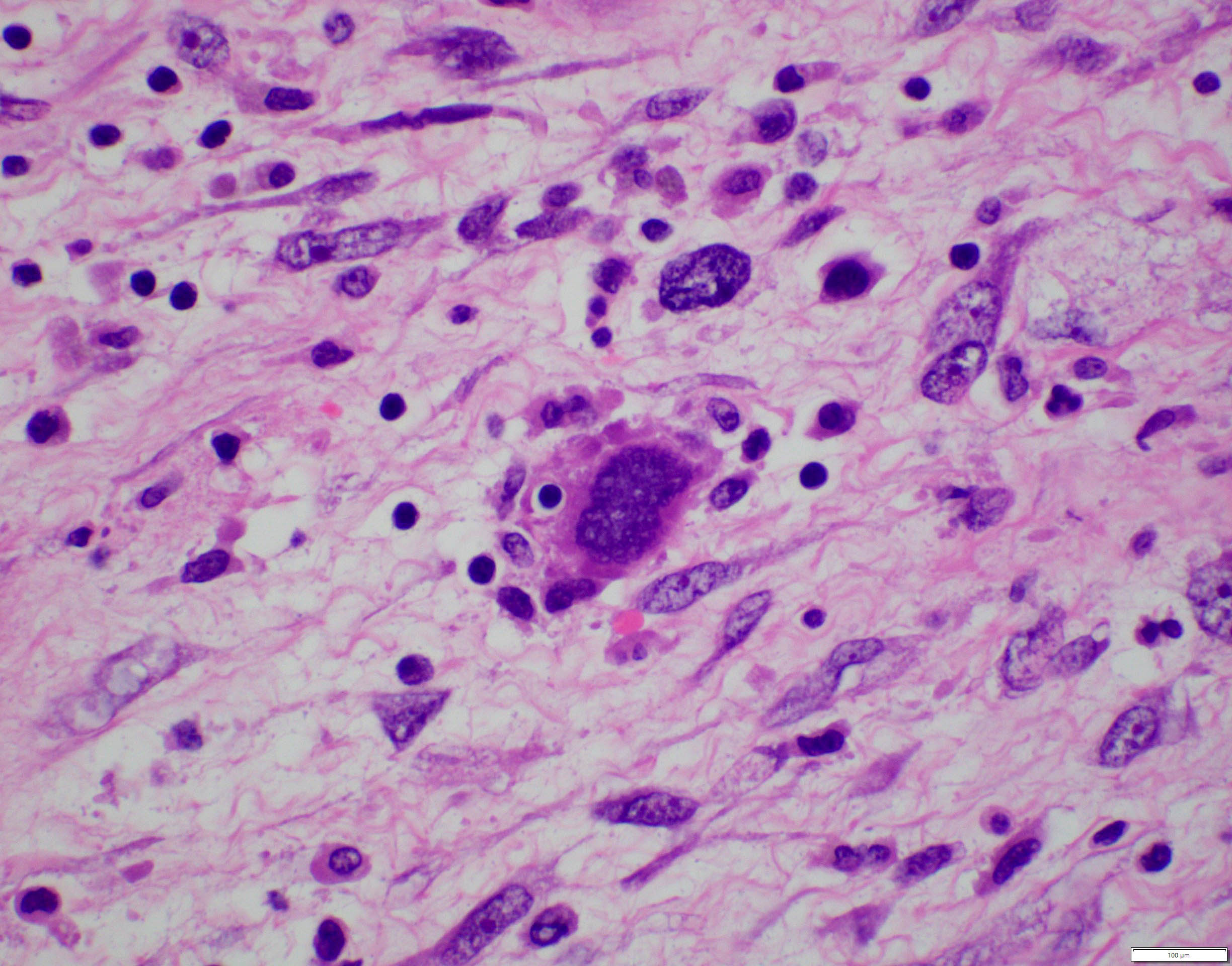
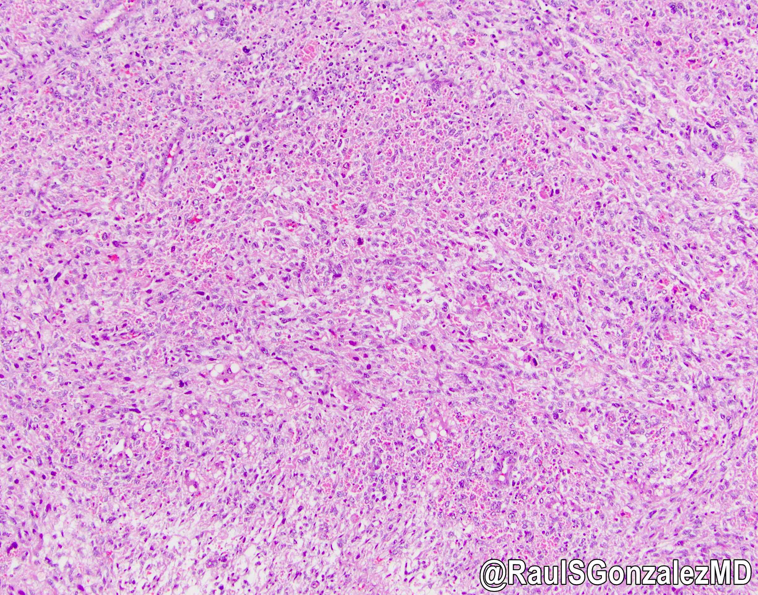
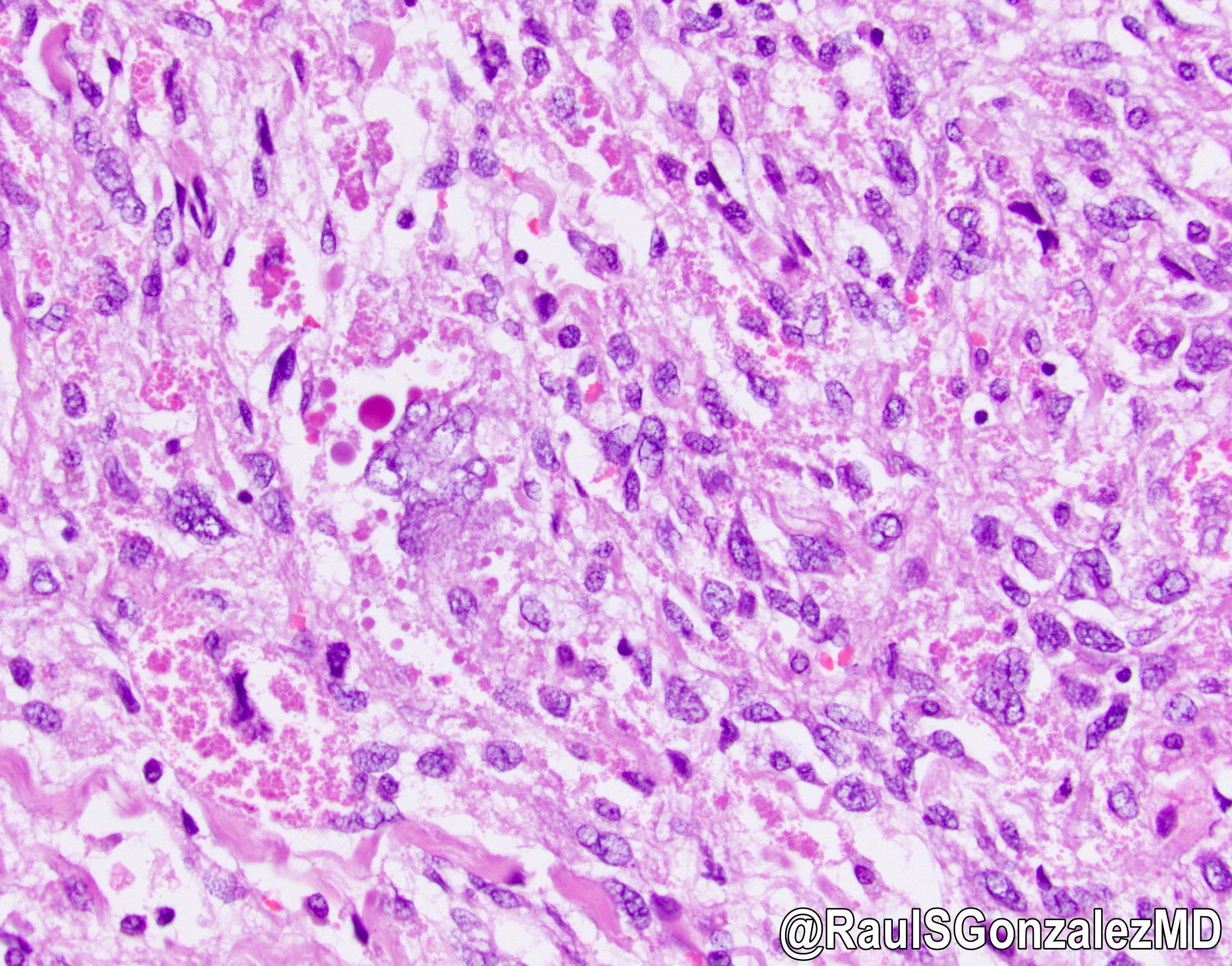
- Variably cellular tumor with anaplastic, spindled / oval cells with prominent hyaline globules and ill defined borders within pseudocapsule
- Nuclei have stippled chromatin, inconspicuous nucleoli
- Variably myxoid stroma with numerous thin walled veins
- Also bizarre tumor cells with prominent eosinophilic cytoplasm and PAS+ diastase resistant hyaline globules
- Extramedullary hematopoiesis common, frequent mitotic activity
- Note that trapped hepatocytes and bile duct structures are present at periphery
- Adults may have partial smooth muscle differentiation (Hum Pathol 2003;34:246)
Microscopic (histologic) images
Positive stains
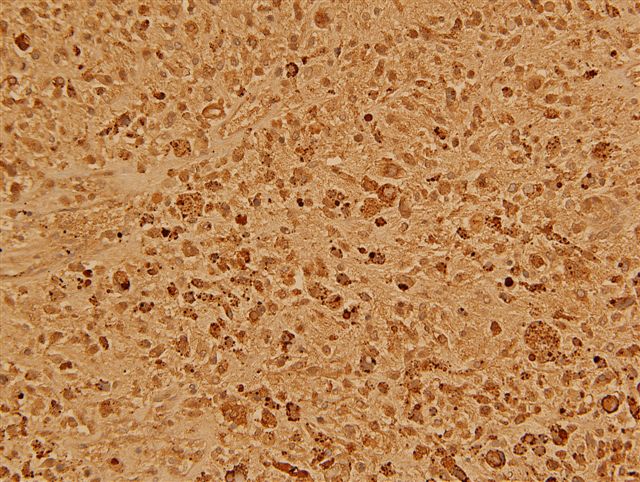
- PAS+ diastase resistant hyaline globules, vimentin, high Ki67 index (Appl Immunohistochem Mol Morphol 2006;14:193)
- Glypican 3 (60%, Hum Pathol 2012;43:695), CD56, paranuclear dot-like CK, BCL2, alpha-1-antitrypsin and alpha-1-antichymotrypsin, variable muscle markers
Negative stains
- Alpha fetoprotein (hyaline globules), keratin, myogenin
Electron microscopy description
- Hyaline globules are lysosomal and possibly apoptotic bodies
Differential diagnosis
- Embryonal rhabdomyosarcoma: usually 2 - 6 years old, myxoid mass extending into bile duct, rhabdomyoblastic differentiation with cytoplasmic cross striations, cambium layer present, no diffuse anaplasia or hyaline globules, myogenin+ and MyoD1+ (Pediatr Dev Pathol 2007;10:89)
- Gastrointestinal stromal tumor: adults, CD117+, DOG1+, CD34+ (usually)
- Hepatoblastoma, mixed form
- Hydatid cyst in endemic areas (J Pediatr Surg 2008;43:E1)
- Mesenchymal hamartoma: usually < 1 year old, cystic, bland tumor cells and no giant cells
- Sarcomatoid hepatocellular carcinoma
- Sclerosing variant of hepatocellular carcinoma: rare in children, has intracellular bile, Mallory-Denk bodies, HepPar1+


