Table of Contents
Definition / general | Essential features | Terminology | ICD coding | Epidemiology | Pathophysiology | Diagrams / tables | Clinical features | Diagnosis | Laboratory | Radiology description | Radiology images | Prognostic factors | Case reports | Treatment | Gross description | Gross images | Microscopic (histologic) description | Microscopic (histologic) images | Positive stains | Negative stains | Videos | Sample pathology report | Differential diagnosis | Board review style question #1 | Board review style answer #1 | Board review style question #2 | Board review style answer #2Cite this page: Abdalla AM, Gonzalez RS. Schistosomiasis. PathologyOutlines.com website. https://www.pathologyoutlines.com/topic/liverschisto.html. Accessed November 28th, 2024.
Definition / general
- Schistosomiasis is a parasitic disease caused by Schistosoma blood flukes
- Liver infection results in fibrosis and obstruction
Essential features
- Most common cause of portal hypertension worldwide
- Adult worms settle in the portal vein and produce thousands of eggs, which leads to inflammation and subsequent periportal fibrosis and portal hypertension (Pathology 2008;40:161)
- Portal fibrosis in schistosomiasis forms thick collagen bundles scattered among fibroblasts
Terminology
- Also known as bilharziasis (first described in 1852 by Theodor Bilharz) and snail fever
ICD coding
- ICD-10: B56.1 - schistosomiasis due to Schistosoma mansoni
Epidemiology
- Schistosomiasis is the second most common human parasitic infection, affecting almost 240 million people worldwide; more than 700 million people live in high risk areas (World Health Organization: Schistosomiasis (Bilharzia) [Accessed 7 August 2024])
- Risk factors include travel to endemic areas and exposure to contaminated freshwater with poor sanitation
- Schistosoma mansoni: Southern and Subsaharan Africa, Nile River Valley in Sudan and Egypt, South America (Brazil, Suriname, Venezuela) and Caribbean
- Schistosoma japonicum: Southeast Asia and China
- Schistosoma mekongi: Cambodia and Laos
- Children are more exposed as they swim in water contaminated with the infectious cercariae
Pathophysiology
- After being excreted in feces into fresh water, Schistosoma eggs hatch and gives rise to ciliated and motile miracidia that penetrate the intermediate host, a snail known as Biomphalaria
- Within the snail, the life cycle progresses through asexual multiplication and the release of cercariae (the infectious forms for humans) into water
- Cercariae penetrate the skin of humans and migrate to the lungs and then the mesenteric veins (J Clin Transl Hepatol 2014;2:212)
- Female worms release eggs, which can travel by portal blood into the liver (Nature 1978;273:609)
- Tissue injury in chronic schistosomiasis is mediated by egg induced granulomas; in the liver, this leads to portal fibrosis (Mem Inst Oswaldo Cruz 2004;99:51)
- Schistosome eggs also release specific substances that directly trigger the production of osteopontin in liver cells
- This protein is known to promote liver fibrosis by activating hepatic stellate cells, causing them to transform into myofibroblasts (Clin Sci (Lond) 2015;129:875)
Diagrams / tables
Clinical features
- Schistosomiasis is the most common cause of portal hypertension worldwide and does not cause cirrhosis
- Within 24 hours after penetration of cercariae, a pruritic rash (swimmer's itch) occurs for 2 - 5 days
- Acute manifestations typically diminish; however, the infection progresses to a chronic phase
- 2 - 8% of patients with heavy chronic infection develop periportal fibrosis (Symmers pipestem fibrosis) and can present with hepatosplenomegaly and esophageal varices, which can lead to gastrointestinal hemorrhage (Int J Parasitol 2014;44:1055)
- Hepatic function is usually preserved
Diagnosis
- Definite diagnosis of schistosomiasis requires identification of schistosome eggs in feces or via tissue sampling
- Stool examination is typically performed with concentration methods (Kato-Katz technique)
- Ultrasound helps determine stage of liver fibrosis (Radiology 1984;153:777)
- Periportal fibrosis appears as an echogenic band surrounding portal vessels extending from the hilum to the periphery of the liver
- In advanced cases, the liver surface may appear nodular
- MRI can also be used (Radiol Case Rep 2016;11:152)
Laboratory
- Direct antigen assay in stool, urine or blood (circulating anodic antigen [CAA] and circulating cathodic antigen [CCA]) (Lancet 2006;368:1106)
- Indirect antibody assay by serology
- Liver function tests are usually normal
Radiology description
- Irregular hepatic surface and septal fibrosis
- Mosaic pattern: echogenic septa outlining polygonal areas of relatively normal liver parenchyma
- Grading of periportal fibrosis thickness on ultrasonography is as follows (Am J Trop Med Hyg 1992;46:403)
- Grade 1: thickness ranges from 3 to 5 mm
- Grade 2: thickness ranges from 5 to 7 mm
- Grade 3: thickness above 7 mm
- S. mansoni usually exhibits portal vein wall thickening with increased echogenicity (bullseye appearance)
Prognostic factors
- Patients with coexisting hepatitis B virus (HBV), hepatitis C virus (HCV), HIV or malaria infections have worse prognoses (Liver 2000;20:281)
- Portal fibrosis may completely regress following curative treatment of the parasites (Mem Inst Oswaldo Cruz 1992;87:129, Am J Trop Med Hyg 1991;44:444)
Case reports
- 26 year old man with massive splenomegaly (Acta Gastroenterol Belg 2018;81:93)
- 38 year old woman with jaundice and a history of tuberculosis (Cureus 2023;15:e35169)
- 39 year old woman with 1 month history of fever and fatigue (Intern Med 2019;58:2495)
- 40 year old woman with intestinal and hepatic schistosomiasis (IDCases 2022;27:e01383)
- 47 year old man with abnormal liver enzymes and ultrasound (Clin Case Rep 2020;8:1522)
Treatment
- Praziquantel is the treatment of choice for all species of schistosomiasis
Gross description
- Liver appears enlarged and nodular but with normal acinar architecture still preserved (Acta Trop 2008;108:79)
- On cut surface, periportal fibrosis (pipestem fibrosis) forms large plaques of hard white fibrous tissue against a background of normal parenchyma
- Portal tract enlargement and a stellate appearance may be observed
Microscopic (histologic) description
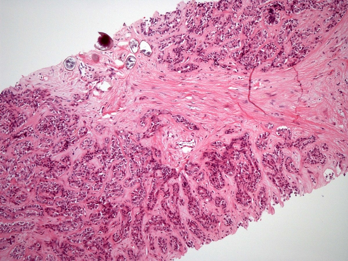
- Early stages show portal inflammation with eosinophils, Kupffer cell hyperplasia and focal hepatocyte necrosis; ova are usually absent
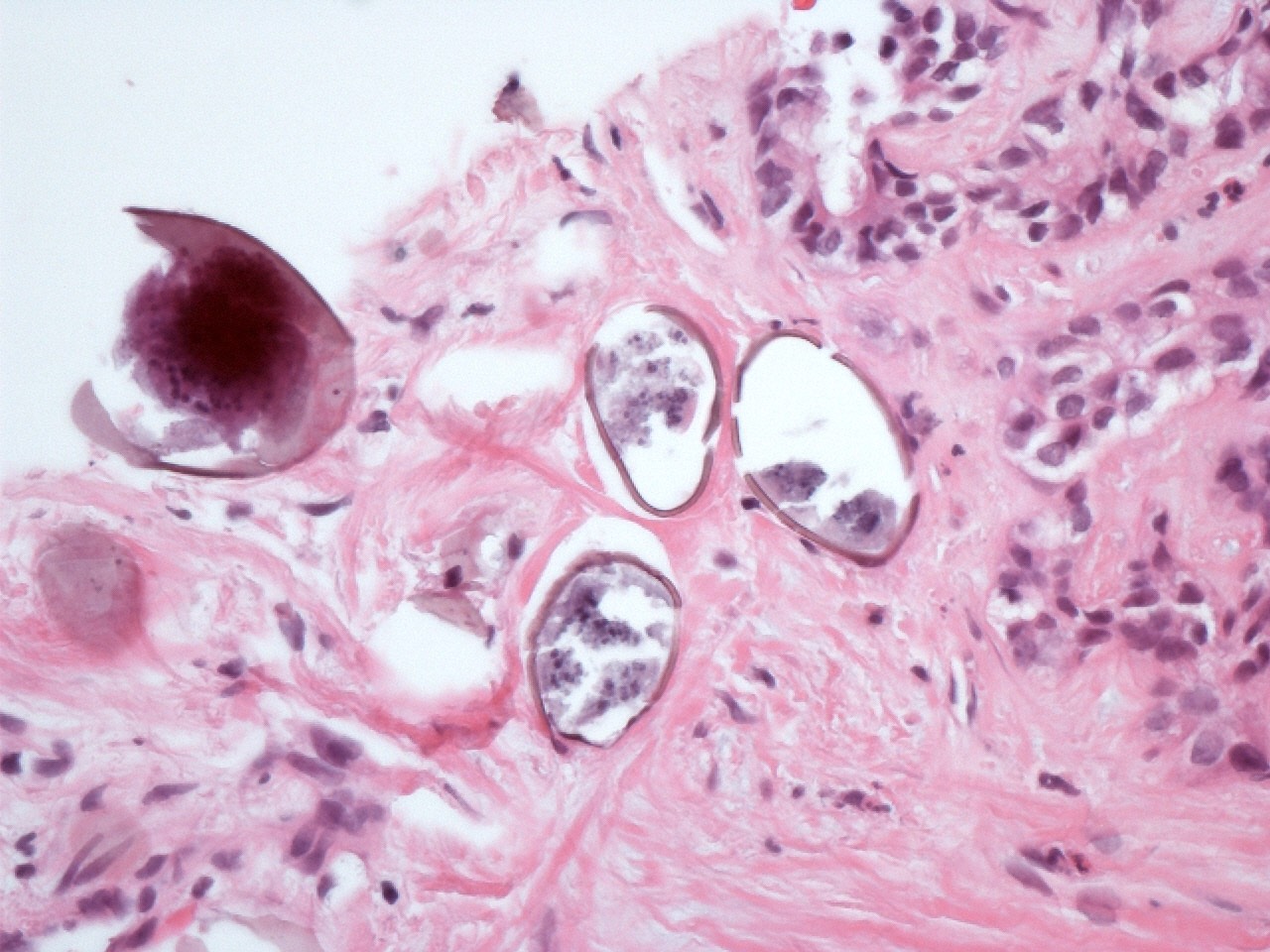
- In chronic disease, there is a granulomatous reaction to the eggs, which may be associated with portal inflammation and giant cells
- Portal tracts become enlarged and densely sclerotic, forming fibrous septa and sometimes sinusoidal fibrosis but cirrhosis does not occur
- Portal veins are obstructed and destroyed, with proliferation of hepatic artery branches
- Fragmentation of muscle fibers of the media of portal veins, most evident on Masson trichrome stain, is a useful indicator for S. mansoni
- Ova may be sparse, rendering their detection difficult; cutting multiple additional tissue sections can assist
- S. mansoni eggs measure 60 x 140 μm and have a lateral spine
- S. japonicum eggs are 70 - 100 μm long and have a minute lateral spine
- Hemozoin pigment (like that in malaria) may be present, particularly in worms within portal veins but also sometimes in macrophages (Burt: MacSween's Pathology of the Liver, 8th Edition, 2023)
Microscopic (histologic) images
Positive stains
- S. mansoni eggs stain positive for acid fast stain Ziehl-Neelsen (Am J Trop Med Hyg 1954;3:1066)
Negative stains
Videos
Schistosomiasis in liver
Sample pathology report
- Liver, orthotopic transplantation:
- Liver with dense portal / periportal fibrosis and rare Schistosoma eggs with associated inflammatory reaction (see comment)
- Negative for malignancy
- Comment: The patient's reported history of noncirrhotic portal hypertension is noted. The findings indicate schistosomiasis as the cause.
Differential diagnosis
- Cirrhosis:
- Patients have portal hypertension clinically and abnormal liver findings on imaging
- Cirrhotic livers demonstrate nodular architecture due to extensive fibrous septa, whereas schistosomiasis does not cause this degree of fibrosis
- Ova are not present
- Hepatoportal sclerosis:
- Patients have portal hypertension
- Portal vein intimal fibrosis and delicate septa with phlebosclerosis and atrophic liver parenchyma
- Ova are not present
- Malaria:
- Hemozoin pigment can be seen in macrophages in malaria infection as well as schistosomiasis
- Ova are not present
- Other trematodes may infect the liver and biliary tract, including Clonorchis sinensis, Opisthorchis species and Fasciola hepatica
- These all have their own distinctive appearance on H&E
Board review style question #1
Which of the following statements is true regarding the microscopic description of liver schistosomiasis?
- Granulomatous inflammation and periportal fibrosis are commonly seen
- It is characterized by the presence of atypical hepatocytes
- It shows diffuse fatty infiltration of hepatocytes
- There is loss of the hepatic lobular architecture
Board review style answer #1
A. Granulomatous inflammation and periportal fibrosis are commonly seen. Liver schistosomiasis is characterized by granulomatous inflammation around the trapped schistosome eggs and periportal fibrosis. Answer B is incorrect because hepatocyte atypia does not occur. Answer C is incorrect because hepatocyte steatosis does not occur. Answer D is incorrect because the hepatic lobular architecture remains intact in this condition.
Comment Here
Reference: Schistosomiasis
Comment Here
Reference: Schistosomiasis
Board review style question #2
Hepatic schistosomiasis is the most common cause worldwide of which of the following clinical findings?
- Abnormal liver appearance on ultrasound
- Excretion of ova
- Gastrointestinal hemorrhage
- Portal hypertension
Board review style answer #2
D. Portal hypertension. Liver schistosomiasis is the most common cause of portal hypertension worldwide, despite not causing cirrhosis. Answers A, B and C are incorrect because while these symptoms can occur in patients with hepatic schistosomiasis, it is not the most common cause for them.
Comment Here
Reference: Schistosomiasis
Comment Here
Reference: Schistosomiasis


