Table of Contents
Definition / general | Essential features | Terminology | Sites | Etiology | Clinical features | Diagnosis | Radiology description | Treatment | Clinical images | Gross description | Microscopic (histologic) description | Microscopic (histologic) images | Positive stains | Sample pathology report | Differential diagnosis | Board review style question #1 | Board review style answer #1Cite this page: Gonzalez RS. Peliosis hepatis. PathologyOutlines.com website. https://www.pathologyoutlines.com/topic/liverpeliosishepatis.html. Accessed April 2nd, 2025.
Definition / general
- Multiple blood filled cysts in liver, with several potential causes
Essential features
- Blood filled hepatic spaces of various sizes, usually discovered by radiology or at autopsy
- Several possible etiologies, including Bartonella henselae or B. quintana infection, which also cause spindle cell / vascular proliferation
Terminology
- Two types:
- Peliosis hepatis refers to bland blood filled spaces of various etiologies
- Bacillary peliosis refers to hepatic spindle cell proliferation with small blood vessels and blood filled spaces secondary to Bartonella infection
- Peliosis should not be confused with lipopeliosis
Sites
- Peliosis may occur in liver or spleen
Etiology
- Associated with steroids (anabolic, contraceptive), danazol, azathioprine, vinyl chloride, Bartonella infection in HIV+ patients (Forensic Sci Int 2005;149:25, N Engl J Med 1997;337:1876)
- May be due to hepatocellular necrosis or veno-occlusive disease
Clinical features
- May be asymptomatic or manifest as jaundice, portal hypertension, liver failure, liver rupture or hepatomegaly
- Patients may also have liver or renal transplant, Castleman disease, nodular regenerative hyperplasia, leukemia / lymphoma (Arch Pathol Lab Med 2004;128:1283)
Diagnosis
- Usually incidental finding at autopsy
Radiology description
- Blood filled spaces visible on imaging
Treatment
- Withdrawal of causative drug, if applicable
- Erythromycin or doxycycline for Bartonella infections
Gross description
- Honeycomb liver with multiple round, red purple, blood filled spaces of varying sizes
Microscopic (histologic) description
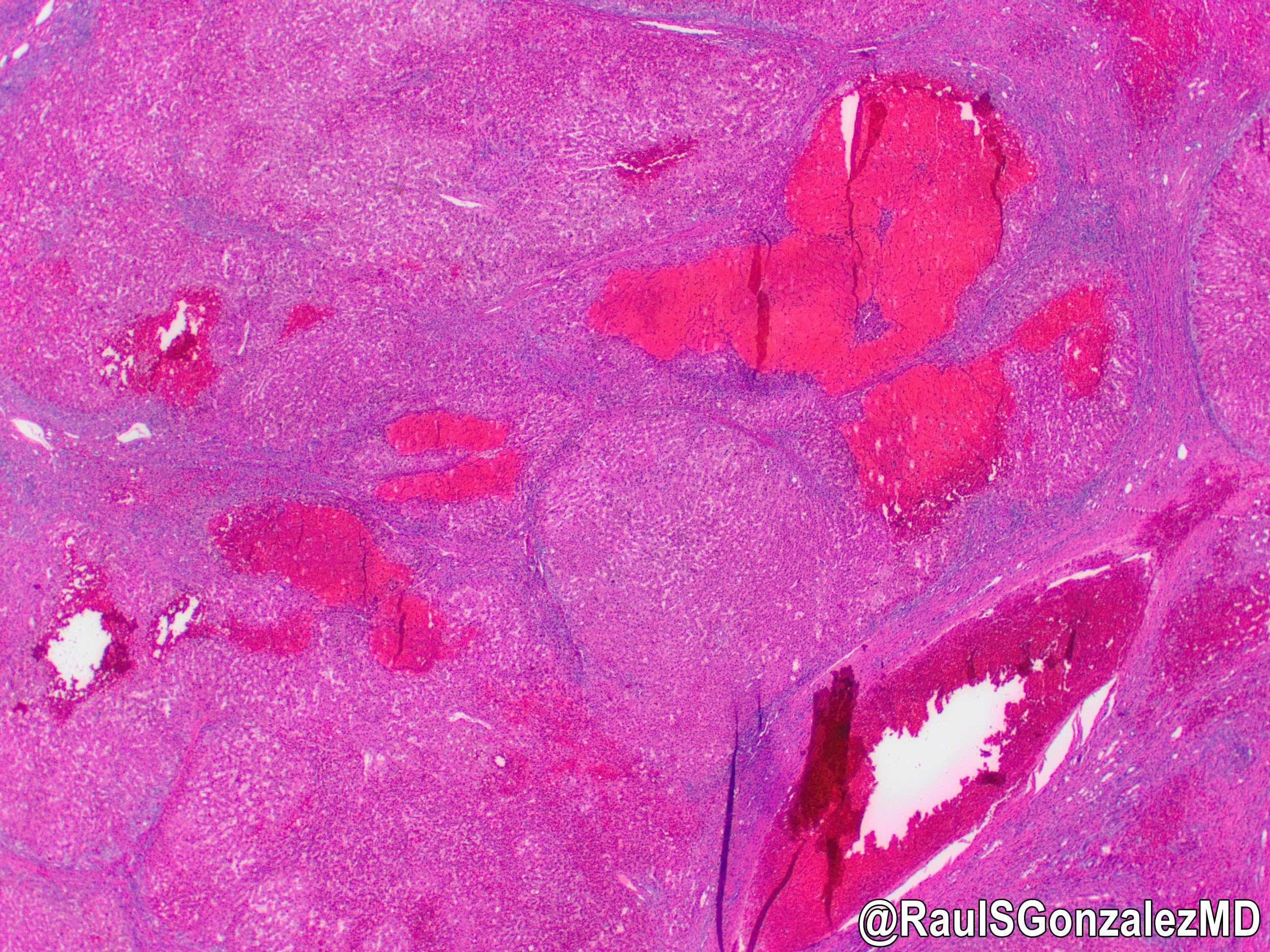
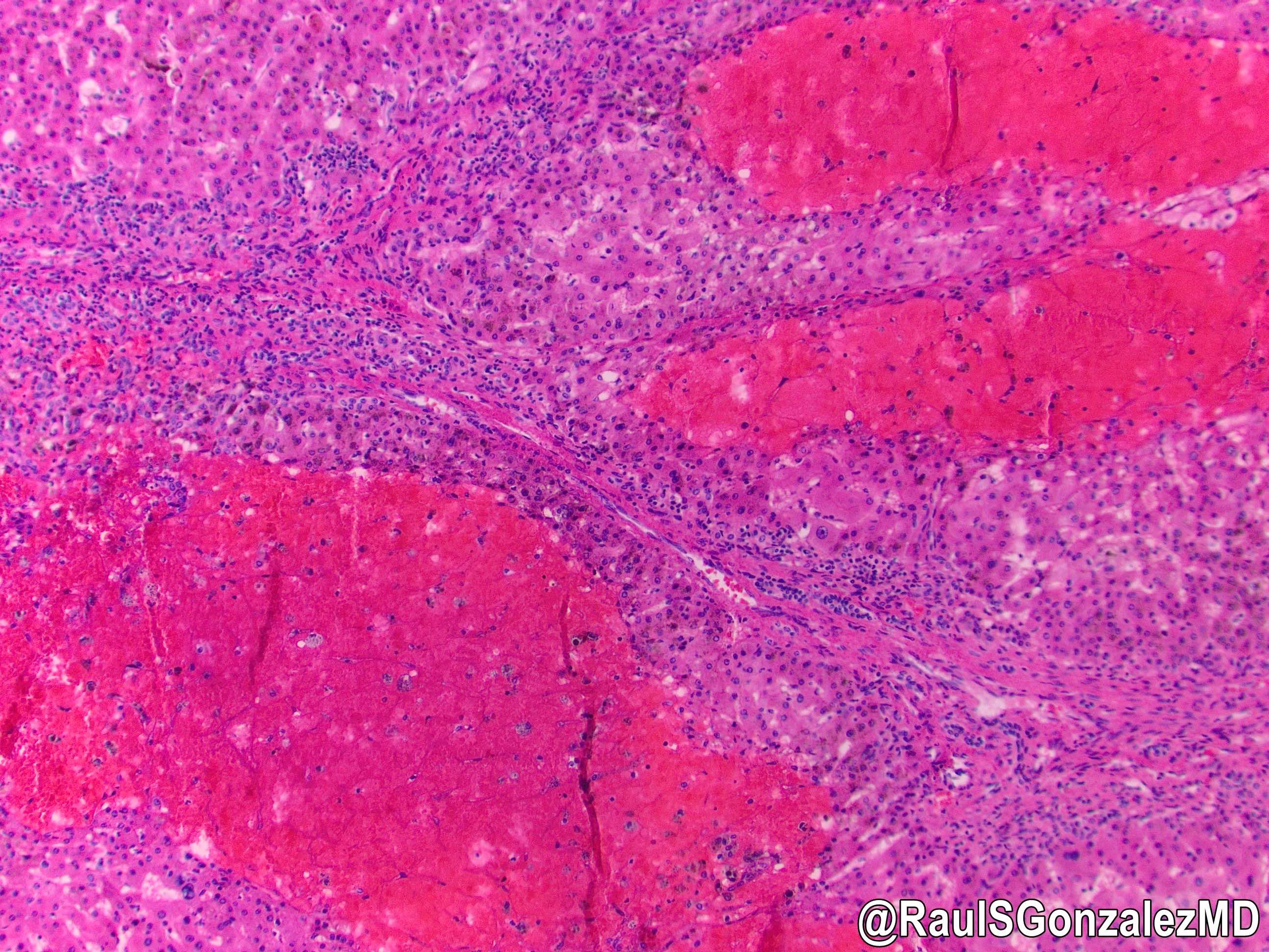
- Blood lakes of various sizes within the liver, which may be continuous
- In phlebectatic subtype, spaces are lined by endothelium and central veins are dilated
- In parenchymal subtype, spaces are not lined and parenchyma has variable hemorrhagic necrosis (Arch Pathol 1964;77:159)
- May have calcifications (J Ultrasound Med 1996;15:257)
- Background liver may show sinusoidal dilation
- Bacillary peliosis may show angiomatosis, with spindle cell process accompanied by blood vessel proliferation
Microscopic (histologic) images
Positive stains
- Warthin-Starry in cases due to Bartonella infection
Sample pathology report
- Liver, biopsy:
- Liver parenchyma with large blood filled cystic spaces and background sinusoidal dilation (see comment)
- Comment: The findings are compatible with peliosis hepatic, which has many potential etiologies. Trichrome and iron stains are unremarkable.
Differential diagnosis
- Hemangioma:
- Discrete lesion with fibrous tissue in background, linking the engorged spaces
- Kaposi sarcoma:
- Can mimic bacillary peliosis but vessels are less well formed
- HHV8 positive
- Peliotic change in hepatocellular carcinoma: background parenchyma clearly malignant (Oncol Lett 2010;1:17)
Board review style question #1
- A 45 year old man presents to the emergency department complaining of abdominal pain but collapses and dies before receiving care. Autopsy shows several findings, including a liver studded with numerous blood filled lakes. Microscopically, these spaces are associated with a vascular / spindle cell proliferation. Which of the following other conditions did the patient likely have?
- Blue rubber bleb nevus syndrome
- Congestive heart failure
- Ganglioneuromatosis
- Hereditary hemorrhagic telangiectasia
- HIV infection
Board review style answer #1


