Table of Contents
Definition / general | Essential features | ICD coding | Epidemiology | Pathophysiology | Diagrams / tables | Clinical features | Diagnosis | Laboratory | Radiology description | Prognostic factors | Case reports | Treatment | Gross description | Microscopic (histologic) description | Microscopic (histologic) images | Positive stains | Electron microscopy images | Molecular / cytogenetics description | Videos | Sample pathology report | Differential diagnosis | Additional references | Board review style question #1 | Board review style answer #1 | Board review style question #2 | Board review style answer #2Cite this page: De Michele S, Lagana SM. COVID-19. PathologyOutlines.com website. https://www.pathologyoutlines.com/topic/liverCOVID19.html. Accessed March 30th, 2025.
Definition / general
- Severe acute respiratory syndrome coronavirus 2 (SARS-CoV-2) primarily causes pulmonary disease but has been implicated in hepatic injury
- Coronavirus disease 2019 (COVID-19) viral hepatitis is defined by the clinical, laboratory and histopathological hepatic changes secondary to SARS-CoV-2 infection
Essential features
- Despite being primarily a pulmonary disease, SARS-CoV-2 infection has been shown to affect extrapulmonary sites, including the liver
- COVID-19 viral hepatitis is defined by the clinical, laboratory and histological liver abnormalities in the setting of real time reverse transcription polymerase chain reaction (RT PCR) proven SARS-CoV-2 infection
- Common laboratory findings include mild elevation of liver function tests: aspartate aminotransferase (AST) > alanine aminotransferase (ALT)
- Histologically, nonspecific findings include macrovesicular steatosis, lobular necroinflammation and portal inflammation
- Viral RNA can be detected in liver tissue by qRT PCR in approximately half of patients who die of COVID-19, although mainly at low levels
ICD coding
- ICD-10:
- ICD-11:
Epidemiology
- Liver injury is more common in severe cases (Lancet 2020;395:497)
- Incidence of elevated serum liver enzymes in hospitalized patients with COVID-19 ranges from 14% to 58% (AASLD: Clinical Best Practice Advice for Hepatology and Liver Transplant Providers During the COVID-19 Pandemic [Accessed 25 May 2022])
- Characteristics of U.S. patients with SARS-CoV-2 and liver enzyme elevation (Hepatology 2020;72:807):
- Median age 65 years
- M > F
- Hispanic / Latino > White > Black
- 5% have underlying chronic liver disease
Pathophysiology
- Angiotensin converting enzyme receptor 2 (ACE2) is the main mode of entry for the virus into the cell
- ACE2 is found on cholangiocytes, endothelial cells and rarely in hepatocytes
- Several possible mechanisms of injury seem to be implicated in SARS-CoV-2 liver injury (Liver Int 2020;40:1278)
- Immune mediated damage secondary to the inflammatory / cytokine response to COVID-19 infection
- Direct cytotoxicity as a result of active viral replication in hepatic cells
- Hypoxic hepatitis
- Drug induced liver injury (DILI)
Clinical features
- Most commonly, respiratory symptoms and fever
- Digestive symptoms, including anorexia, nausea, vomiting and diarrhea in a subset of patients
- Liver involvement manifests as rising liver function tests or acute hepatitis or cholestasis in the setting of COVID-19 infection (Lancet Gastroenterol Hepatol 2020;5:776)
- Common comorbidities: hypertension and diabetes (Hepatology 2020;72:807)
Diagnosis
- RT PCR based SARS-CoV-2 RNA detection from respiratory samples
- Quantitative RT PCR of liver tissue can confirm viral presence in the liver
- SARS-CoV-2 RNA has been detected in the stool of patients with COVID-19 (J Chin Med Assoc 2020;83:521)
- Microscopic examination of liver tissue
Laboratory
- Liver injury:
- Primarily mild elevation of AST and ALT (1 - 2 times upper limit of normal) (Hepatology 2020;72:807, Mil Med Res 2020;7:28)
- AST > ALT > bilirubin > alkaline phosphatase (ALP)
- Gamma glutamyl transferase (GGT) has been reported to be elevated in up to 54% of patients with COVID-19 during hospitalization (Lancet Gastroenterol Hepatol 2020;5:428)
- Other markers (Int J Infect Dis 2020;95:304):
- Lymphopenia, thrombocytopenia, hypoalbuminemia
- Inflammatory markers: elevated D dimer, C reactive protein, IL6, lactate dehydrogenase, creatine kinase and ferritin
Radiology description
- Fatty liver by ultrasound in up to 46% of patients (Mil Med Res 2020;7:28)
Prognostic factors
- Overall risks include:
- Older age
- Underlying diseases
- Secondary infection (Intensive Care Med 2020;46:846)
- Liver specific risk associations include:
- Higher AST, ALT, ALP
- Hypoalbuminemia
- Elevated inflammatory markers (Hepatology 2020;72:807)
Case reports
- 6 month old girl with a history of biliary atresia, status post living donor liver allograft from a COVID-19 positive donor (Arch Pathol Lab Med 2020;144:929)
- 25 year old woman with intermittent fever, 34 year old woman with fever, cough and bilateral lung consolidations and 45 year old previously healthy woman with fever (Hepatology 2004;39:302)
- 25 year old man, 38 year old man and 40 year old woman who developed prolonged and severe cholestasis during recovery from critical cardiopulmonary COVID-19 (Am J Gastroenterol 2021;116:1077)
- 53 year old man, 75 year old woman and 87 year old man with preexisting decompensated cirrhosis (Hepatol Int 2020;14:478)
- 59 year old woman with a chief concern of dark urine (Am J Gastroenterol 2020;115:941)
Treatment
- Liver damage is often transient
- Supportive care
- Patients with abnormal liver function should be closely monitored when using off label lopinavir / ritonavir, chloroquine, hydroxychloroquine and tocilizumab (Lancet Gastroenterol Hepatol 2020;5:776)
- According to the FDA, liver biochemistries should be checked in all patients prior to starting and daily while receiving remdesivir
- Liver transplant might be necessary in the setting of progressive biliary injury and liver failure secondary to COVID-19 cholangiopathy (Am J Gastroenterol 2021;116:1414)
Gross description
- No specific gross pathology
- Varying degrees of steatosis
- Congestion and ischemia (likely perimortem pathology in autopsy cases)
Microscopic (histologic) description
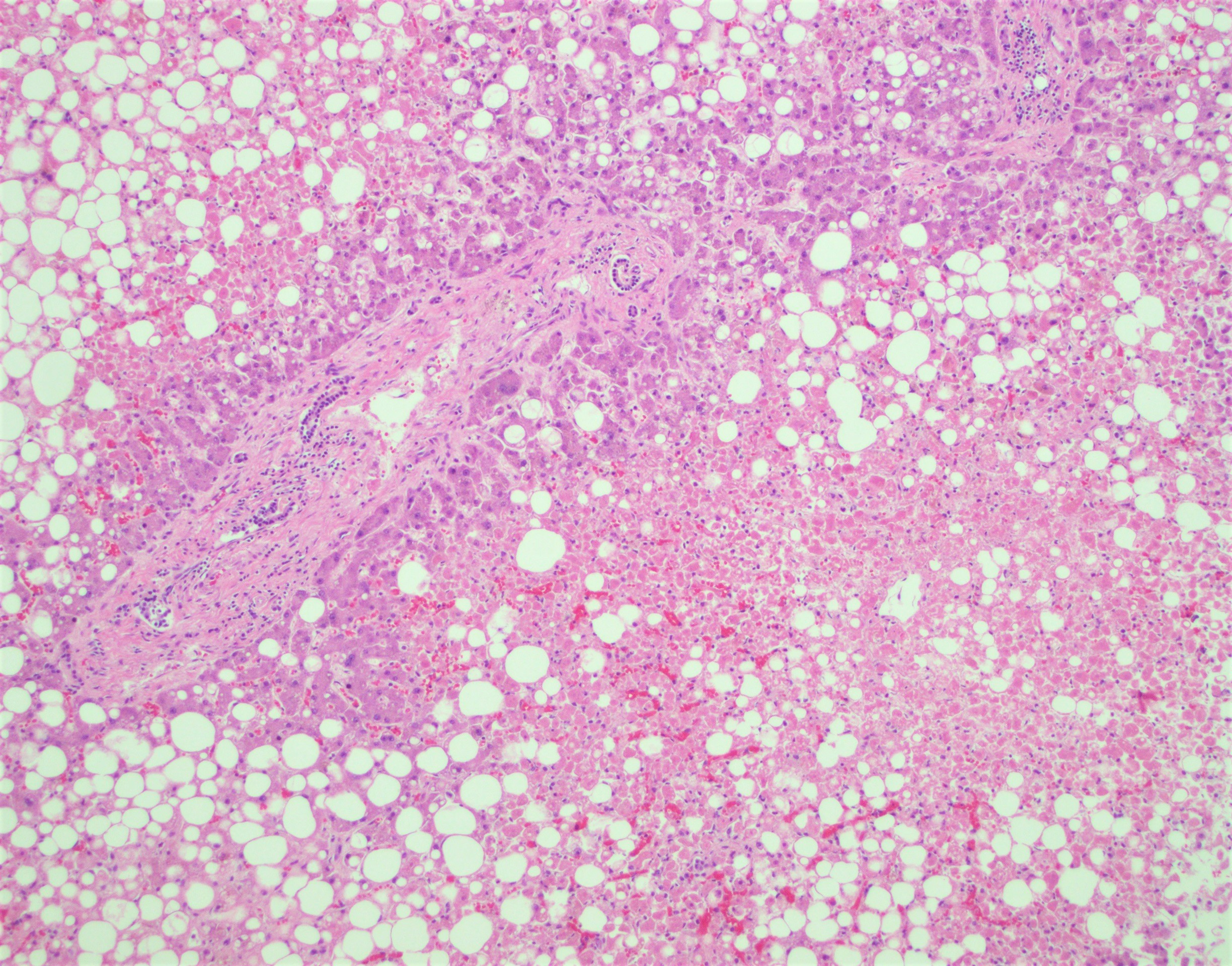
- Steatosis (Liver Int 2020;40:2110, Hum Pathol 2021;109:59)
- Common (up to 75% of patients)
- Predominantly macrovesicular
- Usually mild (involving < 33% of the liver parenchyma) and often azonal
- Usually not associated with ballooning and Mallory-Denk bodies
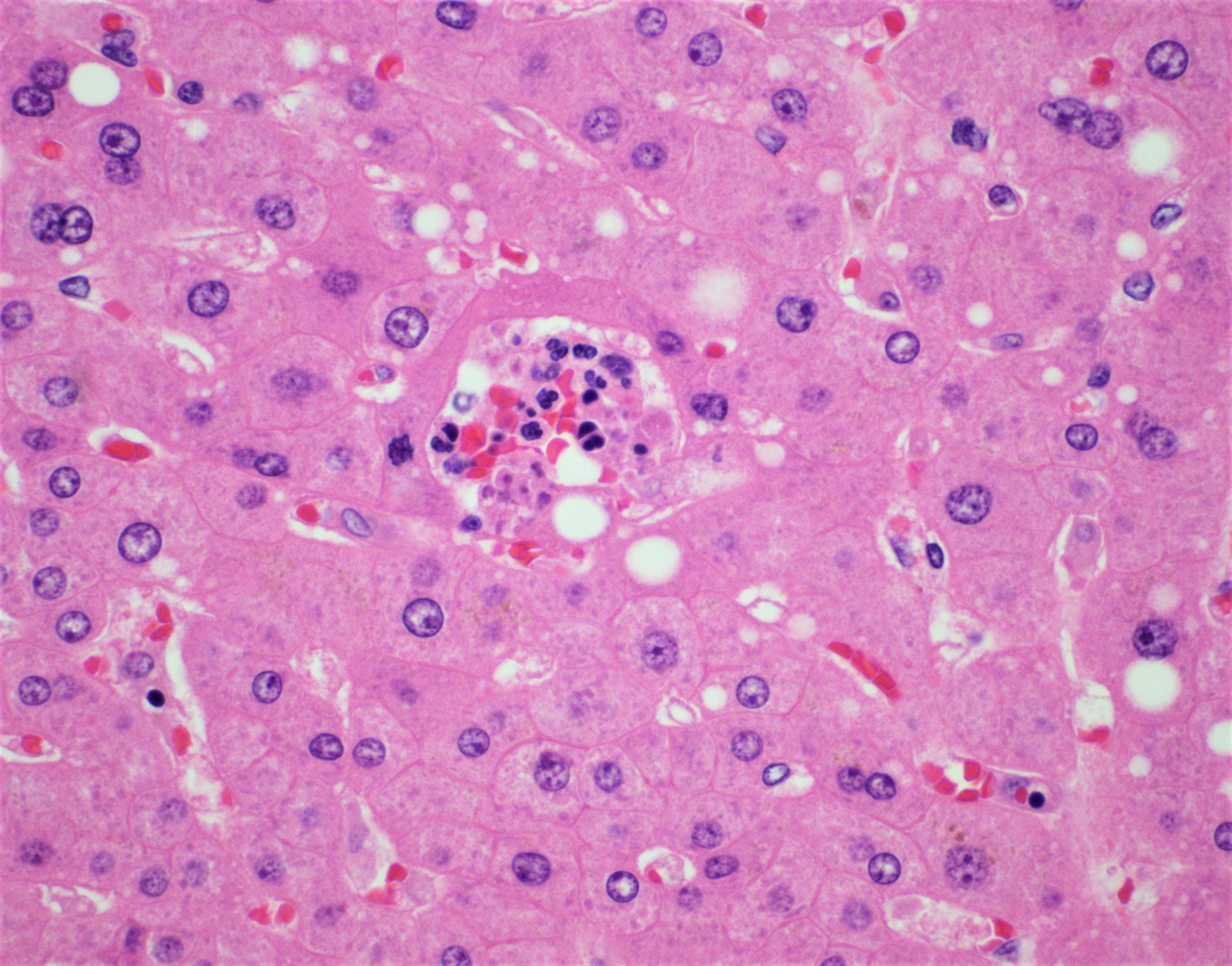
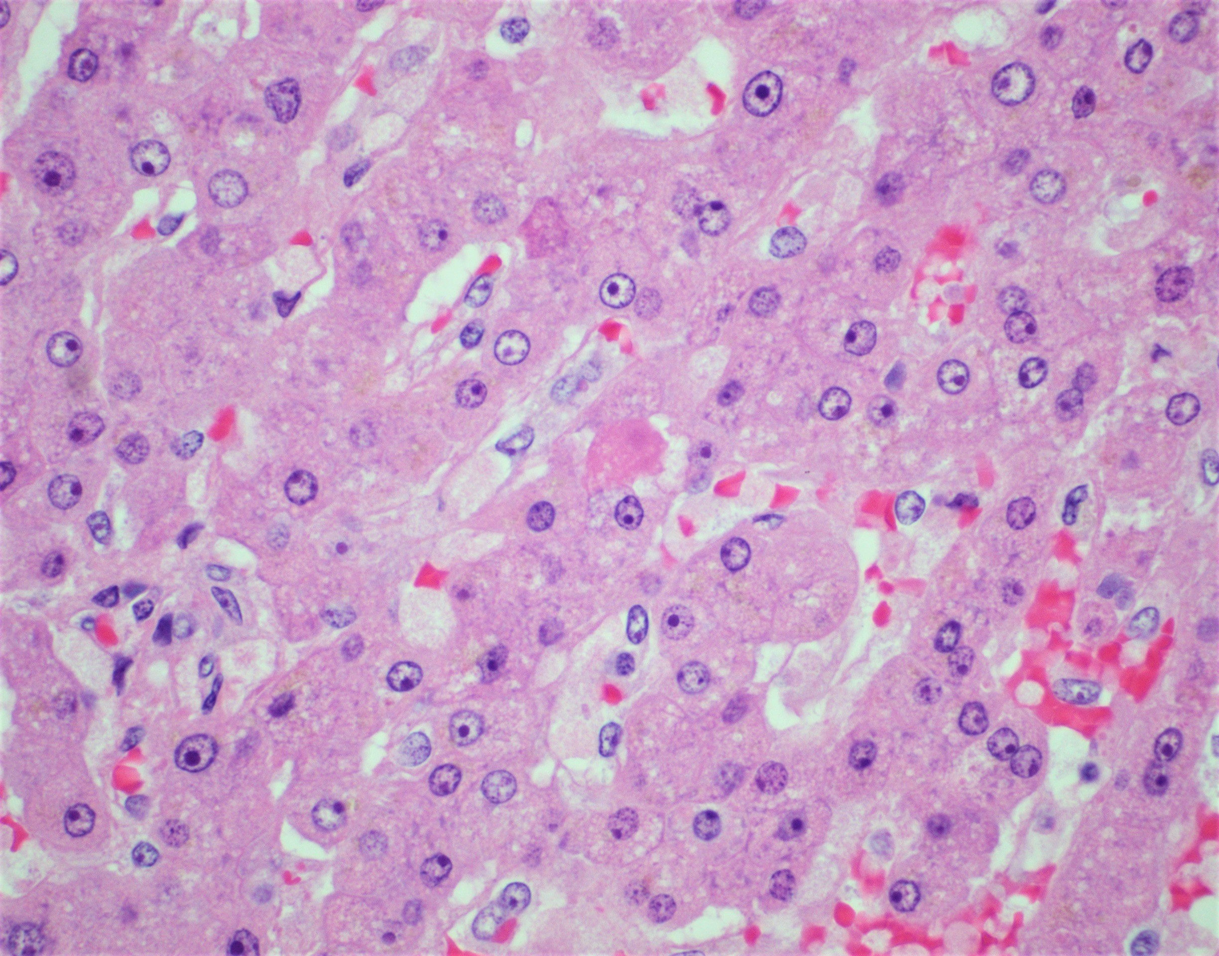
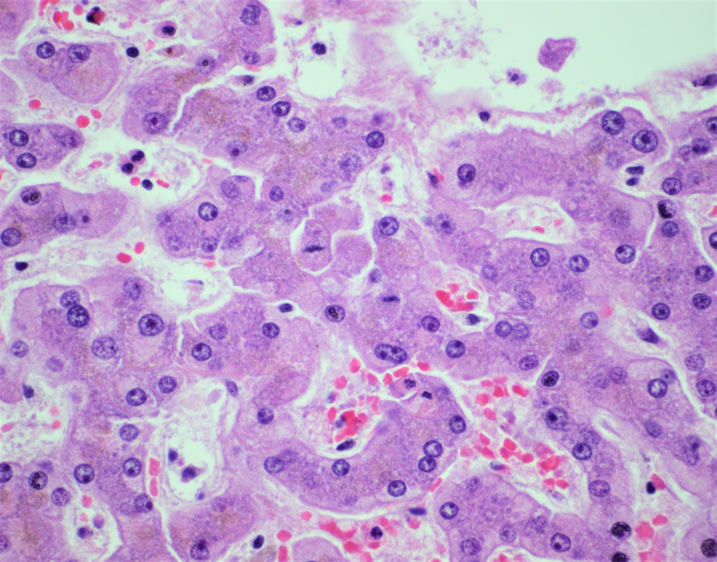
- Acute hepatitis (lobular necroinflammation) (Liver Int 2020;40:2110)
- Present in up to 50% of cases
- Usually mild severity (80% of cases)
- Foci contain apoptotic hepatocytes plus lymphocytes and rare histiocytes without prominent plasma cells
- Lobular mitoses
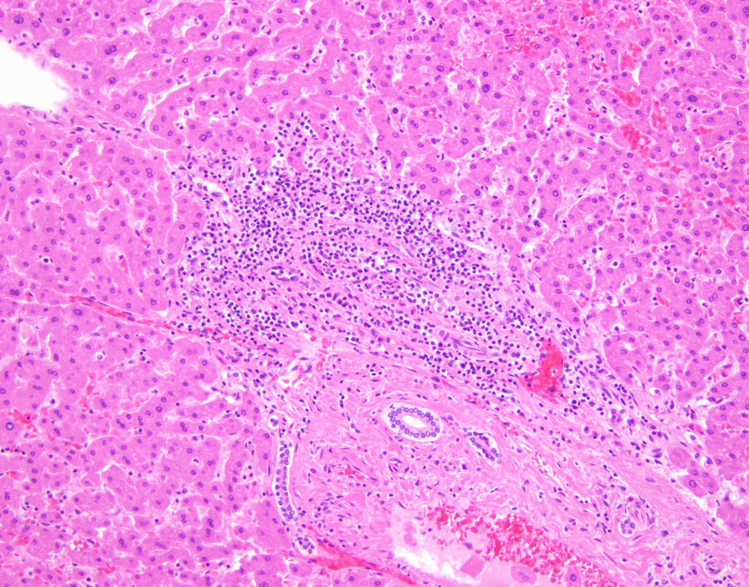
- Portal inflammation (Liver Int 2020;40:2110)
- Up to half of cases
- Minimal to mildly increased portal mononuclear cells (lymphocytes and macrophages)
- Rarely interface hepatitis
- Biliary findings / COVID-19 cholangiopathy
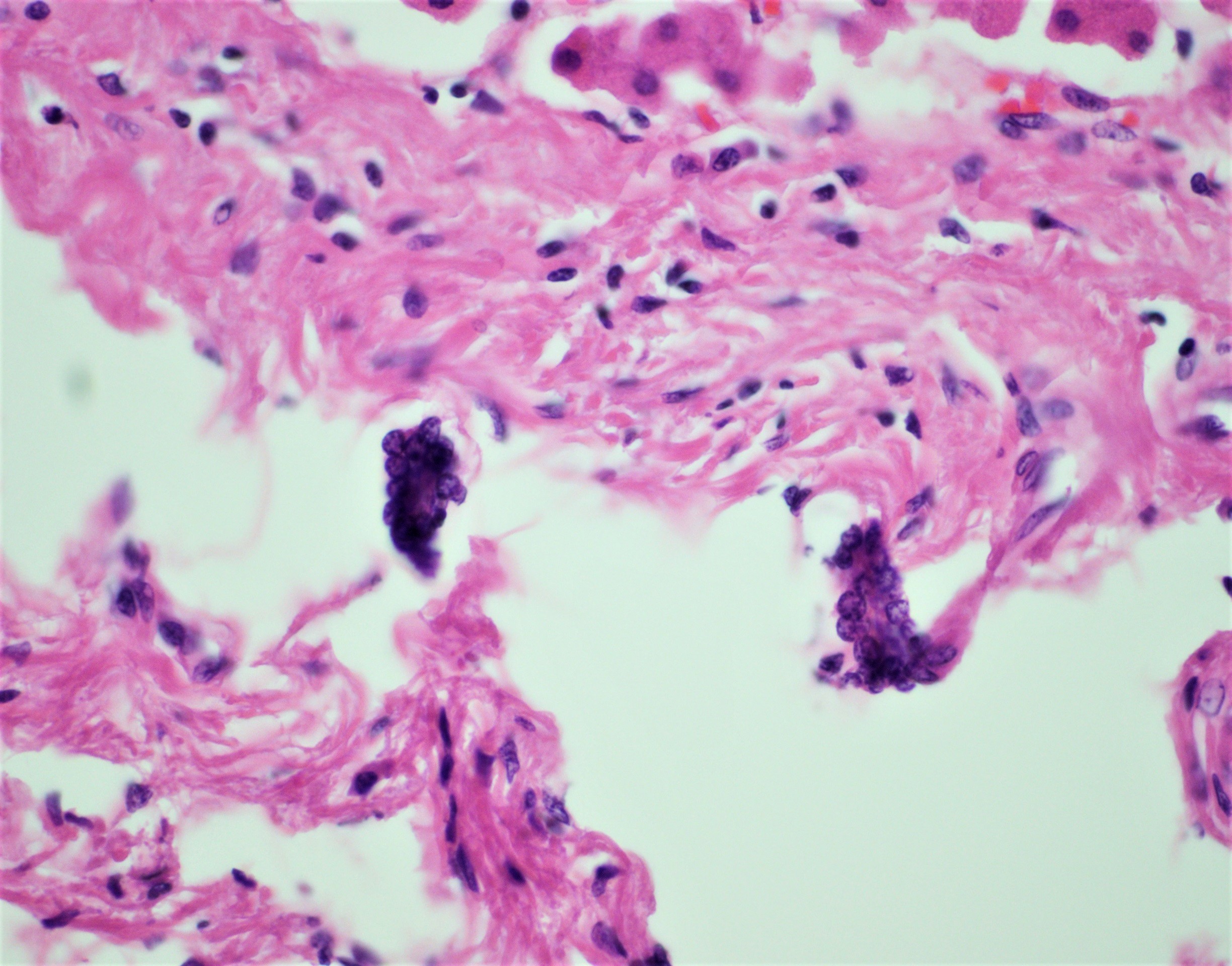
- Mild and focal lobular cholestasis (up to 38% of cases) (Mod Pathol 2020;33:2147)
- Ductopenia, severe cholangiocyte injury, ductular reaction, variable portal inflammation, variable fibrosis (Am J Gastroenterol 2021;116:1077)
- Acute or chronic large duct obstruction without clear bile duct loss (Am J Gastroenterol 2021;116:1414)
- Vascular pathology
- Focal
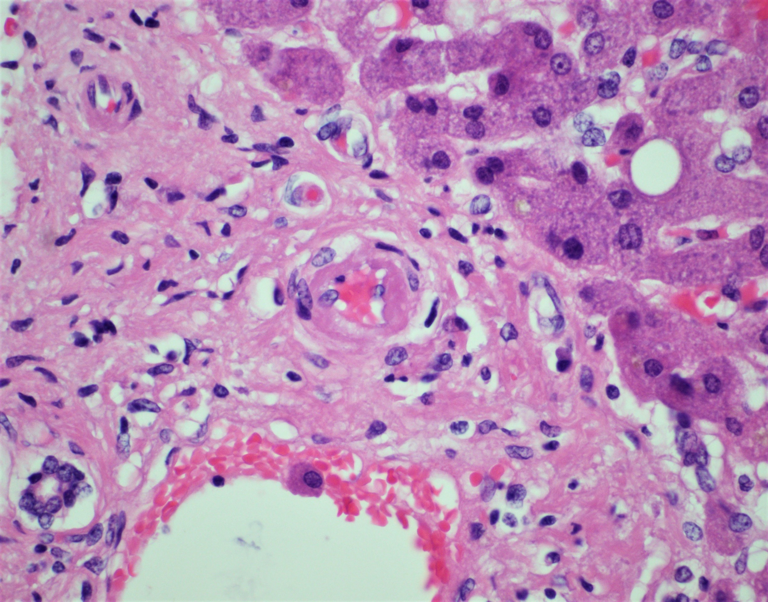
- Phlebosclerosis (reminiscent of veno-occlusive disease), more often with involvement of portal venule (rarely of central vein)
- Abnormalities of portal arterioles:
- Arteriolar muscular hyperplasia
- Hyalinosis and rare fibrinoid necrosis with endothelial apoptosis
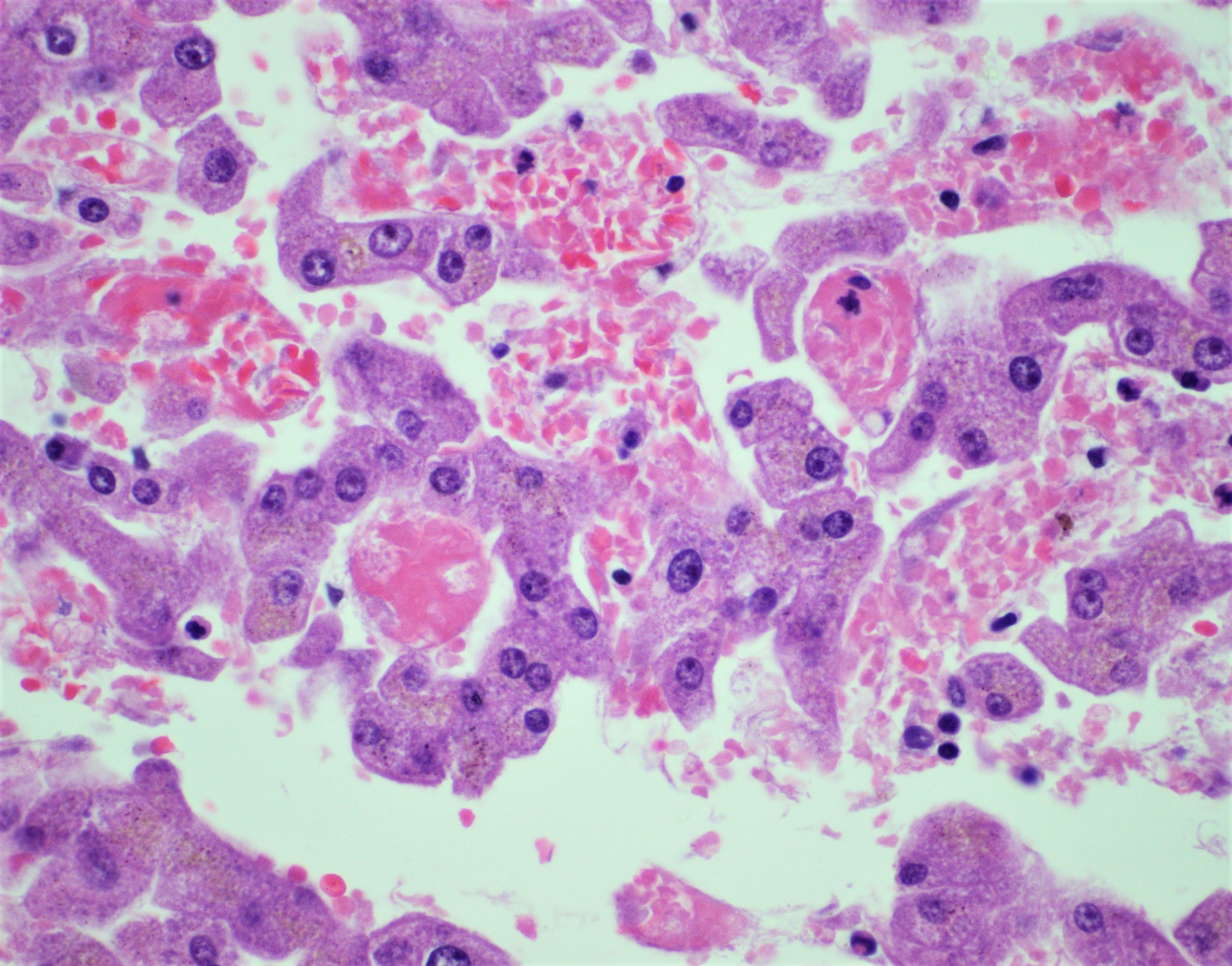
- Sinusoidal erythrocyte aggregation (up to 44%) and platelet microthrombi (up to 70%) (Hepatol Res 2021;51:1000, Hum Pathol 2021;109:59)
- Uncommon findings
- Thrombotic bodies: pale ovoid sinusoidal platelet rich inclusions seen in sinusoidal spaces (CD61+)
- Large megakaryocytes
- Perimortem pathology
- Congestion
- Centrilobular ischemic necrosis
- Reference: Mod Pathol 2020;33:2147
Microscopic (histologic) images
Positive stains
- IHC and ISH for SARS-CoV-2 can be used to detect virus in lung, particularly during the acute phase of diffuse alveolar damage and occasionally in other organs, such as placenta
- Needs validation in the liver
Electron microscopy images
Molecular / cytogenetics description
- RT PCR of liver autopsy samples positive in up to 55% of COVID-19 deceased (Mod Pathol 2020;33:2147, Clin Gastroenterol Hepatol 2021;19:1726)
- Range from 10 copies to 9,254 copies/μl RNA
Videos
Liver histopathological findings in COVID-19 autopsies
Clinical insights: COVID-19 and the liver
Sample pathology report
- Liver, right lobe, core biopsy:
- Acute hepatitis and macrovesicular steatosis consistent with clinical history of SARS-CoV-2 infection
Differential diagnosis
- Reactivation of preexisting liver diseases:
- Correlate with clinical history
- Chronic hepatitis (commonly hepatitis C virus, hepatitis B virus, autoimmune hepatitis):
- Fibrosis implies chronicity, which would not be present in acute COVID-19; however, it has been described in subacute / chronic post COVID-19 biliary injury (Am J Gastroenterol 2021;116:1077, Am J Gastroenterol 2021;116:1414)
- Lymphoid aggregates are common in chronic hepatitis but are not typically encountered in COVID-19
- Preexisting fatty liver disease:
- Clinical risk factors
- Frank steatohepatitis does not seem to be a feature of COVID-19
- Chronic biliary tract disease:
- Features of primary sclerosing cholangitis / secondary sclerosing cholangitis have been reported in post COVID cholangiopathy (Am J Gastroenterol 2021;116:1414)
- Correlate with clinical history
- Drug induced liver injury (Surg Pathol Clin 2018;11:297):
- Clinical history of exposure to hepatotoxic drug
- May show prominent eosinophilia
- May show features reminiscent of autoimmune hepatitis
- Not always possible to distinguish from COVID-19 injury
Additional references
Board review style question #1
The liver sample shown above is from a patient with COVID-19. The histopathological process depicted in the image above is more often associated with which of the following conditions?
- Acetaminophen toxicity
- Autoimmune hepatitis
- COVID-19 associated hepatitis
- Primary biliary cholangitis
- Primary sclerosing cholangitis
Board review style answer #1
B. Autoimmune hepatitis. The finding of interface hepatitis is more common in autoimmune hepatitis but has been described in patients with COVID-19.
Comment Here
Reference: Viral hepatitis - COVID-19
Comment Here
Reference: Viral hepatitis - COVID-19
Board review style question #2
What is the most common histopathologic hepatic finding in patients dying of COVID-19?
- Apoptotic bodies
- Disseminated microthrombi
- Interface hepatitis
- Lobular necroinflammation
- Macrovesicular steatosis
Board review style answer #2


