Table of Contents
Definition / general | Case reports | Microscopic (histologic) description | Microscopic (histologic) images | Positive stains | Molecular / cytogenetics description | Electron microscopy descriptionCite this page: Mihova D. AML with pseudo-Chediak-Higashi anomaly. PathologyOutlines.com website. https://www.pathologyoutlines.com/topic/leukemiapseudochediak.html. Accessed December 23rd, 2024.
Definition / general
- Not part of WHO classification
- Frequent in acute promyelocytic leukemia; up to 25% of AML-M2 (Acta Paediatr Jpn 1990;32:651)
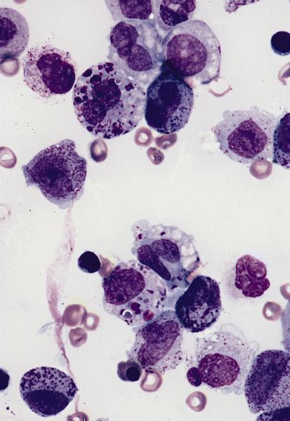
- Giant granules may be due to fusion of primary granules or small dense vesicles
- See also Bone marrow nonneoplastic chapter
Case reports
- 16 year old girl with AML-M5a and t(10;11) (Clin Lab Haematol 2000;22:303)
Microscopic (histologic) description
- Giant cytoplasmic granules
Positive stains
Molecular / cytogenetics description
- May be associated with double minutes (Leukemia 2002;16:152)
Electron microscopy description
- Peroxidase positive granules with a dense matrix but no obvious crystalline structure, may contain membranous lamellae or tubular structures (Cancer Res 1980;40:4473, Sangre (Barc) 1994;39:135)


