
Kidney tumor
Childhood tumors
Nephroblastomatosis / nephrogenic rests
Author: Mandolin S. Ziadie, M.D.

Last author update: 1 December 2011
Last staff update: 4 March 2025 (update in progress)
Copyright: 2003-2025, PathologyOutlines.com, Inc.
PubMed Search: Nephroblastomatosis nephrogenic rests kidney


Table of Contents
Definition / general | Epidemiology | Clinical features | Case reports | Treatment | Gross description | Gross images | Microscopic (histologic) description | Microscopic (histologic) images | Differential diagnosis | Additional referencesCite this page: Ziadie MS. Nephroblastomatosis / nephrogenic rests. PathologyOutlines.com website. https://www.pathologyoutlines.com/topic/kidneytumornephroblastomatosis.html. Accessed April 1st, 2025.
Definition / general
- Nephrogenic rests: persistent foci of embryonal cells seen after the period of normal nephrogenesis
- Nephroblastomatosis: multifocal or diffuse nephrogenic rests
- May be intralobar or perilobar
Epidemiology
- 1% of neonatal kidneys; florid cases associated with congenital anomalies and hypertension
- Intralobar rests have an increased rate of progression to Wilms tumor, are more commonly associated with WT1 mutations, Denys-Drash syndrome and WAGR syndrome
- Perilobar rests are seen in sporadic tumors and are associated with genetic / epigenetic dysregulation at 11p15 (Clin Cancer Res 2008;14:7635), idiopathic hemihypertrophy and Beckwith-Wiedemann syndrome
Clinical features
- Precursor lesions of Wilms tumor, seen in 30% - 44% of kidneys resected for Wilms tumor (WT)
Case reports
- 2 week old 'female' infant with nephrotic syndrome and renal failure (Case of the Week #338)
- Male newborn death with bilateral nephroblastomatosis (Arch Pathol Lab Med 1989;113:729)
- Newborn with lumbosacral ectopic rest (Am J Surg Pathol 2004;28:1389)
- Siblings with universal nephroblastomatosis with nephromegaly (Pediatr Dev Pathol 2009;12:47)
Treatment
- Conservative
Gross description
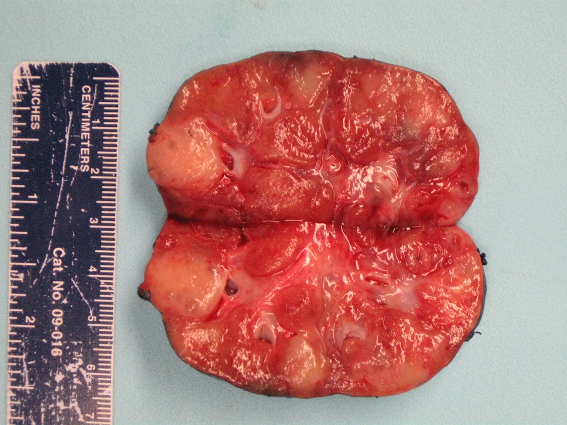
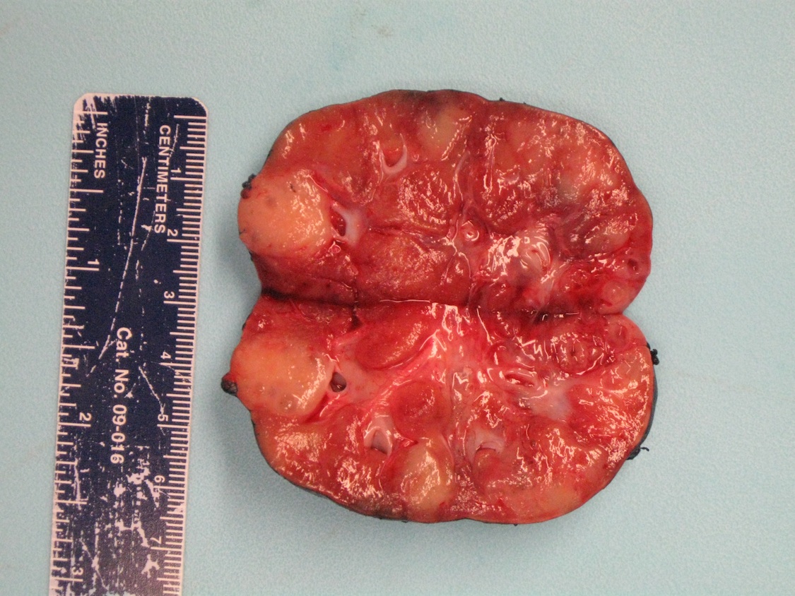
- May be inapparent on gross examination or appear as nodules / foci of capsular thickening / scar beneath the capsule or in the deep cortex / medulla
Gross images
Case of the Week #338

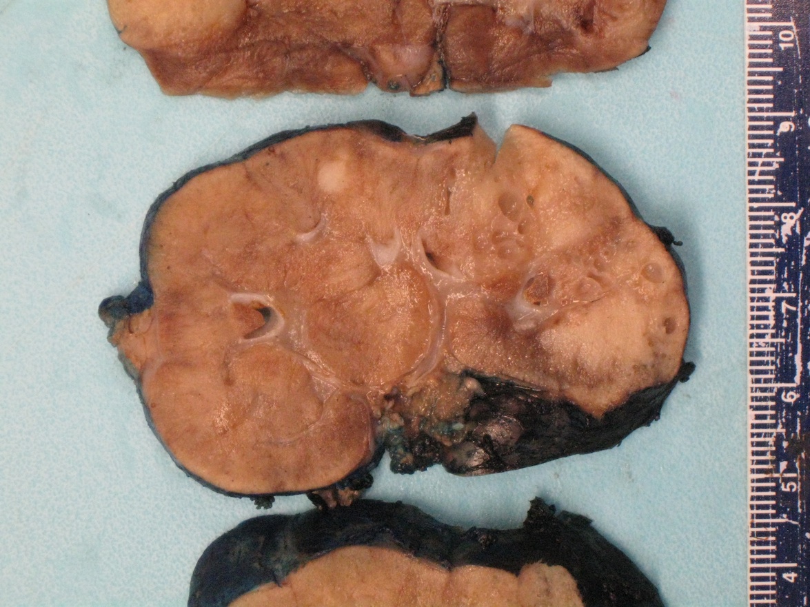
Various images
Microscopic (histologic) description
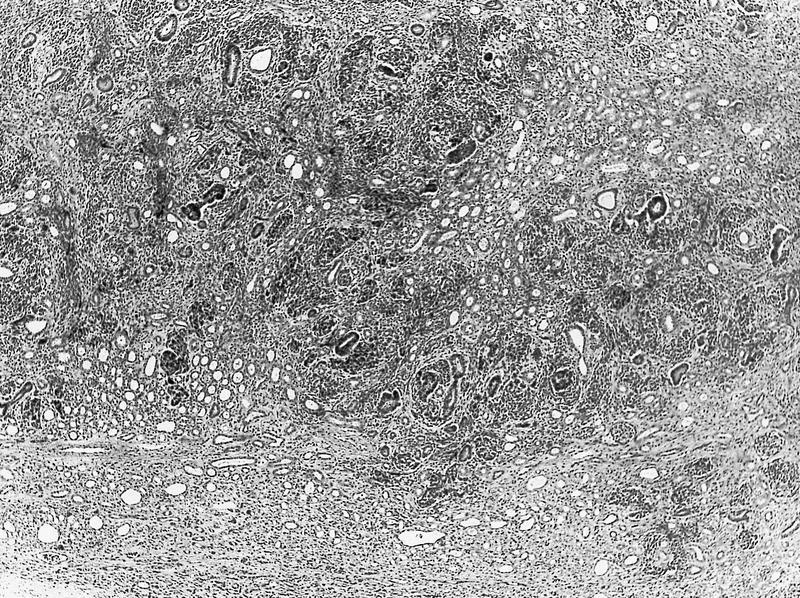
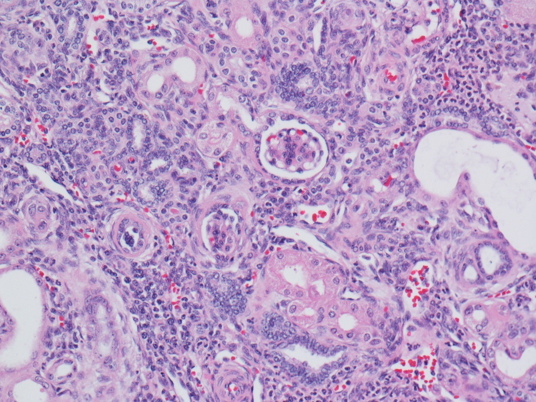
- Tightly packed nests or diffuse sheets of primitive but nonanaplastic blastemal / primitive epithelial cells with scanty stroma
- No cartilage or primitive mesenchyme
- May have a sclerosing or hyperplastic pattern
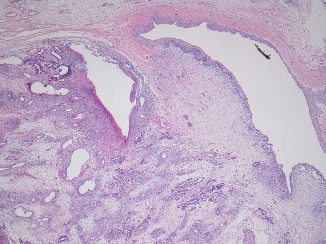
- Intralobar: randomly distributed throughout cortex and medulla with irregular margins, more stroma than blastema or tubules
- Perilobar: peripheral with sharply demarcated margins, composed of blastema and tubules with scanty or sclerotic stroma, often solitary
Microscopic (histologic) images
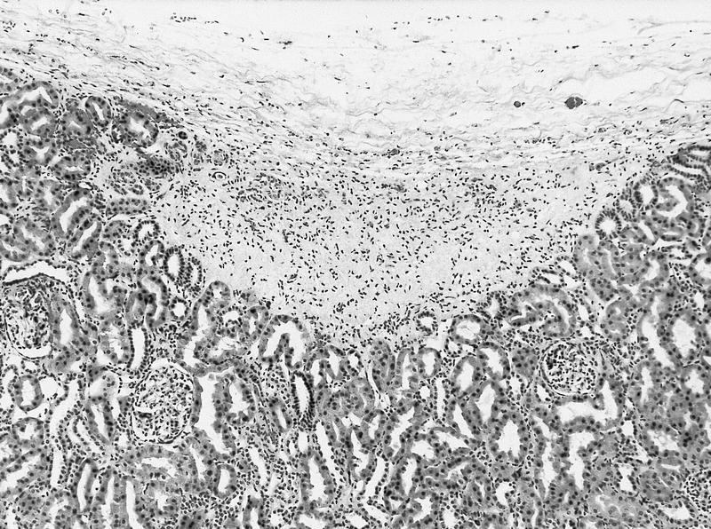
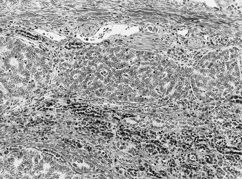
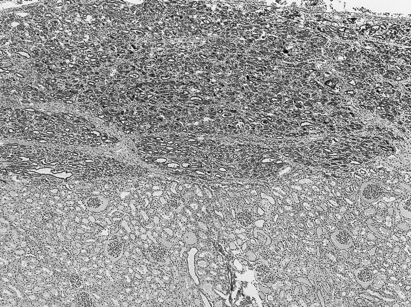
AFIP images
Intralobar

Intralobar nephrogenic rest
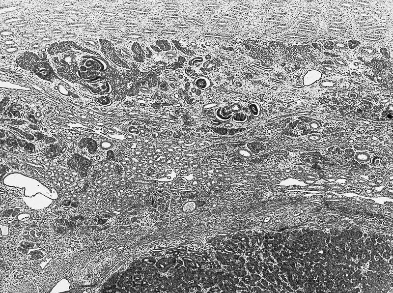
Neoplastic intralobar rest
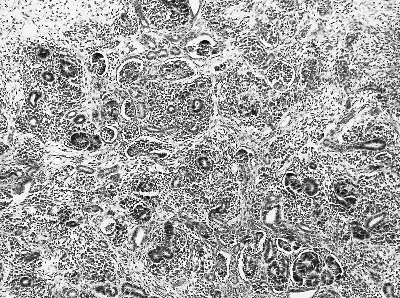
Nephroblastomatosis
Perilobar
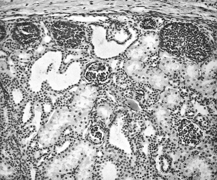

Microscopic rests composed of blastemal cells, are termed "incipient" in neonates and "dormant" otherwise
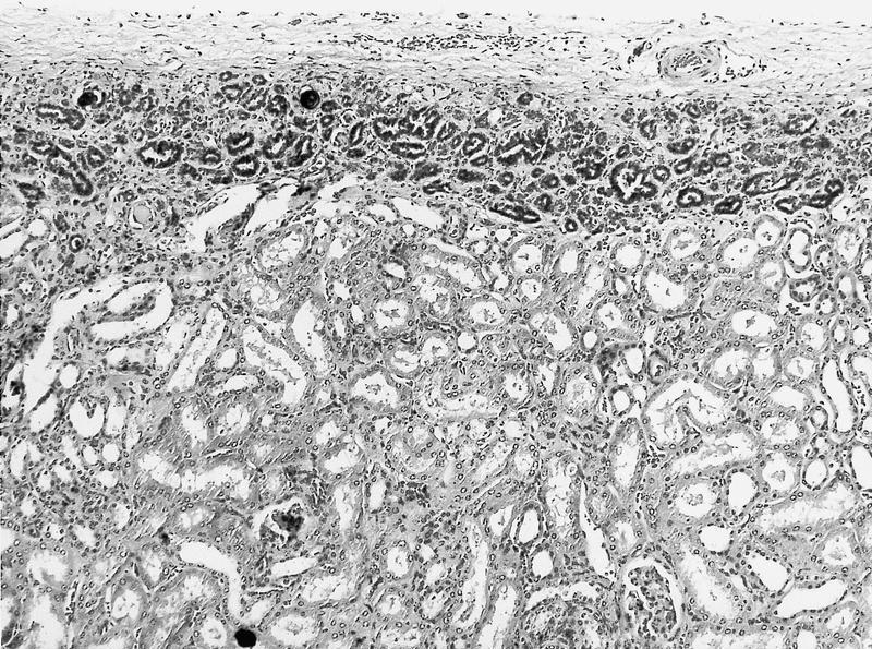
Sclerosing perilobar nephrogenic rest
Focal scarring

Hyperplastic perilobar rest in sclerosing stage
Multifocal hyperplastic perilobar rests

Nearly confluent rind of perilobar rest tissue; thick rind of perilobar tissue

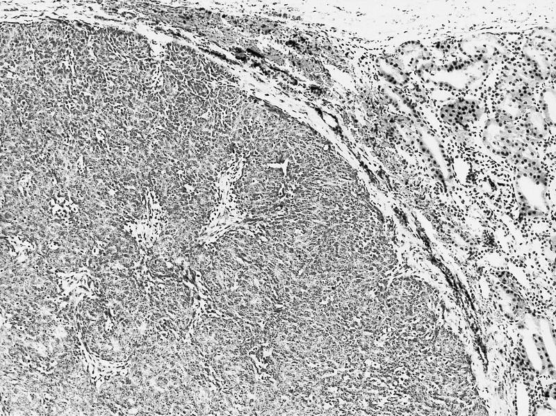
Neoplastic perilobar rest
Case of the Week #338


Various images
Differential diagnosis
Additional references
