Table of Contents
Definition / general | Essential features | Epidemiology | Sites | Clinical features | Prognostic factors | Case reports | Treatment | Microscopic (histologic) description | Microscopic (histologic) images | Positive stains | Negative stains | Differential diagnosis | Additional referencesCite this page: Andeen NK, Tretiakova M. With glandular differentiation. PathologyOutlines.com website. https://www.pathologyoutlines.com/topic/kidneytumormalignanturothelialcarcinomasubtypesglandulardiff.html. Accessed December 27th, 2024.
Definition / general
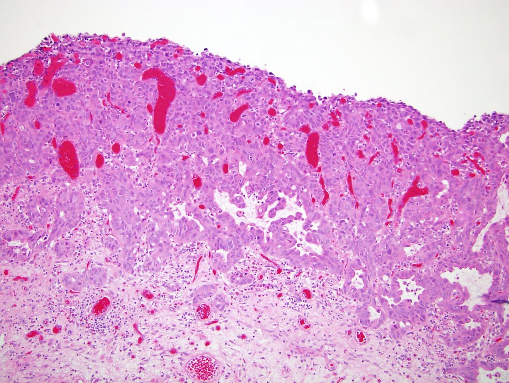
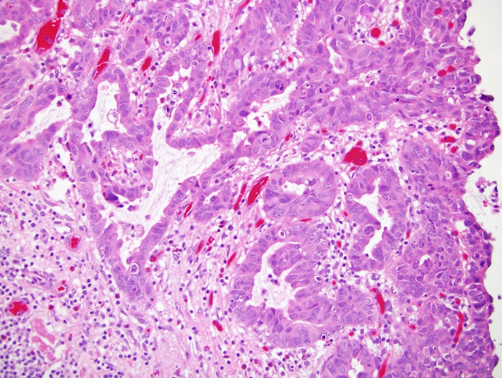
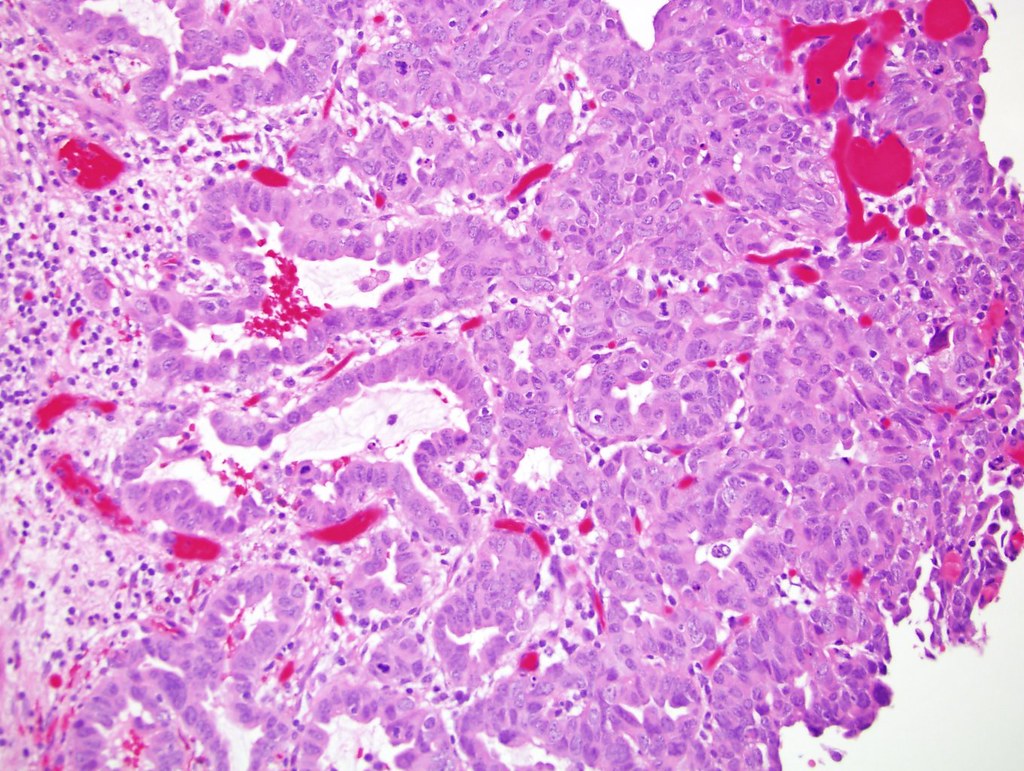
- Glandular differentiation is defined by the presence of true glandular spaces, usually tubular or gland-like lumina, or with morphology similar to enteric adenocarcinomas and variable mucin production
- Rarely may contain signet ring component (Mod Pathol 2009;22:S96, Arch Pathol Lab Med 2007;131:1244)
- Cytoplasmic mucin containing cells are seen in normal urothelium and are not considered to represent glandular differentiation (Arch Pathol Lab Med 2007;131:1244)
Essential features
- Presence of true glandular spaces
- Cytoplasmic mucin is seen in normal urothelium and not diagnostic of glandular differentiation
- No known prognostic significance
Epidemiology
- Foci of glandular differentiation are less common than squamous differentiation, and seen in up to 10% of urothelial carcinomas (Mod Pathol 2009;22:S96)
Sites
- Bladder, kidney
Clinical features
- Similar to other urothelial carcinomas
Prognostic factors
- Prognostic implications of glandular differentiation are unclear; some studies suggest adverse outcome (PLoS One 2014;9:e107027, Mod Pathol 2009;22:S96), others show similar outcomes compared to pure urothelial carcinoma (Urol Oncol 2014;32:117)
Case reports
- Urothelial carcinoma of renal pelvis with gland-like lumina (Biomedical Research 2013;24:175)
Treatment
- Too few cases to establish specific treatment recommendations
Microscopic (histologic) description
- True glandular structures within a conventional urothelial carcinoma
- Glands are of either tubular or enteric type, with a single layer of neoplastic columnar cells radially arranged around a lumen, with or without mucin production
- May resemble enteric adenocarcinomas
- May have signet ring morphology, with neoplastic signet ring cells floating in pools of mucin
Microscopic (histologic) images
Positive stains
- MUC5AC (Virchows Arch 2001;439:609), CK7, CK20, S100P
- Variable with GATA3 and p63 (Hum Pathol 2014;45:1473)
Negative stains
- Villin (Arch Pathol Lab Med 2002;126:1057), CDX2 (Arch Pathol Lab Med 2007;131:1244)
- Uroplakin and thrombomodulin are often negative (Hum Pathol 2014;45:1473)
Differential diagnosis
- Collecting duct carcinoma of the kidney: does not contain conventional urothelial carcinoma, usually positive for PAX8, CD10 and Vimentin
- Cystitis cystica and glandularis: does not have malignant component
- Metastatic adenocarcinoma: clinical history, site specific antibodies
- Microcystic and nested variants of urothelial carcinoma: these may have lumina with central necrotic debris but not true glandular formation
- Urothelial carcinoma of renal pelvis with intratubular spread: smooth contours, limited to pyramids of medulla, no invasion or desmoplasia (Am J Clin Exp Urol 2014;2:102)
Additional references


