Table of Contents
Definition / general | Essential features | ICD coding | Epidemiology | Sites | Pathophysiology | Clinical features | Diagnosis | Radiology description | Radiology images | Prognostic factors | Case reports | Treatment | Gross description | Gross images | Frozen section description | Microscopic (histologic) description | Microscopic (histologic) images | Cytology description | Positive stains | Negative stains | Electron microscopy description | Molecular / cytogenetics description | Videos | Sample pathology report | Differential diagnosis | Additional references | Board review style question #1 | Board review style answer #1 | Board review style question #2 | Board review style answer #2Cite this page: Miller TI, Tretiakova M. Chromophobe eosinophilic variant. PathologyOutlines.com website. https://www.pathologyoutlines.com/topic/kidneytumormalignantrccchromoeosinvar.html. Accessed April 2nd, 2025.
Definition / general
- Histologic variant of chromophobe renal cell carcinoma (ChRCC) with at least 80% of cells with eosinophilic cytoplasm
Essential features
- At least 80% of the neoplastic cells must be eosinophilic
- No difference in prognosis compared with classic / mixed chromophobe renal cell carcinoma
- Must distinguish from benign mimic oncocytoma
ICD coding
Epidemiology
- Chromophobe renal cell carcinoma is ~ 5% of all renal cell carcinomas (Int J Urol 2012;19:894)
- In large study case series, 41 - 51% of ChRCC were eosinophilic variant (Am J Surg Pathol 2008;32:1822, Eur J Surg Oncol 2008;34:687)
- Studies do not mention other epidemiologic differences from classic ChRCC
Sites
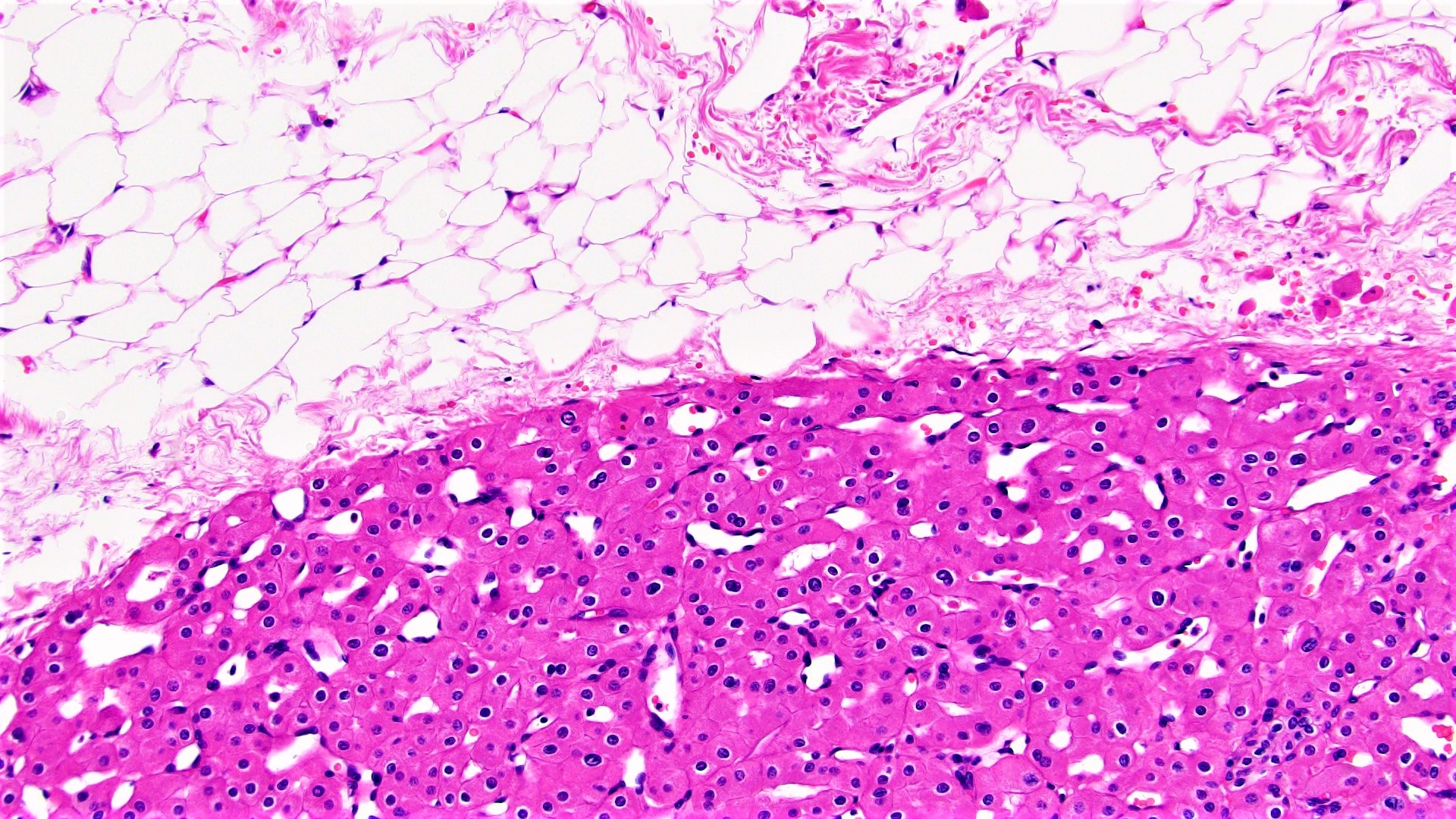
- Kidney in corticomedullary parenchyma
Pathophysiology
- Same as classic ChRCC: neoplasm of intercalated cells of collecting duct (Virchows Arch B Cell Pathol Incl Mol Pathol 1989;56:237)
- Eosinophilic cytoplasm is due to increased mitochondria
Clinical features
- Potential presenting symptoms: hematuria, pain, flank mass, anemia, pyrexia, cachexia, fatigue and weight loss (Int J Urol 2012;19:894)
- Large case series found eosinophilic more likely to be bilateral (11%) and multifocal (22%) (Am J Surg Pathol 2008;32:1822)
- Can be part of Birt-Hogg-Dubé syndrome (BHD) (FLCN mutation) (Am J Surg Pathol 2002;26:1542)
- Fibrofolliculomas, spontaneous pneumothorax, other renal neoplasms
Diagnosis
- Mass seen on imaging with confirmatory renal biopsy or mass resection
- Biopsy may not be diagnostic, especially if limited
Radiology description
- On CT scan, ChRCC more likely to have homogenous enhancement (69%) and can have calcifications (38%) (AJR Am J Roentgenol 2002;178:1499)
- No studies identified differentiating findings in eosinophilic from classic ChRCC
Prognostic factors
- No difference in outcome from classic ChRCC (Eur J Surg Oncol 2008;34:687, Cancers (Basel) 2019;11:1492, Am J Surg Pathol 2008;32:1822)
Case reports
- 31 year old man, long term renal transplant recipient, with 1.8 cm mass in allograft (Case Rep Transplant 2017;2017:4232474)
- 55 year old man with painless hematuria, 9.0 cm upper pole renal mass extending into the pelvicalyceal system and concurrent right atrial myxoma (J Lab Physicians. 2011;3:116)
- 60 year old woman with intermittent painless hematuria, 9.0 cm upper pole renal mass (J Lab Physicians. 2011;3:116)
- 73 year old woman with 1.8 cm upper pole renal mass and 67 year old man with incidental 3.0 cm lower pole renal mass (Proc (Bayl Univ Med Cent) 2015;28:57)
- 80 year old man with incidental 2.5 cm renal mass with partial papillary growth (Int J Clin Exp Pathol 2015 Oct;8:13590)
Treatment
- Typically resection via partial or total nephrectomy, the same as classic ChRCC
Gross description
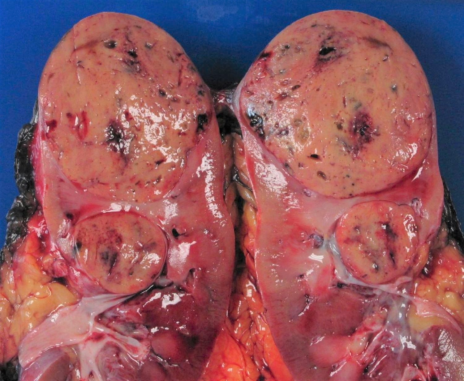
- Usually well circumscribed, lobulated and with beige to brown to yellow coloration
- Eosinophilic variant more likely to have brown coloration compared with classic, which is typically more beige-yellow (Am J Surg Pathol 2008;32:1822)
Gross images
Frozen section description
- Can mimic oncocytoma on frozen section (Biomedicine (Taipei) 2019;9:6)
Microscopic (histologic) description
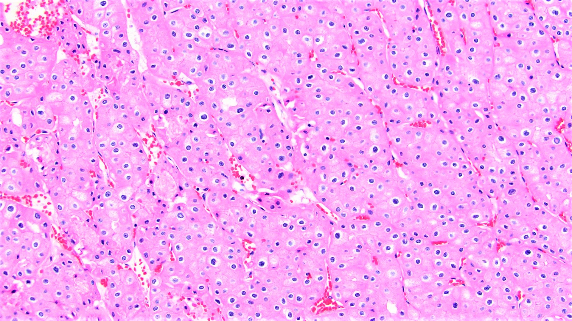
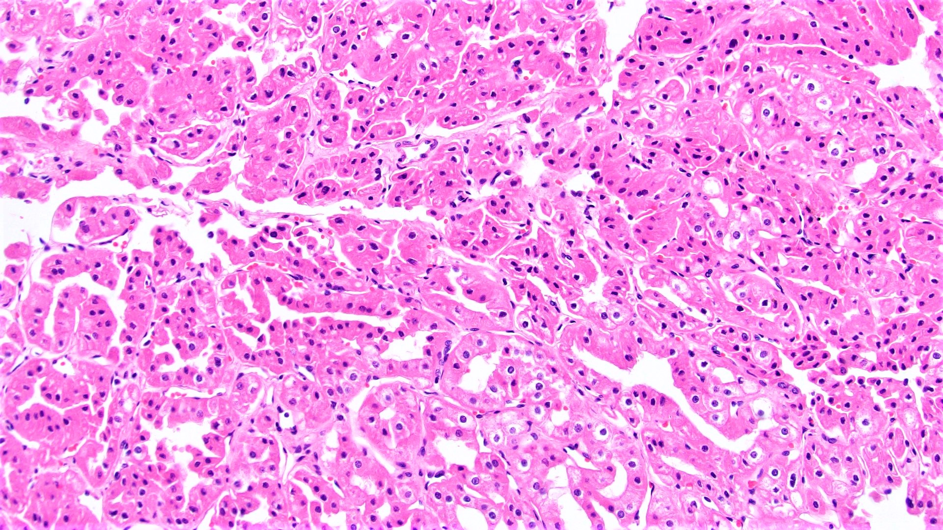
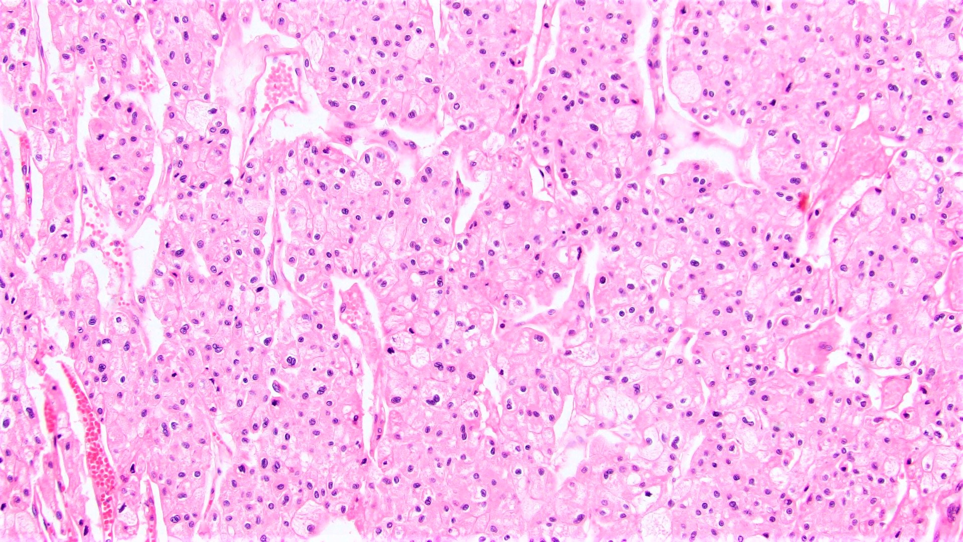
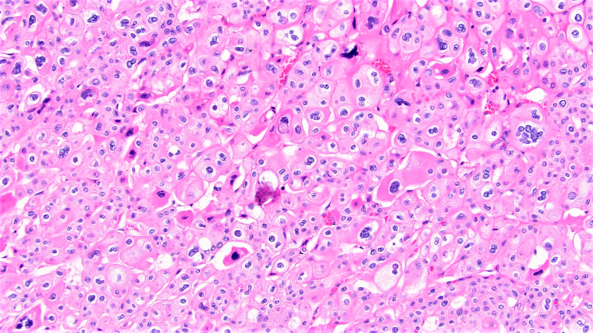
- At least 80% of the chromophobe neoplastic cells must have eosinophilic cytoplasm to classify as eosinophilic variant
- Cells are often smaller than classic chromophobe cells
- Besides eosinophilic cytoplasm and smaller size, have otherwise similar histology to classic ChRCC including
- Well defined / thickened cell border
- Wrinkled, raisinoid nuclei
- Frequent binucleation
- Perinuclear halos / clearing
- Rare mitoses
- Lacking prominent vasculature
- Nested, alveolar or sheet-like architecture
- Nested architecture more common in eosinophilic than classic (Am J Surg Pathol 2008;32:1822)
- Grading is not recommended
- Fuhrman grading system not predictive of outcome and is obsolete (Am J Surg Pathol 2007;31:957)
- WHO / ISUP system has not been validated for ChRCC (Am J Surg Pathol 2013;37:1490)
- Several grading schemes proposed but no overall consensus (Eur Urol 2016;70:93)
Microscopic (histologic) images
Cytology description
- Single or clusters of large cells with moderate pleomorphism
- Abundant granular eosinophilic cytoplasm
- Well defined / accentuated cell border
- Perinuclear clearing / vacuolization
- Binucleation may be seen
Positive stains
- CK7, KIT, Hale colloidal iron (diffuse granular cytoplasmic positivity)
- Claudin 7, EMA, E-cadherin, CK8, CK18, parvalbumin, EpCAM, ESA, ERA, KAI1, FXYD2 (J Clin Pathol 2016;69:661, Mod Pathol 1999;12:310, Am J Surg Pathol 2005;29:747, Mod Pathol 2001;14:760, Bratisl Lek Listy 2020;121:663, Med Mol Morphol 2012;45:98)
- Distinguishing classic versus eosinophilic variants of ChRCC: LMP2 (100% of eosinophilic) (Exp Mol Pathol 2013;94:29)
Negative stains
- Vimentin, CAIX, S100A1, RCC, CD10, GSTa, N-cadherin, amylase α1A, Wnt5a, HNF1β, AMACR, ARPP (Transl Androl Urol 2019;8:S123, Am J Surg Pathol 2000;24:203, Cancers (Basel) 2020;12:602, Bratisl Lek Listy 2020;121:663, Mod Pathol 1999;12:310, Am J Surg Pathol 2013;37:1824, Tumori 2010;96:304, Mod Pathol 2007;20:199)
Electron microscopy description
- Numerous cytoplasmic microvesicles (Am J Surg Pathol 2000;24:1247)
- Mitochondria with tubulocystic cristae (Am J Surg Pathol 2000;24:1247)
- Eosinophilic variant has more abundant mitochondria (Pathol Int 2000;50:872)
Molecular / cytogenetics description
- Compared with classic ChRCC, less frequent losses of chromosomes 1, 2, 6, 10, 13 and 17, suggesting classic has greater chromosomal instability (Cancers (Basel) 2019;11:1492)
- Most common chromosomal losses (in order of most to least frequent): 1, 2, 17, 6, 10, 13 and 21; no chromosomal gains (Adv Anat Pathol 2021;28:8)
- Doubled hypodiploidy by whole genome endoduplication is a common phenomenon in eosinophilic ChRCC (Hum Pathol 2020;104:18)
Videos
Distinguishing eosinophilic chromophobe RCC from oncocytoma
Sample pathology report
- Left kidney, mass, partial nephrectomy:
- Chromophobe renal cell carcinoma, eosinophilic variant (see synoptic report)
Differential diagnosis
- Oncocytoma:
- Round, hyperchromatic nuclei; smooth nuclear border
- Cells arranged in a nested or tubular pattern
- Lacks nuclear pleomorphism, oval nuclei, multiple nucleoli
- CK7 usually negative
- Hale colloidal iron focal positive staining confined to luminal borders
- No copy number alterations or losses of chromosomes 1 and X / Y (Hum Pathol 2020;104:18)
- Hybrid oncocytic / chromophobe tumors (HOCT) (Histol Histopathol 2013;28:1257):
- Can be sporadic but also often found in association with renal oncocytomatosis or Birt-Hogg-Dubé (BHD) syndrome
- Sporadic / oncocytomatosis: solid alveolar pattern, scattered cells with perinuclear halos but with no raisinoid nuclei
- BHD: admixed areas of oncocytoma and ChRCC, scattered chromophobe cells, intracytoplasmic vacuoles
- No aggressive behavior
- Usually parvalbumin, antimitochondrial antigen and CK7 positive
- Sporadic cases do not exhibit mutations in genes that are recurrently mutated in oncocytoma or ChRCC; syndromic cases with folliculin gene mutations (Mod Pathol 2019;32:1698)
- Eosinophilic variant of clear cell renal cell carcinoma:
Additional references
Board review style question #1
Board review style answer #1
A. CK7+, KIT+, vimentin-, CAIX-
The image shown is chromophobe renal cell carcinoma (ChRCC), eosinophilic variant. Histologic clues to the diagnosis of ChRCC are the raisinoid nuclei, perinuclear clearing / halos and well defined cell borders. Furthermore, > 80% of the cells have an eosinophilic cytoplasm, thus making it the eosinophilic variant of ChRCC. The immunoprofile of ChRCC is typically CK7+, KIT+, vimentin- and CAIX-. The other entity to consider is oncocytoma, but in this case the degree of nuclear pleomorphism (with enlarged, wrinkled and oval nuclei) favors ChRCC. Answer D (CK7-, KIT+, vimentin-, CAIX-) would be the immunohistochemical profile most typical of oncocytoma, although vimentin can often be focally positive. Answer E is the immunoprofile most typical of clear cell renal cell carcinoma; while there is an eosinophilic variant of this condition, the perinuclear clearing and raisinoid nuclei are still most characteristic of ChRCC (Pathol Res Pract 2015;211:303).
Comment Here
Reference: Chromophobe eosinophilic variant
Board review style question #2
Which of the following is true regarding chromophobe renal cell carcinoma (ChRCC), eosinophilic variant compared with the classic type of ChRCC?
- No differences in cytogenetics have been found between the eosinophilic variant and classic ChRCC
- The classic variant is more likely to have a brown coloration grossly
- The classic variant typically has smaller cells than the eosinophilic variant
- The eosinophilic variant must have 100% eosinophilic cells
- The prognosis of the eosinophilic variant is the same as classic ChRCC
Board review style answer #2
E. The prognosis of the eosinophilic variant is the same as classic ChRCC
Studies have shown no difference in prognosis between the eosinophilic variant of ChRCC and classic ChRCC. Answer A is wrong as a recent study showed that the eosinophilic variant frequently has less chromosomal instability compared with classic ChRCC and is often characterized by doubled hypodiploidy (Eur J Surg Oncol 2008;34:687, Cancers (Basel) 2019;11:1492, Am J Surg Pathol 2008;32:1822, Cancers (Basel) 2019;11:1492, Hum Pathol 2020;104:18, Am J Surg Pathol 2008;32:1822). Answer B is wrong as the eosinophilic variant is more likely to be brown in coloration grossly. Answer C is wrong as the eosinophilic variant typically has smaller cells compared with classic ChRCC. Answer D is wrong because, by definition, the eosinophilic variant only needs to have > 80% eosinophilic cells.
Comment Here
Reference: Chromophobe eosinophilic variant


