Table of Contents
Definition / general | Procedure | Sections to obtain | Tips | Frozen section | Gross description | Gross images | Sample gross description report | Board review style question #1 | Board review style answer #1Cite this page: Zynger D. Grossing & frozen section. PathologyOutlines.com website. https://www.pathologyoutlines.com/topic/kidneytumormalignantgrossing.html. Accessed April 3rd, 2025.
Definition / general
- This topic describes how to gross specimens obtained from partial and radical nephrectomy procedures for suspected renal cortical tumors
- It does not describe nephroureterectomy specimens removed for renal pelvic and ureteral tumors
- Essential clinical history: none
Procedure
- Weigh the specimen
- Measure the overall specimen and renal vein and ureter if present
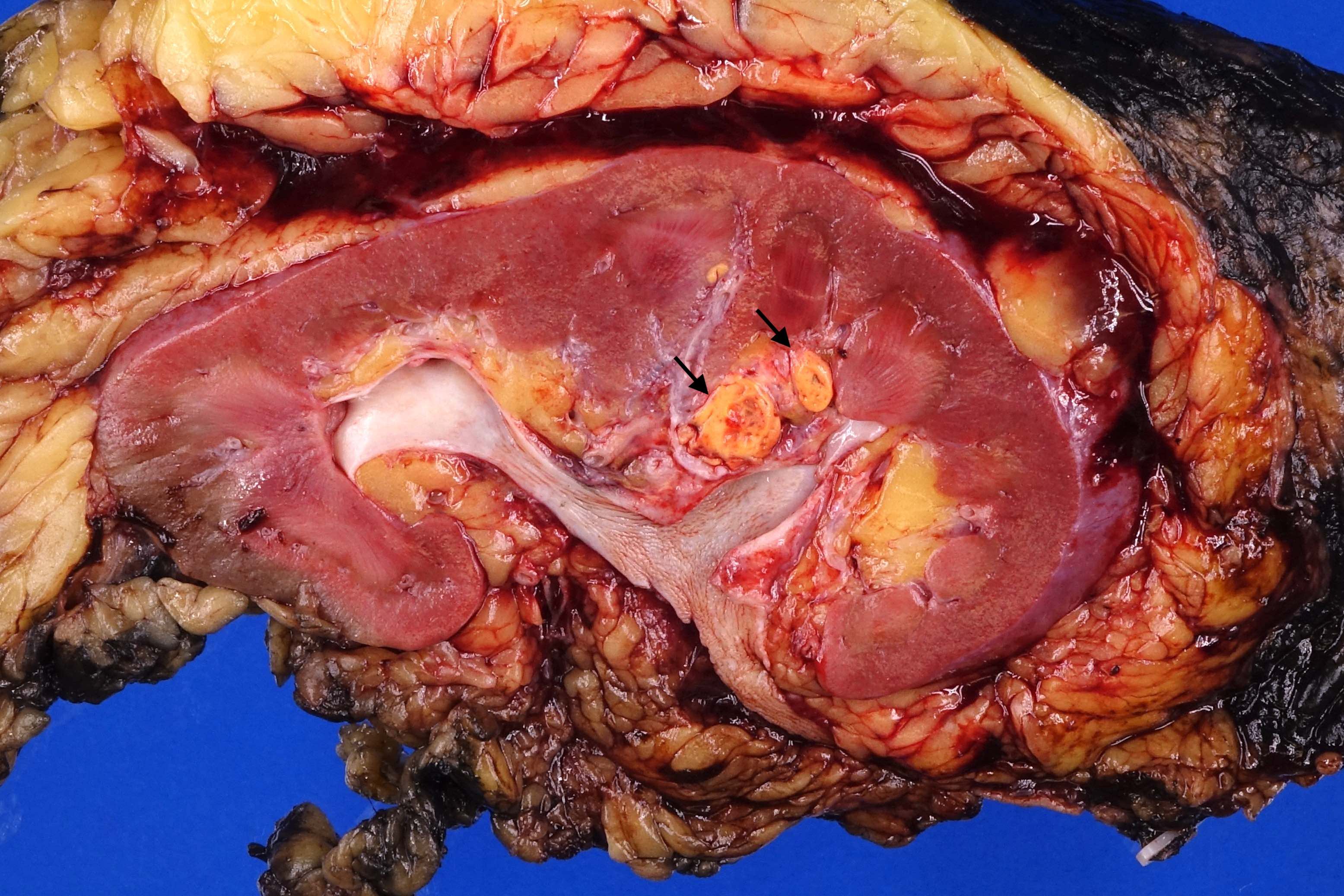
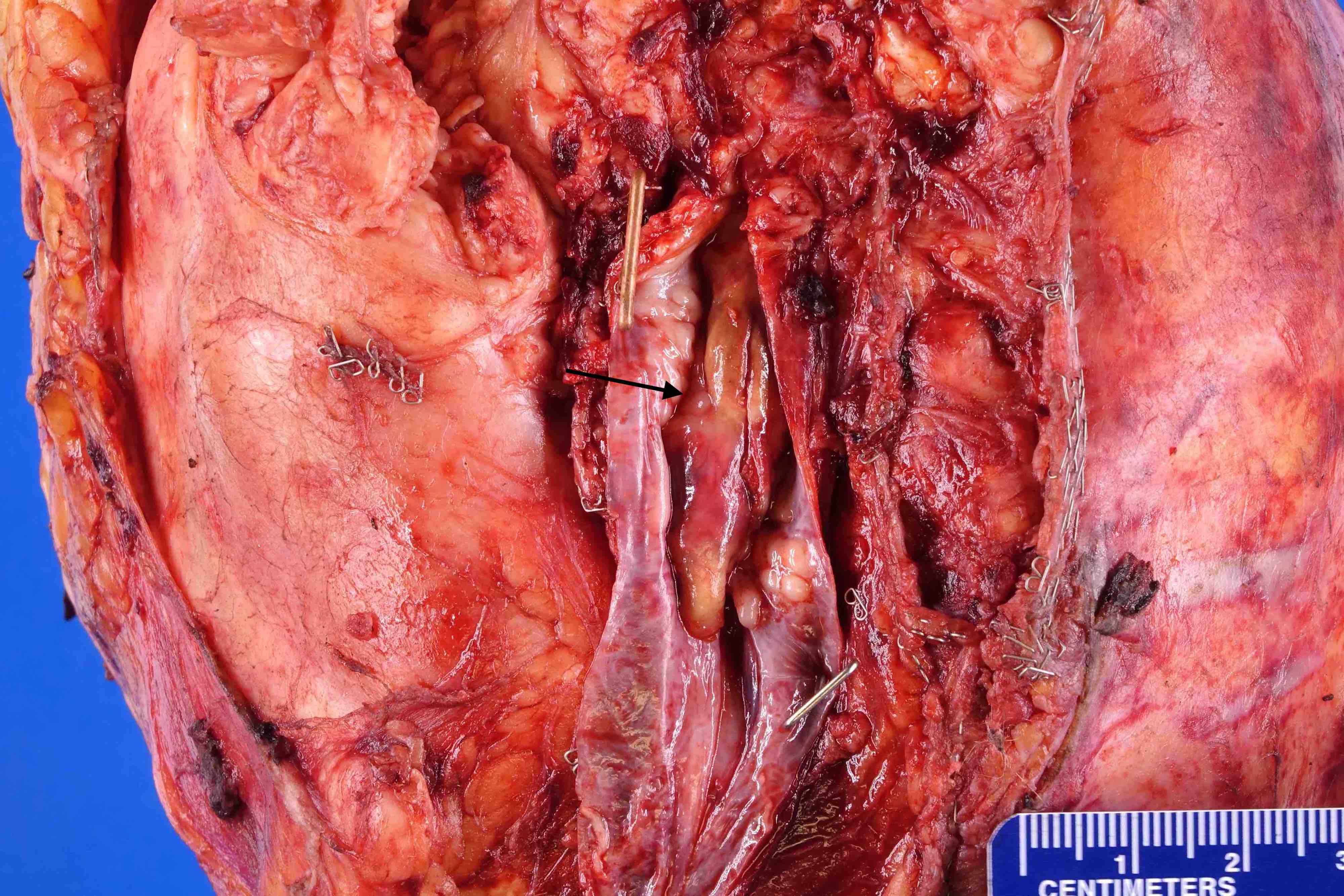
- Assess the renal vein and margin for presence of tumor
- Measure the distance of tumor to the renal vein margin
- Longitudinally cut open the renal vein and continue cutting the vein branches within the kidney to search for intraluminal tumor
- If tumor is present near or protruding through the renal vein margin, measure the distance of tumor to the renal vein margin
- Partial nephrectomy:
- Examine the parenchymal resection margin and capsular / perirenal margin and note any tumor present
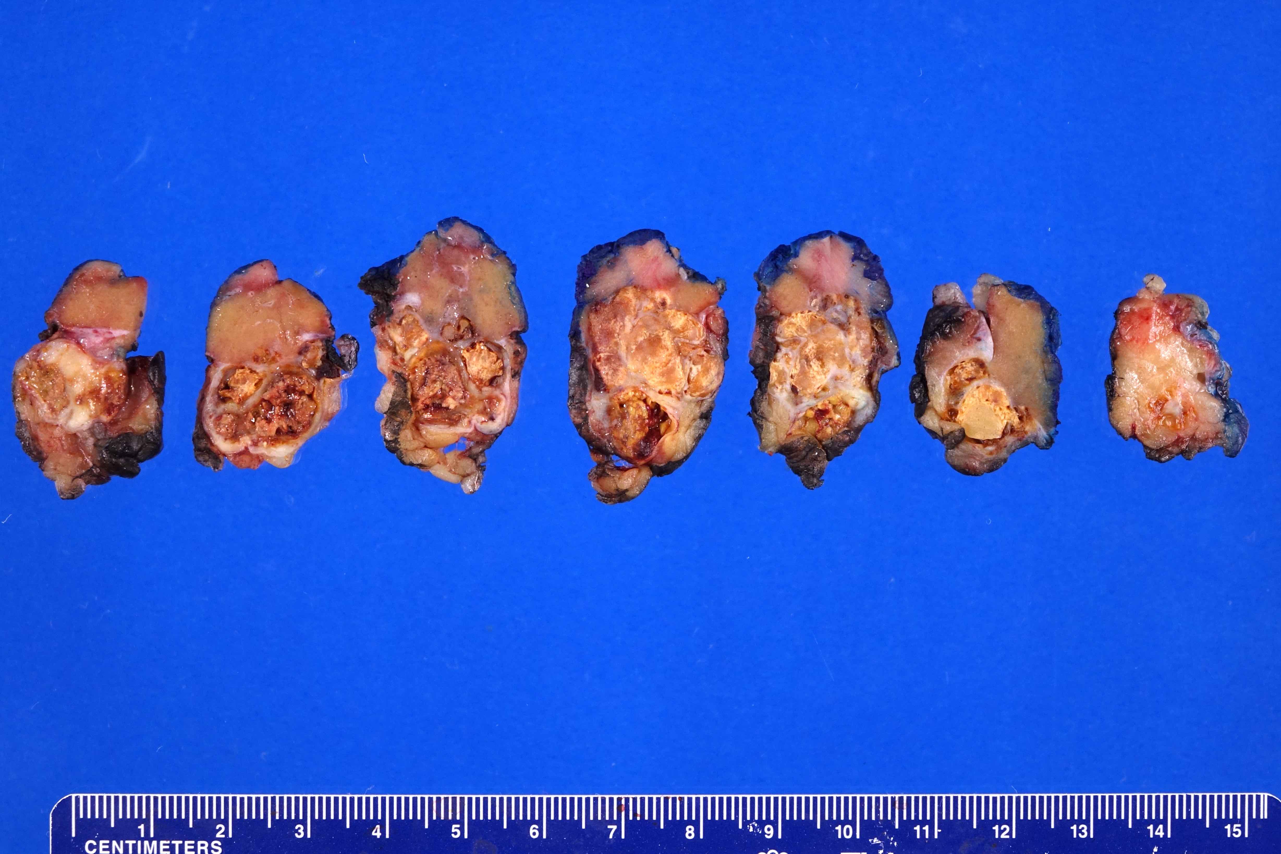
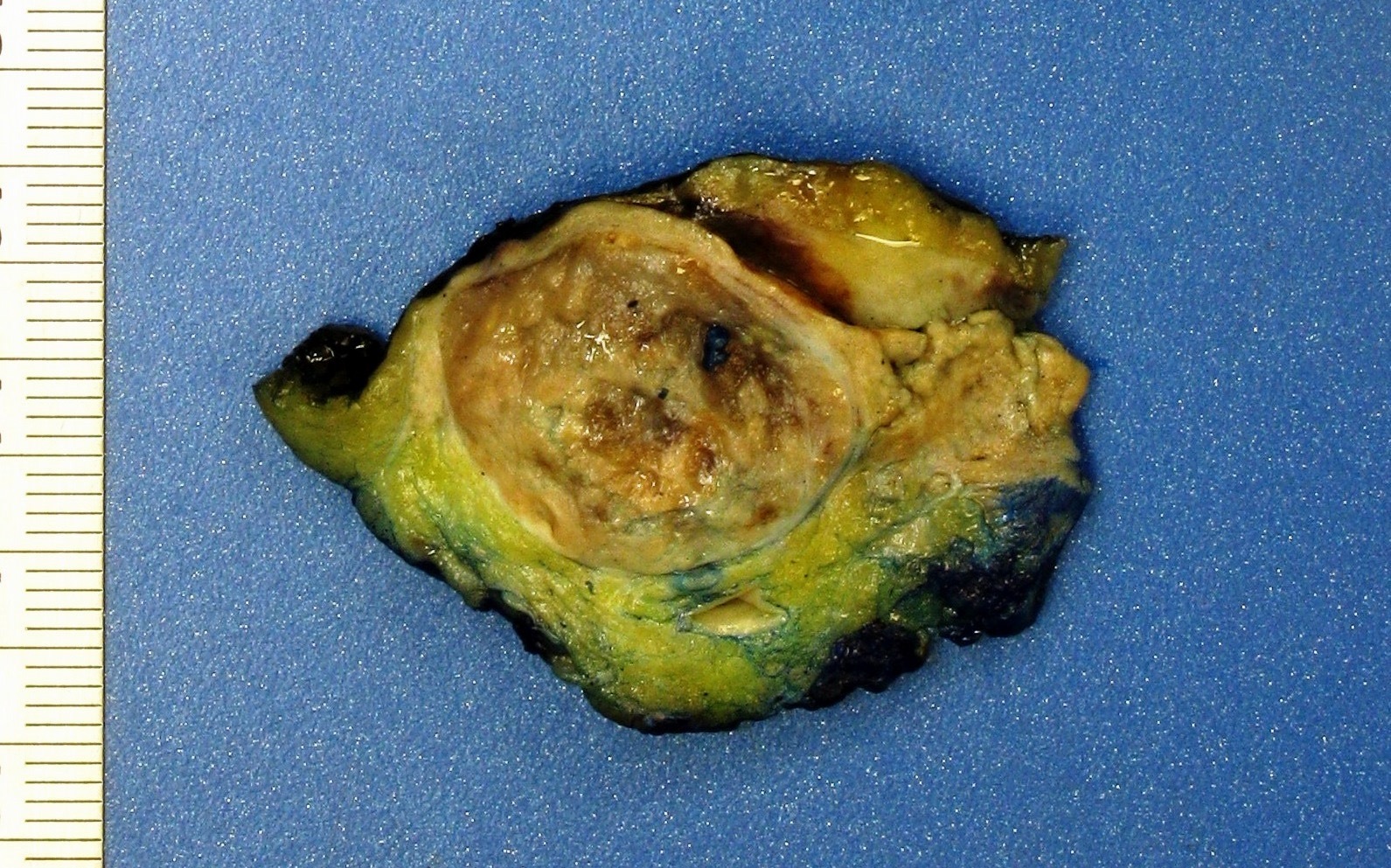
- Ink the entire surface of the renal parenchymal surgical resection margin blue
- Ink the outer surface of the capsule / perinephric adipose margin black
- Make a cut with a long knife perpendicular to the parenchymal margin to bivalve the specimen (i.e. to open like a book)
- Serially section the specimen
- Measure the distance of the tumor to the closest renal parenchymal surgical margin and to the inked outer surface / capsule
- Radical nephrectomy:
- Palpate the specimen to locate the tumor and examine the outer surface for evidence of involvement of Gerota fascia
- Ink the outer surface surrounding the tumor black
- Assess the inked outer surface (Gerota fascia)
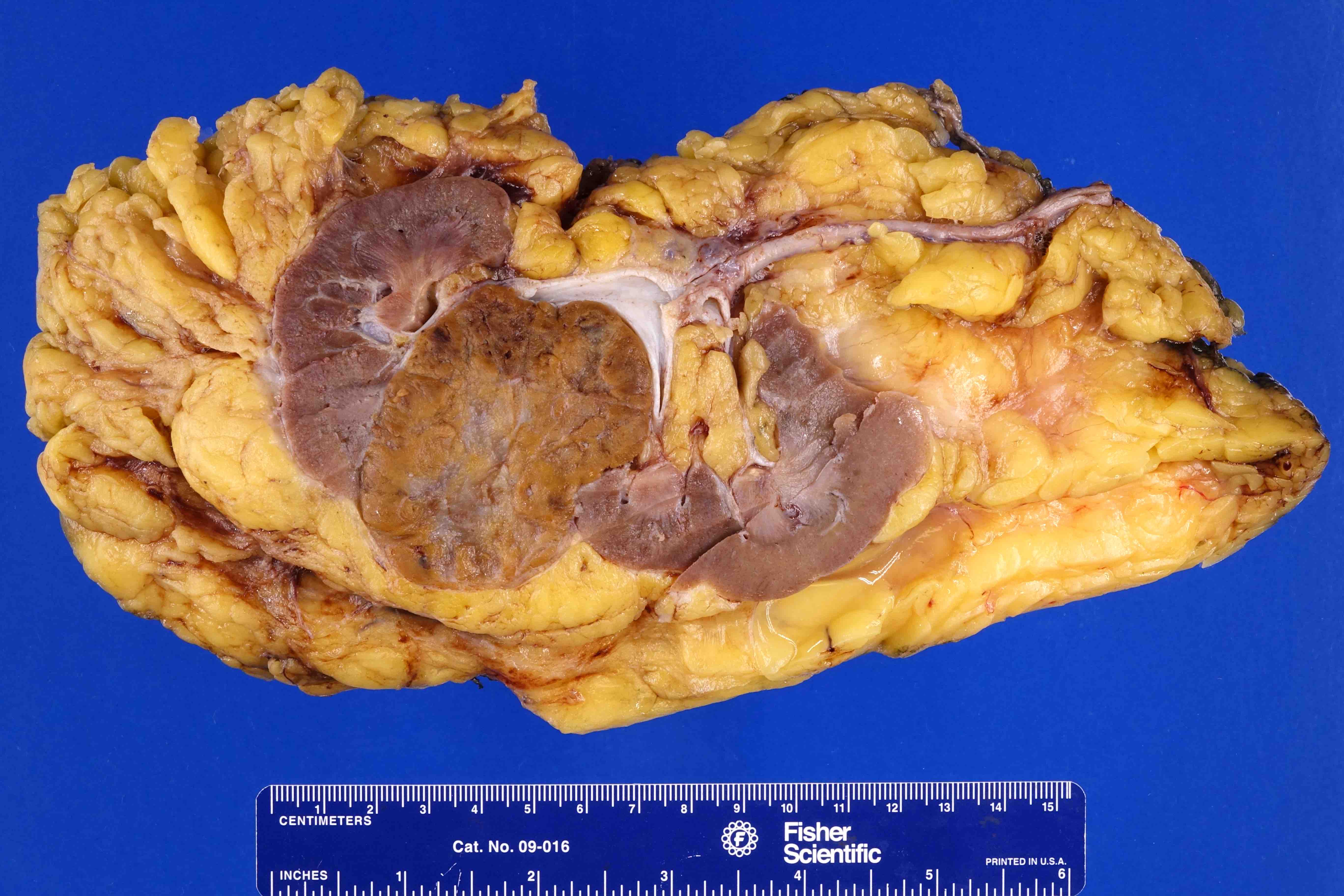
- Make a longitudinal cut with a long knife along the long axis of the kidney to bivalve the specimen (i.e. open like a book) either from medial to lateral or vice versa
- Serially section the entire specimen, including perinephric fat, at 1 cm intervals either along the same plane as the first cut creating "pages in a book" (anterior to posterior) or using a "bread loaf" technique (superior to inferior)
- Dictate the pole (upper, mid, lower) in which the tumor is located
- Measure the distance of the nearest tumor to the nearest inked outer surface
- Measure the tumor to 1/10 of a centimeter
- Describe the tumor (color, consistency)
- Assess and document the percentage of necrosis
- For multiple discrete tumors that do not connect to each other and have a similar gross appearance, measure and state the location of at least the largest 3 tumors
- If additional similar tumors are present, provide a range of sizes of these tumors and state which poles of the kidney are involved
- For additional tumors that do not appear grossly similar to the main tumor, measure and state the location
- Assess for areas where the tumor appears to be invading perinephric fat, renal sinus fat or the pelvicaliceal system, as this impacts pT classification, and document this in the gross description
- Measure the distance of the tumor to the nearest perinephric fat, renal sinus fat and pelvicaliceal system if grossly uninvolved
- Assess the nonneoplastic renal parenchyma
- Search the superior perirenal fat for an adrenal gland and serially section if identified
- Examine the adipose tissue for lymph nodes, particularly the perihilar area
Sections to obtain
- Renal vein with no tumor thrombus, artery and ureter:
- If there is no tumor near the renal vein margin, take shave doughnut margins of ureter, renal vein and artery and place them in 1 cassette
- Renal vein with tumor thrombus, artery and ureter:
- Carefully trim as close as possible to the renal vein margin using scissors and place the trimmings in 1 cassette
- Place the ureter and renal artery in a second cassette
- Take 2 representative cross sections (doughnuts) of the renal vein / branch with thrombus and submit in 2 separate cassettes
- Main tumor, partial nephrectomy:
- Submit 1 block of the tumor per 1 cm of maximum tumor dimension up to 15 blocks maximum
- Include sections that contain tumor to the nearest inked capsule / perirenal adipose tissue, necrosis and adjacent normal parenchyma
- If invasion of perinephric fat, renal sinus fat or pelvicaliceal system is present, submit sections
- If a block of tumor is submitted containing fat, specify if the fat is peripelvic or perirenal in the cassette summary
- Main tumor, radical nephrectomy:
- Representatively submit tumor for a total of 1 block per 1 cm of maximum tumor dimension up to 15 blocks maximum
- About 1/3 of the submitted tumor blocks should be of the tumor to the nearest renal sinus fat
- For areas that are suspicious for gross invasion of peripelvic fat, 1/3 of the tumor blocks should be of tumor to peripelvic fat but at a minimum submit 3 blocks of tumor to peripelvic adipose
- About 1/3 of the submitted tumor blocks should be of the tumor to the nearest perirenal fat
- If the tumor is too far away (greater than 2 cm) from the perirenal or peripelvic fat, submit 1 representative section labeled "perirenal fat closest to tumor" or "peripelvic fat closest to tumor" which will not count as a representative tumor block
- If a block of tumor is submitted containing fat, specify if the fat is peripelvic or perirenal in the cassette summary
- If the inked surface appears to be involved by tumor, submit 2 representative blocks of this area
- If the inked surface is uninvolved, submit 1 block of Gerota fascia with the nearest tumor, which can be counted as part of the representative blocks submitted of the tumor to perirenal adipose
- If the tumor is too far away (greater than 2 cm) from the nearest inked Gerota fascia, submit a representative block labeled "Gerota fascia closest to tumor" which will not count as a representative tumor block
- The remaining 1/3 of the tumor blocks should be of tumor to the nearest pelvicaliceal system (if not present in the peripelvic fat sections), additional sections of tumor (particularly from firm to fleshy gray-white areas suspicious for sarcomatoid differentiation) and representative tumor with necrosis
- Additional tumors:
- For multiple discrete tumors that do not connect to each other and have a similar gross appearance, measure and sample at least the largest 3 tumors
- Additional tumors that do not appear grossly similar to the main tumor should be sampled
- Uninvolved renal parenchyma:
- Take 1 section of normal kidney as far from the tumor as possible and submit in 1 block
- Adrenal gland:
- If the adrenal gland is unremarkable, submit 1 block
- For an adrenal gland that appears to be involved by tumor renal in origin, submit 2 blocks of adrenal gland with tumor, 2 blocks of fat between the adrenal gland and the kidney and 1 block of normal adrenal, in order to differentiate between direct extension of tumor (pT4) versus metastasis (pM1)
- For an adrenal lesion that appears to be adrenal in origin (adrenal cortical adenoma, adrenal cortical hyperplasia), submit 1 block per 1 cm of tumor in addition to 2 blocks of fat between the adrenal gland and the kidney and 1 block of normal adrenal
- Lymph nodes:
- Submit all lymph nodes identified
Tips
- Renal vein margin:
- Tumor may protrude several centimeters past the renal vein margin yet the margin may not have adherent tumor and should therefore be described as grossly negative
- Tumor size:
- 4.0, 7.0 and 10.0 cm are size cutoffs for pT classification
- Measurement should include tumor within the perinephric and peripelvic adipose
- Measurement should not include tumor within the renal vein
- Larger tumors frequently have peripelvic fat invasion (more than 90% of clear cell renal cell carcinomas over 7 cm have peripelvic fat invasion)
- Adrenal gland:
- Always note the presence or absence in the dictation
- Tumor submission example:
- If the tumor is 6 cm and does not appear to invade the peripelvic adipose, submit 2 cassettes of tumor with nearest peripelvic fat and pericaval system, 2 cassettes with perirenal fat and 2 additional cassettes of tumor
Frozen section
- Click here for the frozen section procedure topic
Margins:
- Assessment of surgical margins is often performed in partial nephrectomy cases (Urology 2006;67:923, Scand J Urol Nephrol 2005;39:222)
- However, clinical significance may be limited, as only rare patients harbor residual disease in a follow up radical nephrectomy specimen or repeated partial resection of margin
- Recurrence due to positive margin is uncommon (Urology 2011;77:1400, J Urol 2005;173:385)
- Routine use is controversial due to a relatively high false negative rate, controversy over the prognosis of a positive margin and its inconsistent impact on intraoperative management (BJU Int 2015;116:868)
- Other indications (although no consensus exists) are:
- Solid renal mass in a diffusely cystic kidney or other unusual setting
- Synchronous renal and extrarenal masses
- Cystic renal lesions
- Multiple renal masses (Arch Pathol Lab Med 2005;129:1585, J Urol 2008;179:461)
- Cystic and spindle cell tumors are often misdiagnosed at frozen section
- Clear cell renal cell carcinoma can be confused with inflammatory lesions, particularly those containing foamy histiocytes
Gross description
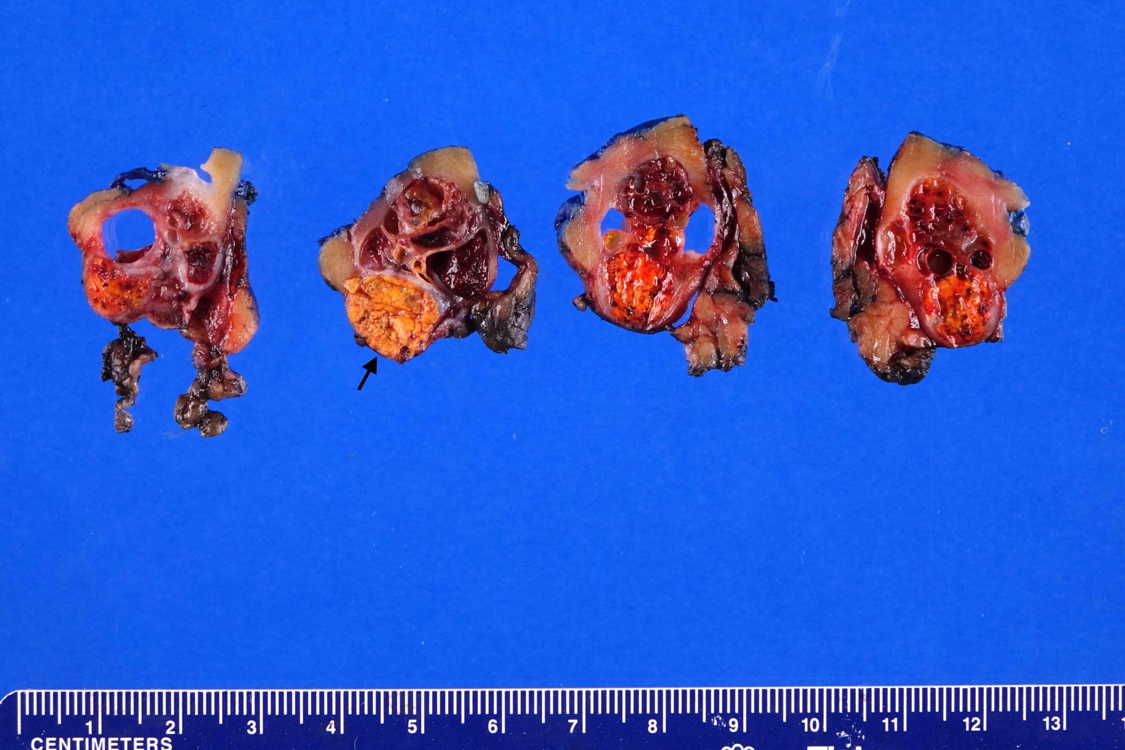
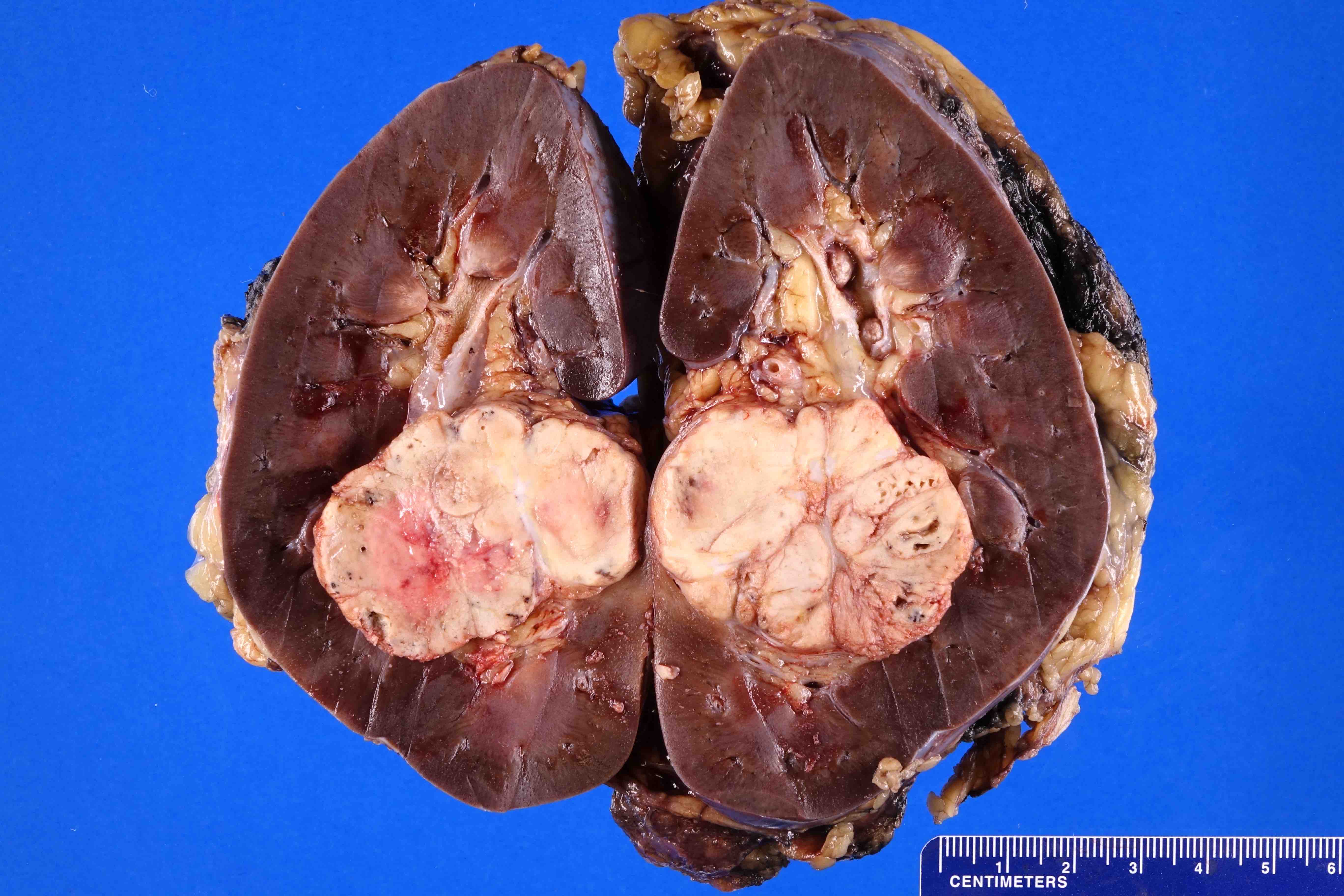
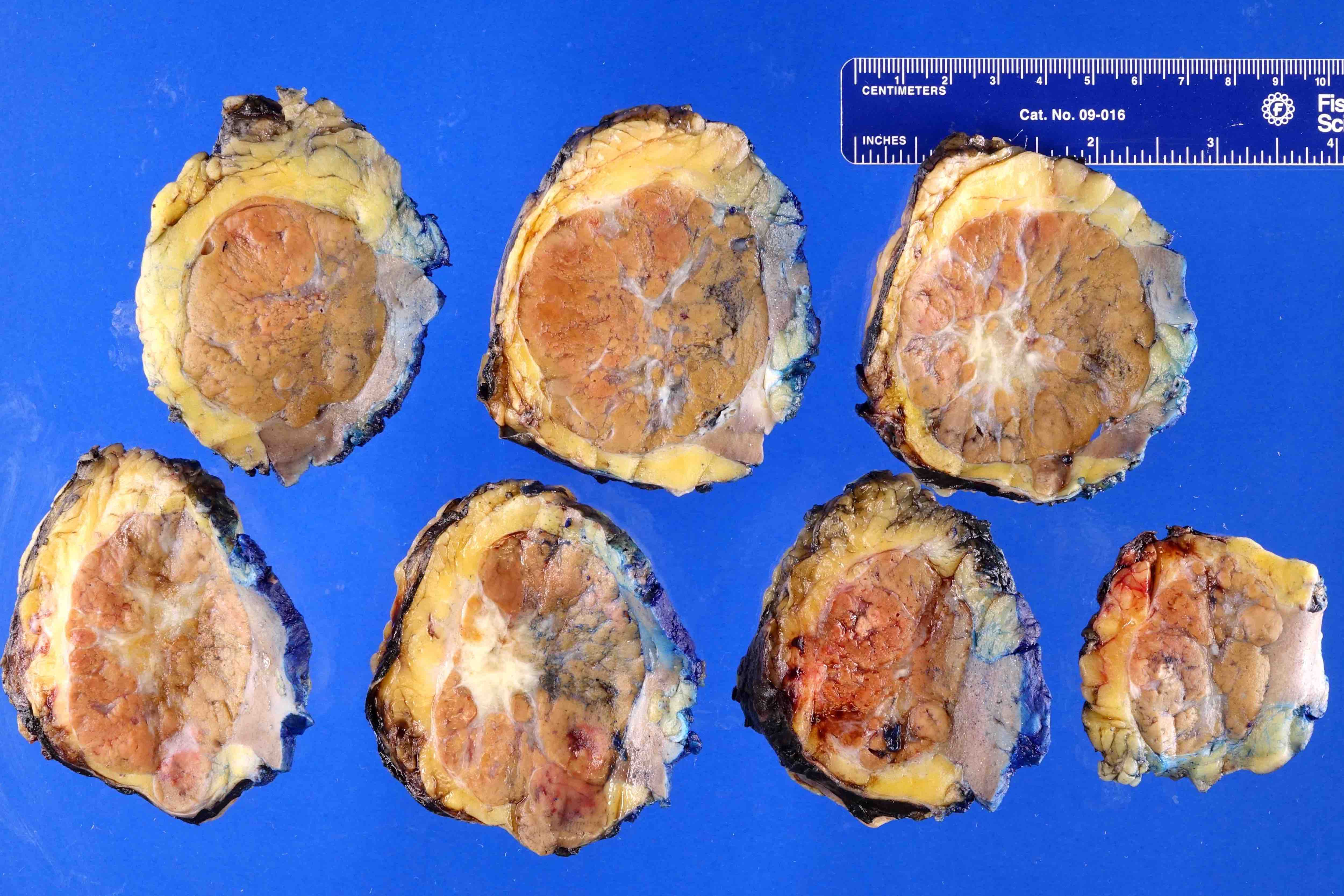
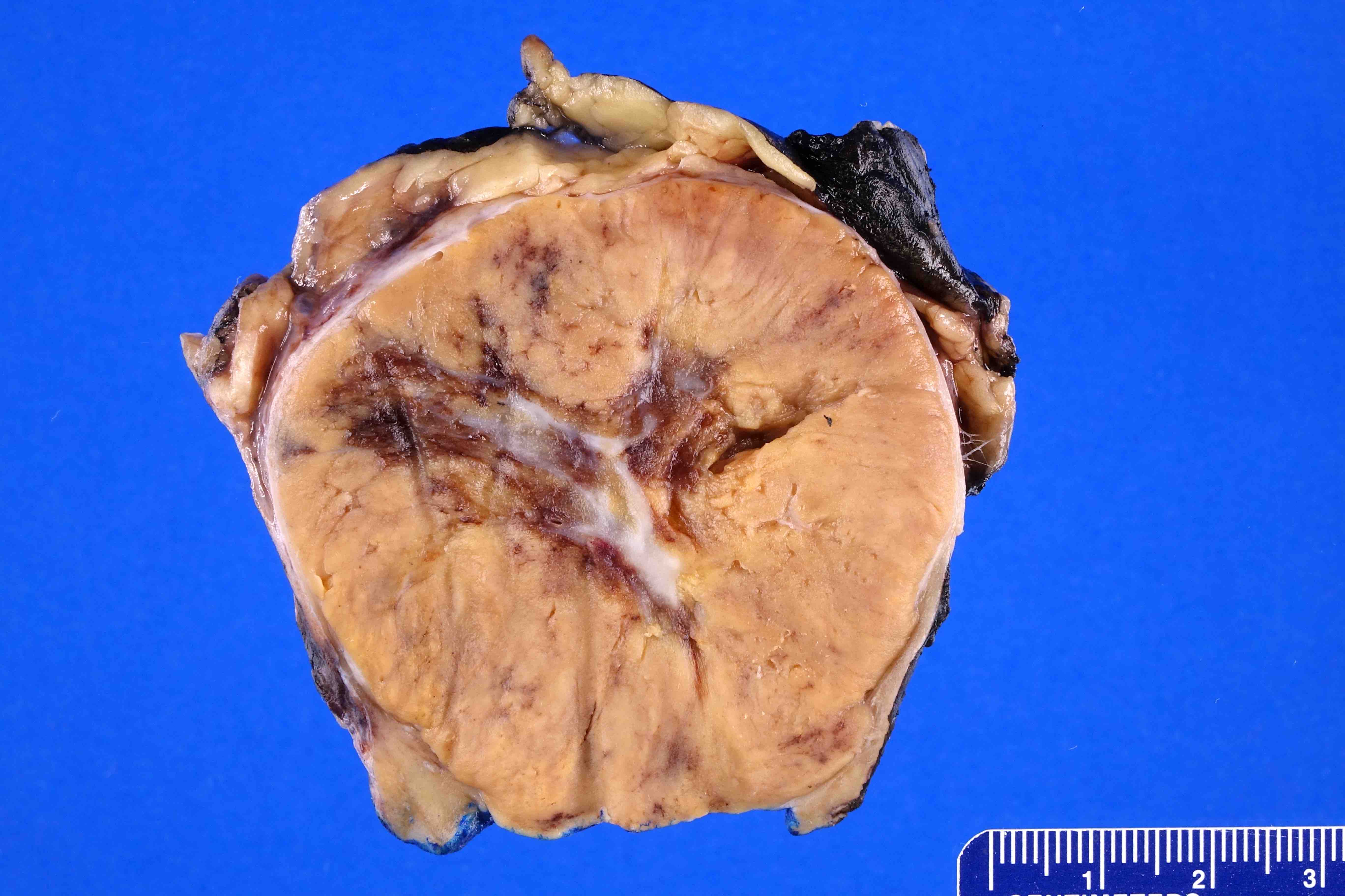
- Renal cell carcinoma, clear cell type:
- Most common overall and most common to be large with invasion of surrounding adipose
- Yellowish, solid to cystic
- May have hemorrhage and necrosis
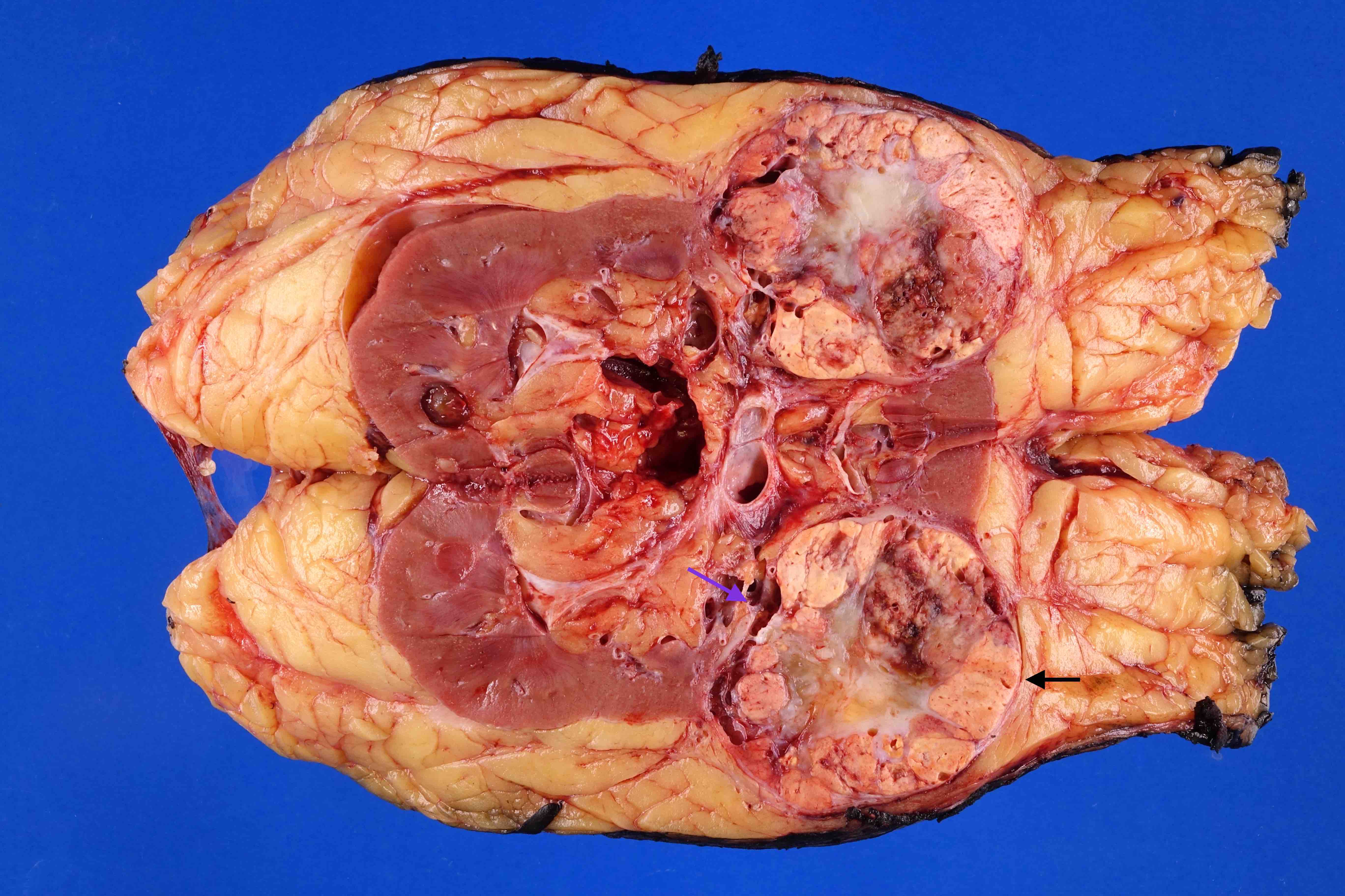
- Renal cell carcinoma, papillary type:
- Unencapsulated, tan
- Renal cell carcinoma, clear cell papillary carcinoma:
- Tan, usually cystic
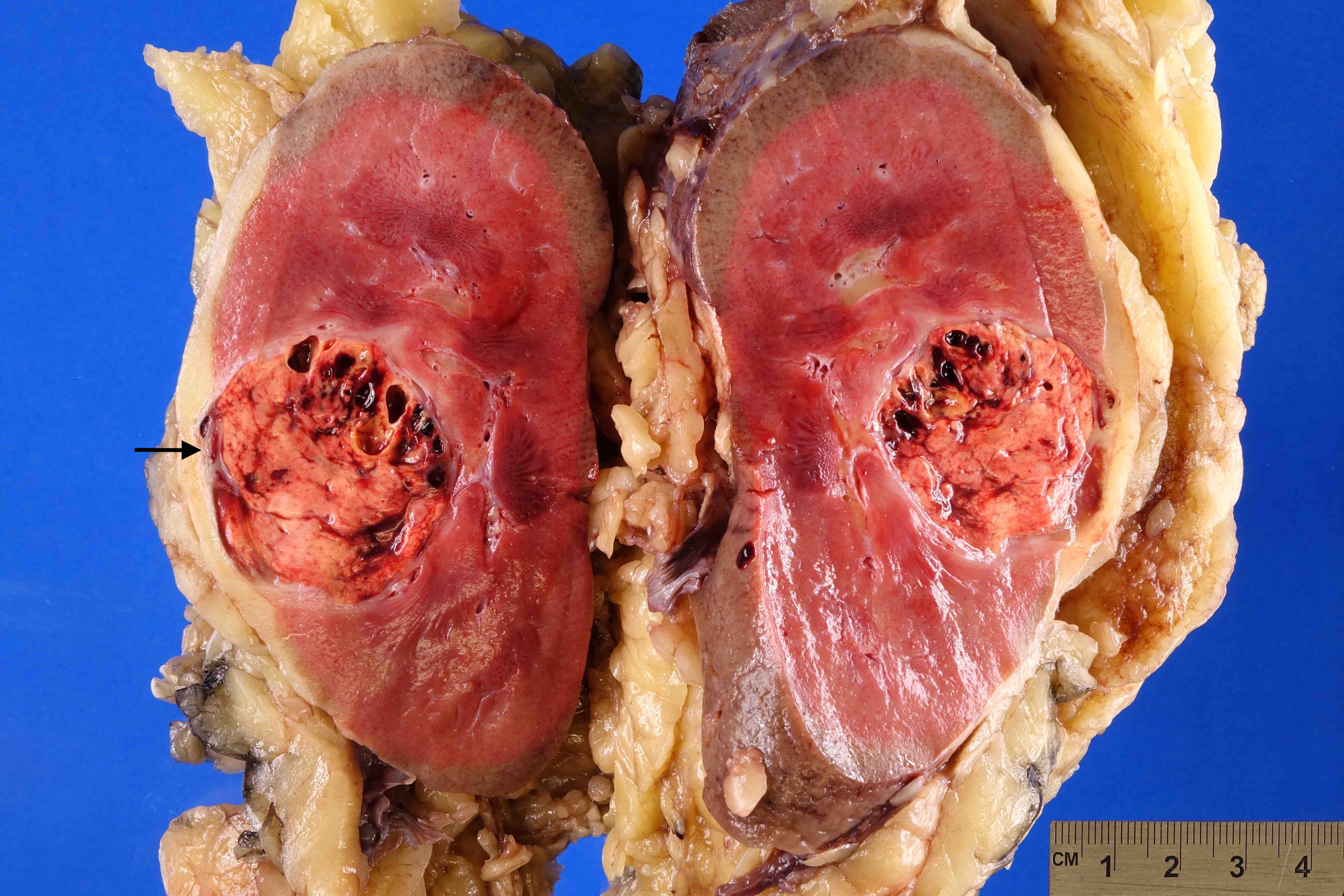
- Renal cell carcinoma, chromophobe type:
- Well circumscribed, tan
- Renal cell carcinoma with sarcomatoid differentiation:
- White, solid
- Oncocytoma:
- Well circumscribed, unencapsulated, mahogany brown
- May have central scar
- Renal medullary interstitial cell tumor:
- Within medulla
- Small (< 2 cm), white
Gross images
Sample gross description report
- Partial nephrectomy:
- The specimen is received in a properly labeled container with the patient's name and accession number, designated as "right partial nephrectomy" and consists of a portion of kidney that weighs 150 g and measures 6.2 x 4.3 x 3.1 cm. The renal parenchymal margin is inked blue and the renal capsular margin is inked black. Cut section of the specimen reveals a yellow-white, well circumscribed tumor that measures 4.7 x 3.3 x 2.1 cm. Necrosis is not present. The tumor is 0.3 cm from the renal parenchymal margin. The tumor is 0.5 cm from the inked capsule and perinephric fat. The uninvolved renal parenchyma is unremarkable.
- A1, tumor with nearest renal parenchymal margin (perpendicular); A2, tumor with nearest inked perinephric adipose; A3, firm area of tumor with white discoloration; A4, tumor with hemorrhage; A5, center of tumor; A6, uninvolved renal parenchyma
- Radical nephrectomy:
- The specimen is received in a properly labeled container with the patient's name and accession number, designated as "right nephrectomy" and consists of a kidney and perirenal fat with an attached segment of ureter, renal vein and artery that weighs 350 grams and measures 12.2 x 8.3 x 4.1 cm. The renal vein is 3.2 cm in length by 1.1 cm in diameter. The outer surface surrounding the tumor is inked black. Tumor is not present within the renal vein or its branches. Located in the upper pole is a yellow-tan-white, well circumscribed tumor that measures 6.2 x 5.3 x 4.3 cm. The tumor does not appear to involve the renal sinus adipose or pelvicaliceal system. The tumor does not appear to involve the perinephric adipose. The tumor does not appear to involve the inked outer surface. Nearest tumor is 0.4 cm from the perinephric adipose, 0.3 cm from the renal sinus adipose and 0.6 cm from the inked outer surface. The tumor contains hemorrhage and 10% necrosis. The uninvolved renal parenchyma is unremarkable. The perinephric fat does not contain an adrenal gland. Examination of the renal hilar fat does not reveal lymph nodes.
- A1, margins of ureter, vein and artery (shave); A2-3, tumor with nearest renal sinus fat and pelvicaliceal system; A3, tumor with nearest perinephric fat and inked Gerota fascia; A4-5, tumor with nearest perinephric fat; A6, tumor with necrosis; A7; tumor with firm, white, indurated areas; A8, uninvolved renal parenchyma
Board review style question #1
Board review style answer #1
D. Renal cell carcinoma, clear cell type. None of the other tumors are typically golden yellow in color.
Comment Here
Reference: Kidney tumor - Grossing & frozen section
Comment Here
Reference: Kidney tumor - Grossing & frozen section


