Table of Contents
Definition / general | Essential features | Terminology | ICD coding | Epidemiology | Sites | Pathophysiology | Etiology | Clinical features | Radiology description | Prognostic factors | Case reports | Treatment | Gross description | Gross images | Microscopic (histologic) description | Microscopic (histologic) images | Positive stains | Negative stains | Electron microscopy description | Molecular / cytogenetics description | Molecular / cytogenetics images | Sample pathology report | Differential diagnosis | Board review style question #1 | Board review style answer #1 | Board review style question #2 | Board review style answer #2Cite this page: Tretiakova M. ALK translocation. PathologyOutlines.com website. https://www.pathologyoutlines.com/topic/kidneytumorALKtranslocationrcc.html. Accessed April 1st, 2025.
Definition / general
- Renal cell carcinoma (RCC) associated with anaplastic lymphoma kinase (ALK) gene rearrangement (chromosome 2p23)
- Affects children with sickle cell trait or adults without sickle cell trait
- In children, resembles renal medullary carcinoma: medulla centric with diffuse infiltrating growth, lymphoplasmacytic infiltrate, large polygonal discohesive or spindled cells, cytoplasmic vacuoles and vesicular nuclei
- In adults, heterogeneous solid architecture, mucinous cribriform, signet ring and solid rhabdoid patterns, high grade eosinophilic cells with intracytoplasmic lumina (Pol J Pathol 2018;69:109, Semin Diagn Pathol 2015;32:90, Histopathology 2019;74:31)
Essential features
- Architecturally heterogeneous eosinophilic tumor with polygonal, rhabdoid, signet ring and spindled cells, mucin production and cytoplasmic vacuoles
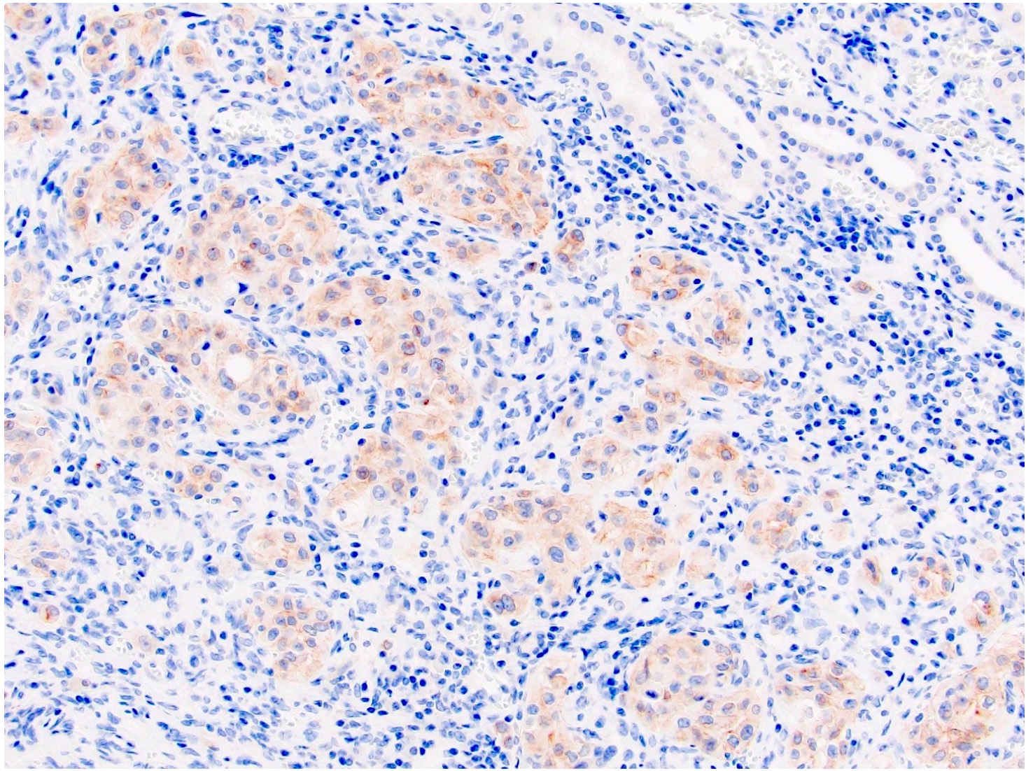
- Diffuse ALK protein expression
- Detection of ALK gene rearrangement by FISH, RT-PCR or RNA sequencing
Terminology
- First 2 cases of VCL-ALK fusion RCC described in 2010 (Mod Pathol 2011;24:430)
- Synonyms: ALK rearrangement associated RCC, ALK translocation RCC, ALK-RCC
- Provisional entity in the WHO classification (2016)
ICD coding
- ICD-10: C64 - malignant neoplasm of kidney, except renal pelvis
Epidemiology
- Rare; < 25 cases described
- Bimodal age distribution:
- Children with sickle cell trait, often African descent (range: 6 - 19 years)
- Adults without sickle cell trait (range: 33 - 61 years)
- Incidence: 3.5 - 3.8% of pediatric renal cancers, 0.4 - 0.5% of adult renal cancers (Cancer 2018;124:3381, Histopathology 2017;71:53, Genes Chromosomes Cancer 2016;55:442, Cancer 2012;118:4427, Histopathology 2019;74:31)
Sites
- Solitary kidney mass
- Most commonly in renal medulla, renal pelvis or mid kidney
Pathophysiology
- ALK belongs to the insulin receptor tyrosine kinase superfamily and is normally expressed at a low level in the central nervous system
- ALK rearrangement represents an oncogenic driver mutation that probably occurs during the early stage of carcinogenesis (Histopathology 2019;74:31)
Etiology
- In children, related to sickle cell trait in a subset of patients (Am J Surg Pathol 2014;38:858)
Clinical features
- Majority with hematuria, flank, abdominal or periumbilical pain (Pol J Pathol 2018;69:109, Histopathology 2017;71:53)
- Incidental in ~33% of cases
- Most pT1a or pT1b
Radiology description
- Ultrasound sonography demonstrates a hypoechoic medulla centric mass
- Simple computed tomography scan shows an isodense mass
- Contrast computed tomography scan shows a slightly enhancing or heterogeneous enhancing mass (Pol J Pathol 2018;69:109)
Prognostic factors
- Children with VCL-ALK: no recurrence or distant metastasis reported to date
- Adult non-VCL-ALK RCC: ~33% have adverse prognosis (Histopathology 2017;71:53, Histopathology 2019;74:31)
- Small number of cases and short followup in the majority of them precludes from accurate assessment of risk factors
Case reports
- 16 year old boy with novel HOOK1-ALK fusion (Genes Chromosomes Cancer 2016;55:814)
- 19 year old woman with ALK-RCC and Hodgkin lymphoma (Pathol Int 2017;67:626)
- 33 year old woman and 38 year old man with RCC harboring novel STRN-ALK fusion (Am J Surg Pathol 2016;40:761)
- 55 year old woman with ALK-TPM3 rearrangement and loss of chromosome 3 (Pol J Pathol 2018;69:109)
- Increased ALK1 copy number and RCC (Virchows Arch 2014;464:241)
Treatment
- Primary tumor, early stage: radical nephrectomy or nephroureterectomy
- Metastatic ALK-RCC responds to ALK inhibitor alectinib (Eur Urol 2018;74:124)
- Possibly responds to crizotinib, similar to ALK rearrangement lung cancer (Semin Diagn Pathol 2015;32:90)
Gross description
- Pediatric patients: medulla centric, irregularly shaped solid tumor mass with infiltrative borders (Genes Chromosomes Cancer 2016;55:442, Pol J Pathol 2018;69:109)
- Adult patients: well demarcated solid tumor in mid kidney, tan to brown or white to gray-white color, cystic change or hemorrhage may be present but pseudocapsule is absent
- Size: 3 - 7 cm
Gross images
Microscopic (histologic) description
- Pediatric patients (Genes Chromosomes Cancer 2016;55:442, Cancer 2018;124:3381):
- Diffuse sheet-like infiltrating growth pattern
- Lymphoplasmacytic infiltrate and intravascular sickling
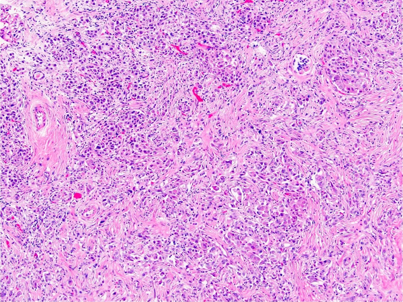
- Round, oval and polygonal tumor cells with abundant vaguely granular eosinophilic cytoplasm and frequent intracytoplasmic lumina
- Abundant background mucin and intracytoplasmic mucin
- Moderately polymorphic, predominantly vesicular nuclei with small nucleoli, occasional grooves and rare vacuoles
- Adult patients (Histopathology 2019;74:31, Semin Diagn Pathol 2015;32:90):
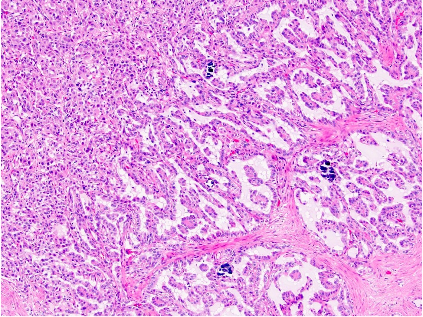
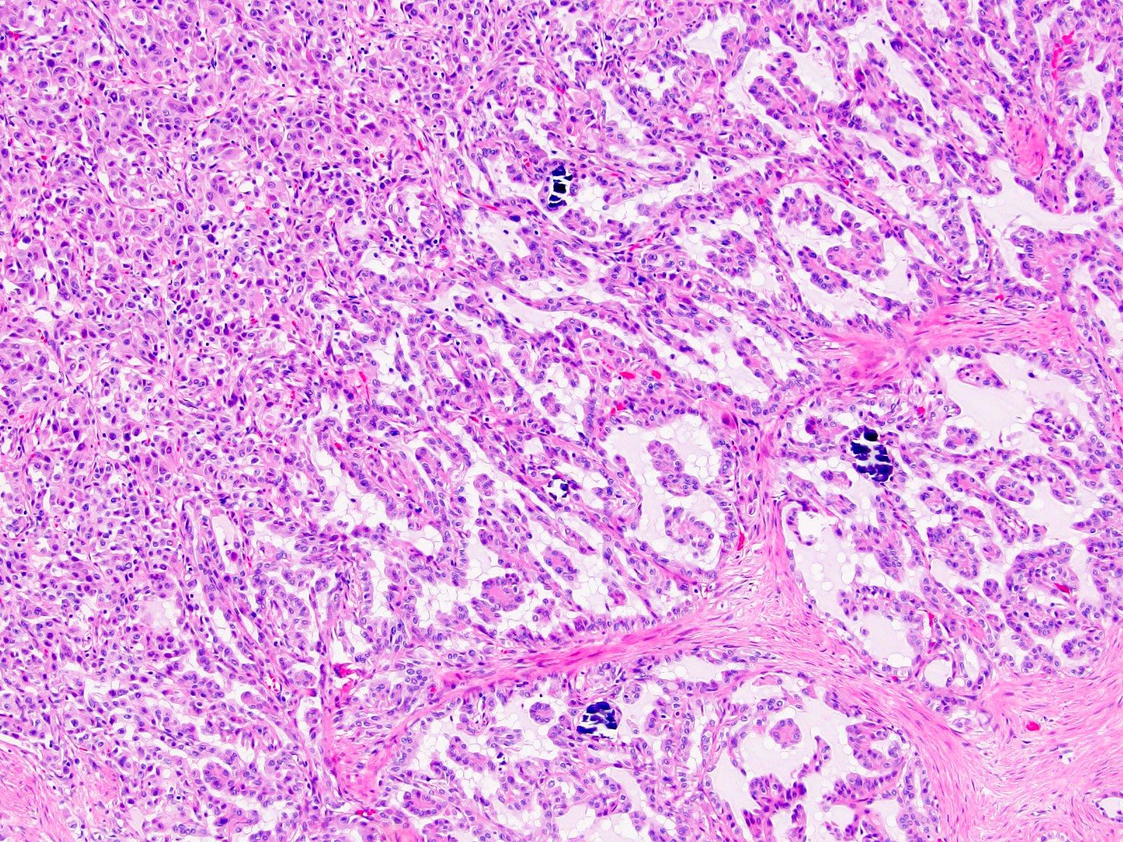
- More heterogeneous architecture: solid, mucinous cribriform, reticular, tubular, papillary growth; discohesive sheets or infiltrating single cells
- Eosinophilic polygonal cells, rhabdoid or signet ring due to intracytoplasmic vacuolization
- May contain psammoma bodies and foamy macrophages
Microscopic (histologic) images
Positive stains
Negative stains
- RCC, CD117 / KIT, S100, HMB45, MelanA, cathepsin K, WT1
- Ki67 very low (< 5%)
Electron microscopy description
- Tumor cells with bundles of tonofilaments, intercellular junctions, desmosomes, intracytoplasmic lumina lined by microvilli and lipofuscin-like lysosomal structures (Genes Chromosomes Cancer 2016;55:442)
Molecular / cytogenetics description
- VCL-ALK fusion (strong association with sickle cell trait) (Genes Chromosomes Cancer 2011;50:146)
- TPM3-ALK fusion (no association with sickle cell trait) (Genes Chromosomes Cancer 2016;55:442)
- Other less common fusions: STRN-ALK, EML4-ALK, HOOK1-ALK (Am J Surg Pathol 2016;40:761, Pol J Pathol 2018;69:109)
- Clonal inversion involving 2p23 (Mod Pathol 2011;24:430)
- No fusion but increased copy number of ALK1 (Virchows Arch 2014;464:241)
- ALK copy number gain could be identified in up to 10% of clear cell RCC and associated with worse cancer specific survival (Mod Pathol 2012;25:1516)
Molecular / cytogenetics images
Sample pathology report
- Left kidney, nephrectomy:
- Renal cell carcinoma with ALK gene rearrangement (see comment and synoptic report)
- Comment: The sections show an infiltrating epithelioid neoplasm in the kidney, arranged as lobules, small nests and ducts, some with blue mucinous material in the lumina. Rare, dispersed apparent mucin containing cells are present. The neoplastic cells have an eosinophilic cytoplasm and slightly pleomorphic, variably enlarged and hyperchromatic nuclei, which have a generally low grade cytology. Many nuclei have intranuclear vacuoles. Cells with rhabdoid and signet ring features are noted but there are virtually no mitoses. Our differential diagnosis of this infiltrating high grade adenocarcinoma is very broad, including collecting duct carcinoma, translocation carcinoma, urothelial carcinoma, metastatic carcinoma, unclassified renal cell carcinoma and renal medullary carcinoma. Immunohistochemical stains showed that the tumor cells are positive for keratin, PAX8, INI1 and negative for GATA3 and TTF1, which is characteristic of a primary renal cell carcinoma. Additionally performed immunostaining with ALK was diffusely positive. FISH study showed evidence of a translocation or rearrangement involving the ALK gene (100% split apart signals). These findings are consistent with ALK rearranged RCC, a rare subtype of renal cell carcinoma.
Differential diagnosis
- Very broad, thus immunostaining with ALK antibody and molecular studies confirming ALK translocation are critical for diagnosis
- Renal medullary carcinoma:
- Collecting duct carcinoma:
- Also in medullary location but usually larger size, predominant tubular growth, stromal desmoplasia, high Ki67
- MiT family translocation renal cell carcinoma:
- Also TFE3 positive and can have similar morphology
- Can be positive for melanocytic markers and cathepsin K
- Papillary renal cell carcinoma and mucinous tubular and spindle cell carcinoma:
- Also has tubulopapillary architecture, mucin production, psammoma bodies, foamy macrophages and overlapping immunoprofile
- Lack ALK immunohistochemical expression
- Unclassified renal cell carcinoma:
- ALK immunohistochemistry negative
- Metastases, especially ALK rearranged lung adenocarcinoma:
- Usually positive for TTF1 / napsin A and negative for PAX8
- Also check clinical history (Semin Diagn Pathol 2015;32:90, Pol J Pathol 2018;69:109)
Board review style question #1
Which is true about ALK translocation renal cell carcinoma?
- Can be easily distinguished from other renal carcinomas due to heterogeneous solid, tubular, cribriform and rhabdoid morphology
- Detection of ALK rearrangement is critical for diagnosis
- It affects only pediatric patients
- Typically has aggressive course
Board review style answer #1
B. Detection of ALK rearrangement is critical for diagnosis. ALK rearranged renal cell carcinoma (ALK-RCC) has been recently added as a provisional entity into the 2016 World Health Organization classification. ALK-RCC is characterized by fusion of a variety of genes with the anaplastic lymphoma kinase (ALK) gene occurring in children with sickle cell trait and adults without sickle cell trait. It is a rare tumor affecting patients from 6 to 61 years old (mean 30 years). In children, ALK-RCC are medulla based and morphologically resemble medullary renal carcinomas. In adults, ALK-RCC is very heterogeneous with solid, tubular mucinous, rhabdoid, cribriform and signet ring histology. Tumor cells could be amphophilic, eosinophilic, polygonal rhabdoid and often contain cytoplasmic vacuoles. Immunostaining with ALK is positive (focally or diffusely), whereas INI1 protein is intact. ALK gene rearrangement should be confirmed by FISH or molecular studies. The majority of reported tumors are indolent; however, some cases may pursue an aggressive clinical course.
Comment Here
Reference: ALK translocation
Comment Here
Reference: ALK translocation
Board review style question #2
Board review style answer #2
C. TFE3, ALK, CK7. Microscopic image shows tumor with solid and nested areas transitioning to tubulopapillary areas with psammoma bodies and abundant extracellular mucin. These features in various proportions could be seen in papillary RCC, MiT family translocation RCC, mucinous tubular and spindle cell carcinoma, collecting duct and renal medullary carcinoma. All of the above panels will be helpful in narrowing down the differential diagnosis but only in answer C was an antibody for ALK rearrangement RCC added. Young patient age and high morphologic variability of tumor lacking specific features of more common renal cancer subtypes (unclassifiable RCC) should raise suspicion of this rare tumor and warrant ordering of ALK immunostaining and FISH study.
Comment Here
Reference: ALK translocation
Comment Here
Reference: ALK translocation


