Eye

General

Staging-uvea melanoma


Editor-in-Chief: Debra L. Zynger, M.D.
Martin Hyrcza, M.D., Ph.D., M.Sc.

Last author update: 6 May 2019
Last staff update: 12 December 2024 (update in progress)

Copyright: 2003-2024, PathologyOutlines.com, Inc.

PubMed Search: TNM staging uveal melanoma[TI]

See Also: Uveal melanoma

Martin Hyrcza, M.D., Ph.D., M.Sc.
Cite this page: Hyrcza M. Staging-uvea melanoma. PathologyOutlines.com website. https://www.pathologyoutlines.com/topic/eyeuveatnmuvealmelanoma.html. Accessed December 23rd, 2024.
Definition / general
  • Based on AJCC 8th edition staging system
  • Uveal tract is the middle layer of the eye, between sclera and retina
  • Uveal tract consists of the iris, ciliary body and the choroid
Essential features
  • AJCC 7th edition staging was sunset on December 31, 2017; as of January 1, 2018, use of the 8th edition is mandatory
  • There are 2 separate pT classification systems for uveal melanomas: one for the iris and one for the ciliary body and choroid melanomas
  • Tumors with < 50% of their volume located in the iris should be categorized using the ciliary body and the choroid pT classification
  • Clinical and pathological T categories are the same
  • Some patients with iris melanoma experience a secondary open angle glaucoma caused by the tumor cells and tumor pigment laden macrophages becoming trapped in and obstructing the trabecular meshwork
ICD coding
  • ICD-10: C69.3 - Choroid
  • ICD-10: C69.4 - Ciliary body and iris
Diagrams / tables

Contributed by Martin Hyrcza, M.D., Ph.D., M.Sc.

Tumor size category

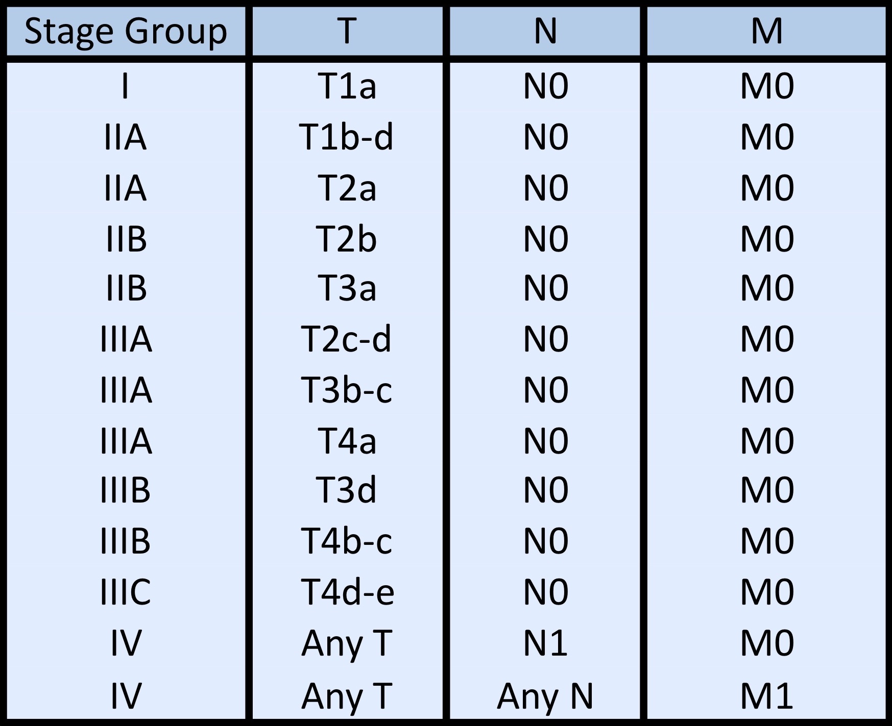
Stage groups

Primary tumor (pT)
Iris:
  • pTX: primary tumor cannot be assessed
  • pT0: no evidence of primary tumor
  • pT1: tumor limited to the iris
    • pT1a: tumor limited to the iris, not more than 3 clock hours in size
    • pT1b: tumor limited to the iris, more than 3 clock hours in size
    • pT1c: tumor limited to the iris, with secondary glaucoma
  • pT2: tumor extending into the ciliary body, choroid or both
    • pT2a: tumor extending into the ciliary body, without secondary glaucoma
    • pT2b: tumor extending into the ciliary body and choroid, without secondary glaucoma
    • pT2c: tumor extending into the ciliary body, choroid or both, with secondary glaucoma
  • pT3: tumor extending into the ciliary body, choroid or both, with scleral extension (but without extrascleral extension)
  • pT4: tumor with extrascleral extension
    • pT4a: tumor with extrascleral extension ≤ 5 mm in largest diameter
    • pT4b: tumor with extrascleral extension > 5 mm in largest diameter

Ciliary body and choroid:
  • pT1: tumor size category 1
    • pT1a: tumor size category 1 without ciliary body involvement or extraocular extension
    • pT1b: tumor size category 1 with ciliary body involvement
    • pT1c: tumor size category 1 without ciliary body involvement but with extraocular extension ≤ 5 mm in largest diameter
    • pT1d: tumor size category 1 with ciliary body involvement and extraocular extension ≤ 5 mm in largest diameter
  • pT2: tumor size category 2
    • pT2a: tumor size category 2 without ciliary body involvement or extraocular extension
    • pT2b: tumor size category 2 with ciliary body involvement
    • pT2c: tumor size category 2 without ciliary body involvement but with extraocular extension ≤ 5 mm in largest diameter
    • pT2d: tumor size category 2 with ciliary body involvement and extraocular extension ≤ 5 mm in largest diameter
  • pT3: tumor size category 3
    • pT3a: tumor size category 3 without ciliary body involvement or extraocular extension
    • pT3b: tumor size category 3 with ciliary body involvement
    • pT3c: tumor size category 3 without ciliary body involvement but with extraocular extension ≤ 5 mm in largest diameter
    • pT3d: tumor size category 3 with ciliary body involvement and extraocular extension ≤ 5 mm in largest diameter
  • pT4: tumor size category 4 or any tumor with extraocular extension > 5 mm
    • pT4a: tumor size category 4 without ciliary body involvement or extraocular extension
    • pT4b: tumor size category 4 with ciliary body involvement
    • pT4c: tumor size category 4 without ciliary body involvement but with extraocular extension ≤ 5 mm in largest diameter
    • pT4d: tumor size category 4 with ciliary body involvement and extraocular extension ≤ 5 mm in diameter
    • pT4e: any tumor size category with extraocular extension > 5 mm in largest diameter

Notes:
  • Largest basal diameter and tumor thickness are used to determine size category (see table above), required to assign pT category to melanomas of the ciliary body and choroid
  • Proper grossing technique is required to obtain largest basal diameter and tumor thickness in enucleation specimens
  • Globe should be transilluminated with strong light source to map out the tumor shadow on the sclera in order to determine the best orientation of pupil-optic nerve section
  • Pupil-optic nerve section should capture the largest basal diameter based on the shadow
  • In clinical practice, largest basal diameter may be estimated in optic disc diameters (dd) (average 1 dd = 1.5 mm) and tumor thickness may be estimated in diopters (average 3 diopters = 1 mm)
Regional lymph nodes (pN)
  • pNX: regional lymph nodes cannot be assessed
  • pN0: no regional lymph node metastasis
  • pN1: regional lymph node metastasis or discrete orbital tumor deposits
    • pN1a: metastasis in one or more regional lymph nodes
    • pN1b: discrete orbital tumor deposits discontinuous with the globe without regional lymph node metastases

Notes:
  • Regional lymph nodes include preauricular, submandibular and cervical
Distant metastasis (pM)
  • pM0: no distant metastasis
  • pM1: distant metastasis
    • pM1a: largest diameter of the largest metastasis is ≤ 3 cm
    • pM1b: largest diameter of the largest metastasis is 3.1 - 8.0 cm
    • pM1c: largest diameter of the largest metastasis is > 8 cm
Prefixes
  • y: preoperative radiotherapy or chemotherapy
  • r: recurrent tumor stage
AJCC prognostic stage groups
Stage group I: T1a N0 M0
Stage group IIA:  T1b - d  N0 M0
Stage group IIA: T2a N0 M0
Stage group IIB: T2b N0 M0
Stage group IIB: T3a N0 M0
Stage group IIIA: T2c - d N0 M0
Stage group IIIA: T3b - c N0 M0
Stage group IIIA: T4a N0 M0
Stage group IIIB: T3d N0 M0
Stage group IIIB: T4b - c N0 M0
Stage group IIIC: T4d - e N0 M0
Stage group IV: any T N1 M0
Stage group IV: any T any N M1
Registry data collection variables
  • Patient age, gender
  • Prior treatment
  • Tumor site: iris, ciliary body, choroid
  • Largest basal diameter and thickness
  • Scleral and optic nerve invasion
  • Extraocular extension
  • Histologic type: spindle, mixed, epithelioid
  • Mitotic count
  • Vascular invasion
  • Extravascular matrix pattern
  • Molecular results (if known):
    • Chromosome 3 and 8 loss / gain
    • BAP1 status
    • Gene expression profile
    • Multiplex ligation dependent probe amplification (MLPA) analysis
Histologic grade (G) / histopathologic type
  • G1: spindle (> 90% spindle cells)
  • G2: mixed (> 10% epithelioid cells and < 90% spindle cells)
  • G3: epithelioid (> 90% epithelioid cells)

Notes:
  • There is a lack of agreement on the proportions needed for the diagnosis of spindle cell and epithelioid types; 10% and 90% are provided for guidance only
Gross images

AFIP images
Missing Image

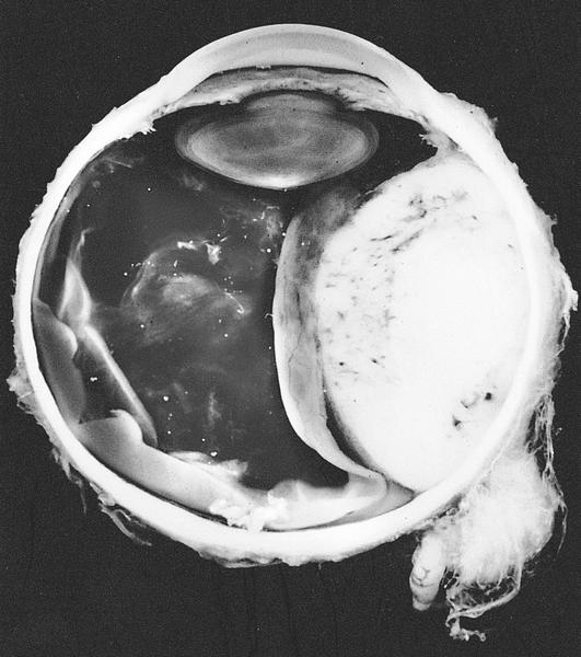
Gross extraocular extension

Whole mount images

AFIP images
Missing Image

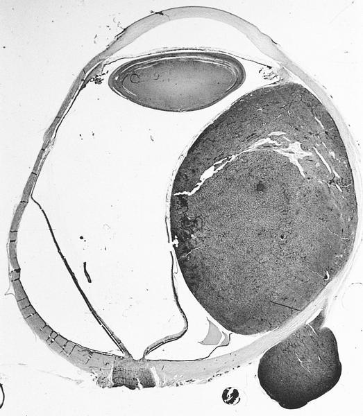
Choroidal melanoma with extraocular extension

Microscopic (histologic) images

Contributed by Martin Hyrcza, M.D., Ph.D., M.Sc.

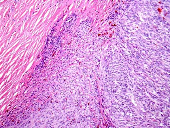
Scleral invasion

Extrascleral extension

Board review style question #1
Uveal melanoma involving 2 mm of iris, all of ciliary body and 9 mm of the choroid has a thickness of 12 mm and largest basal diameter of 16 mm. There is no extraocular extension present and no glaucoma. The pT category is

  1. pT2b
  2. pT2d
  3. pT3b
  4. pT3d
Board review style answer #1
Back to top
Image 01 Image 02