Table of Contents
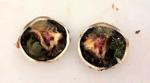
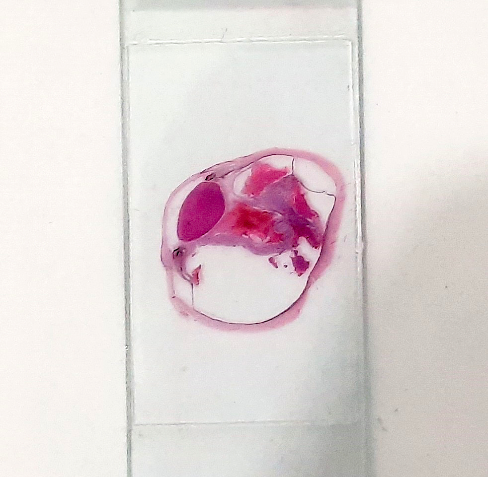
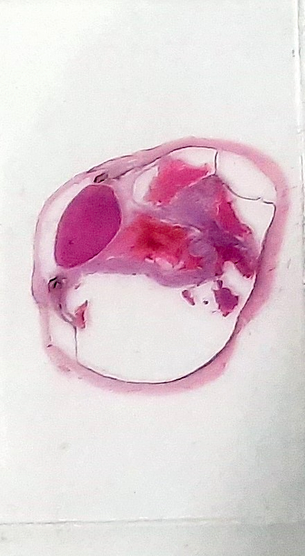
Definition / general | Gross images | Whole mount images | Microscopic (histologic) description | Microscopic (histologic) imagesCite this page: Pernick N. Detachment of retina. PathologyOutlines.com website. https://www.pathologyoutlines.com/topic/eyeretinaretinaldetachment.html. Accessed December 23rd, 2024.
Definition / general
- Separation of neurosensory retina (rods and cone and more superficial layers) from retinal pigment epithelium
- Rhegmatogenous ("due to a rupture or fracture") detachment: associated with full thickness retinal defect, such as collapse of vitreous, causing traction on retinal internal limiting membrane, causing tears and seepage of vitreous between neurosensory layer and retinal pigment epithelium; treated by relieving vitreous traction
- Nonrhegmatogenous detachment: no retinal break; due to significant exudates or conditions causing leakage of fluid from choroidal circulation beneath the retina, such as choroidal tumors and malignant hypertension
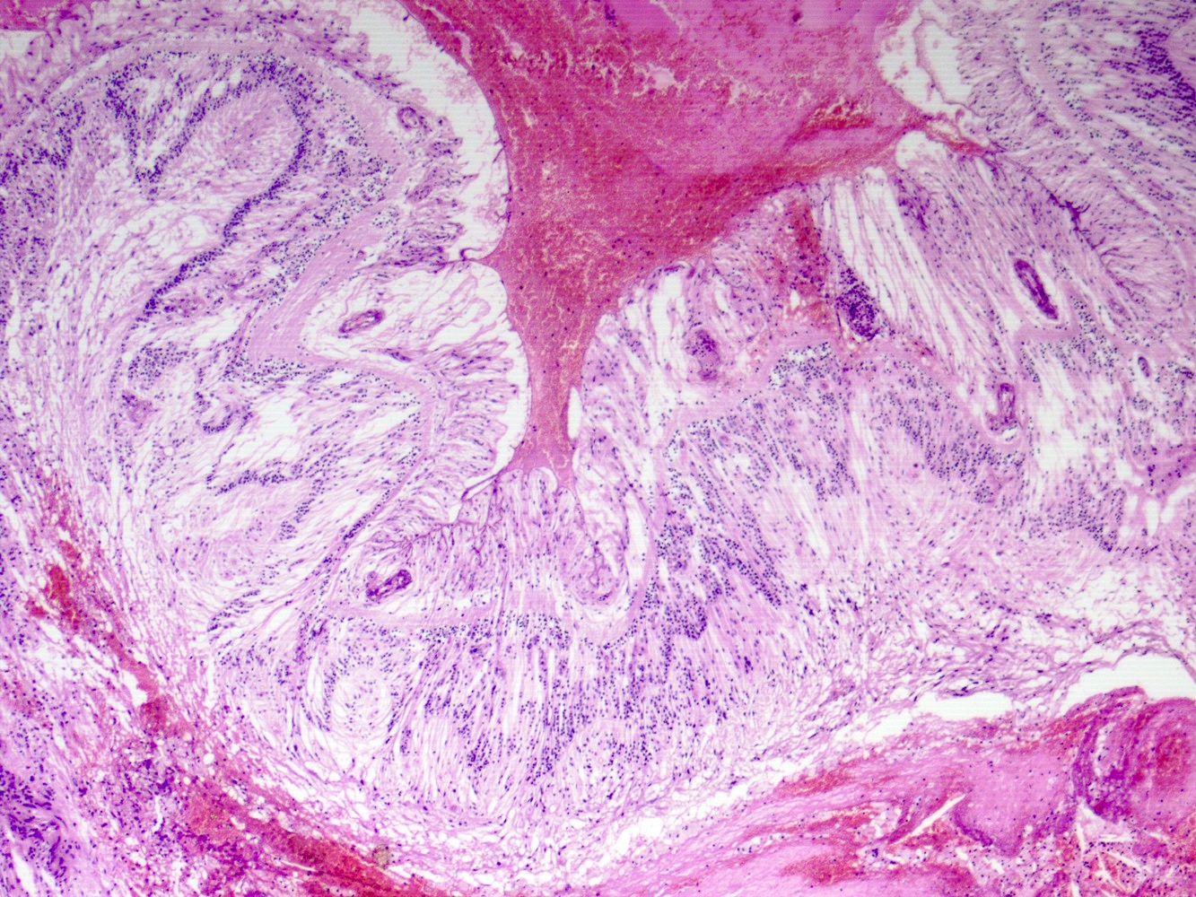
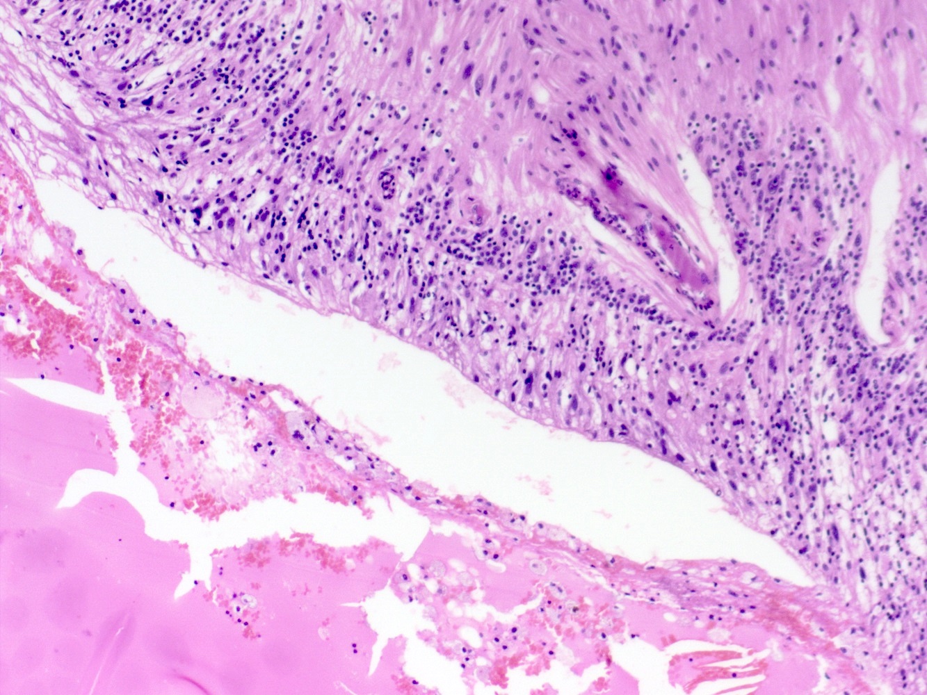
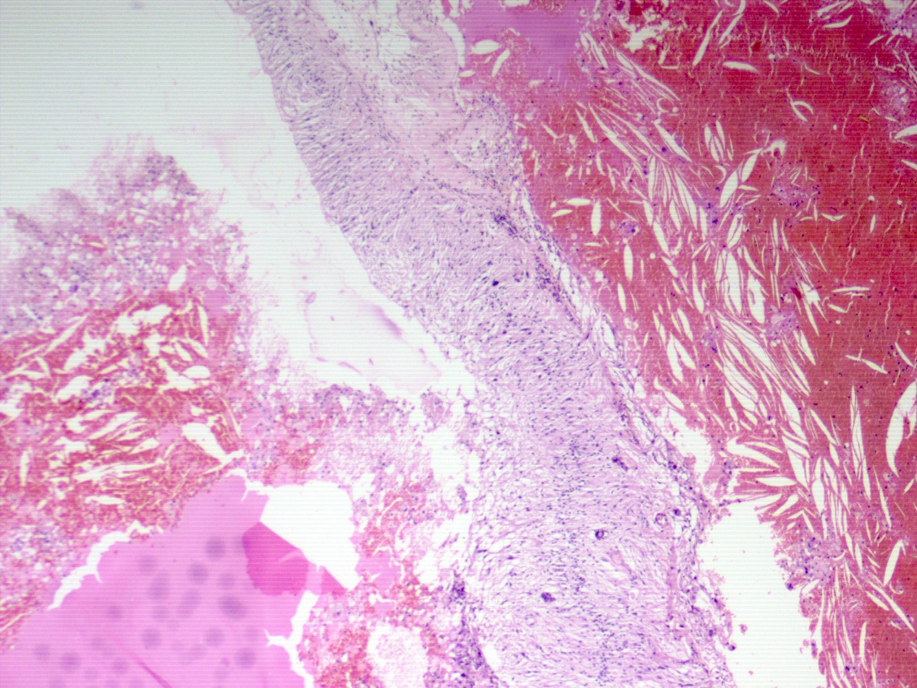
- Chronic retinal detachment may cause loss of photoreceptor outer segments, gliosis and development of microcystic spaces in detached retina
Microscopic (histologic) description
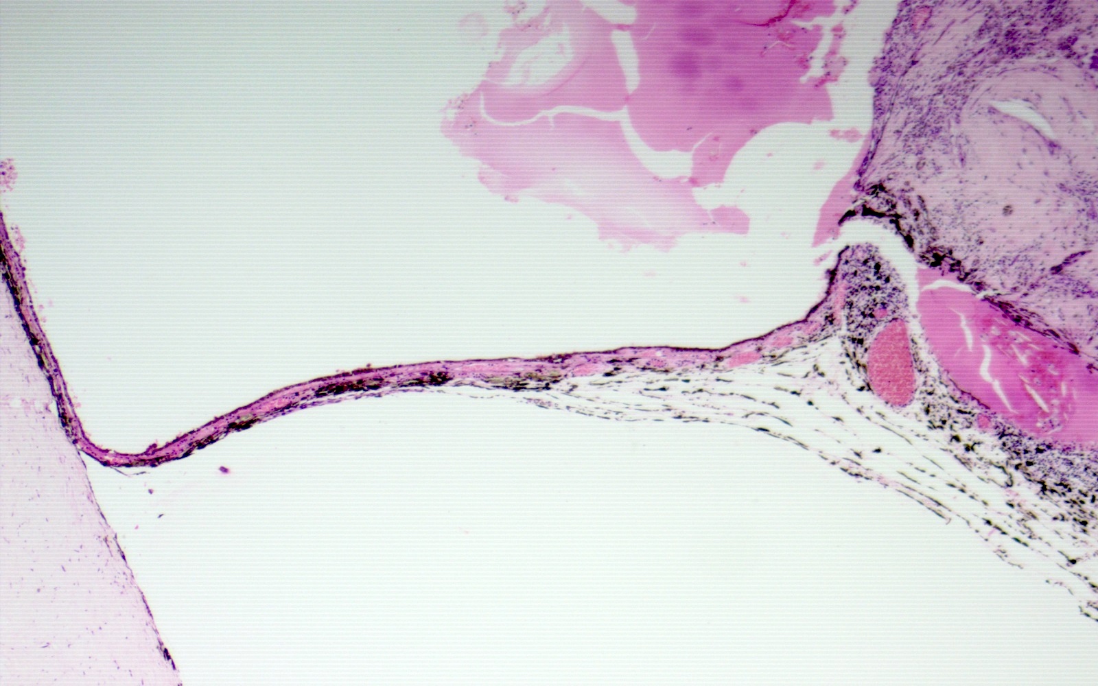
- Early changes are degeneration of outer retinal layers and photoreceptors with subretinal exudates
- Late changes are disruption and atrophy of retinal architecture with marked gliosis and proliferative vitreoretinopathy


