Table of Contents
Definition / general | Radiology images | Clinical images | Whole mount images | Microscopic (histologic) description | Microscopic (histologic) images | Cytology images | Positive stains | Negative stainsCite this page: Pernick N. Rhabdomyosarcoma-orbit. PathologyOutlines.com website. https://www.pathologyoutlines.com/topic/eyeorbitrms.html. Accessed December 23rd, 2024.
Definition / general
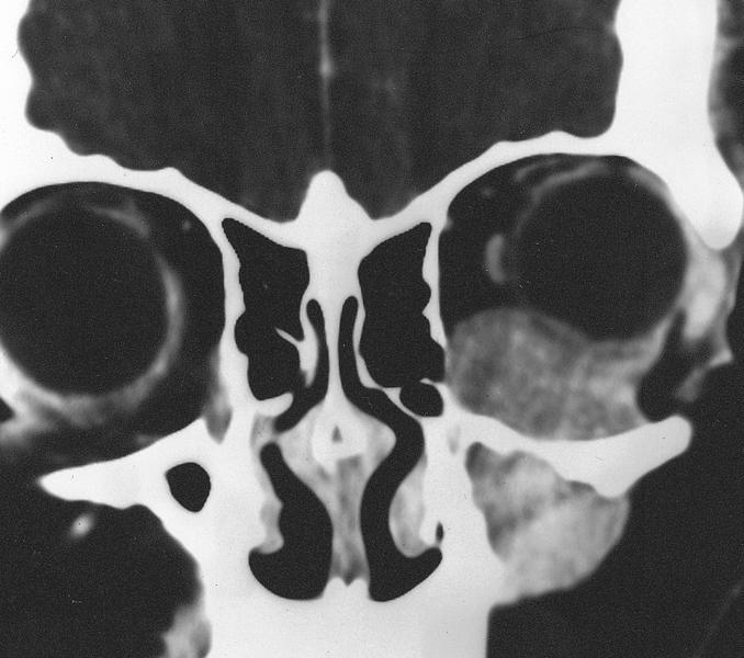
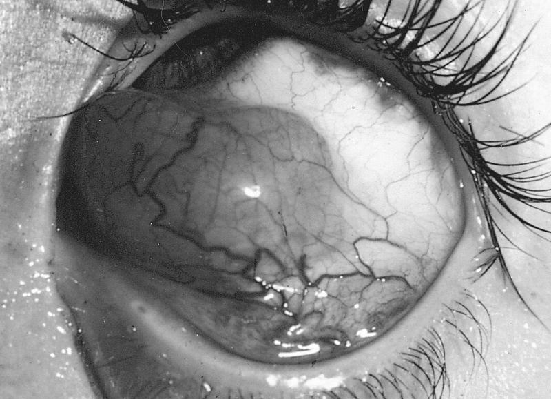
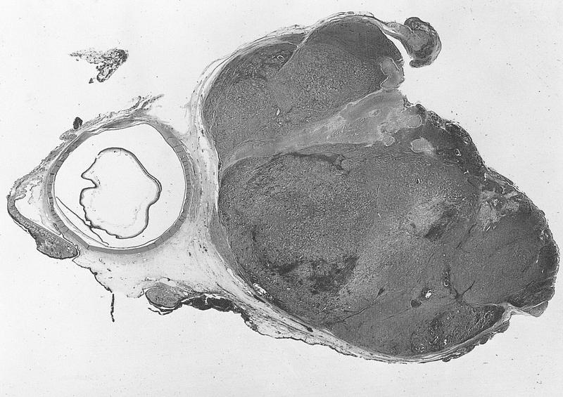
- Most common orbital sarcoma in childhood
- Usually embryonal and alveolar subtypes (alveolar more aggressive)
- Often rapid onset of unilateral proptosis
- May occur after radiation therapy for retinoblastoma, close to previously irradiated fields
- Tumors in retinoblastoma patients may have rosette-like structures
- References: Am J Surg Pathol 1998;22:1351 (postradiation therapy for bilateral retinoblastoma)
Microscopic (histologic) description
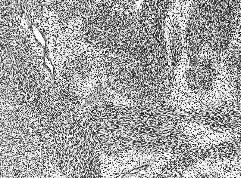
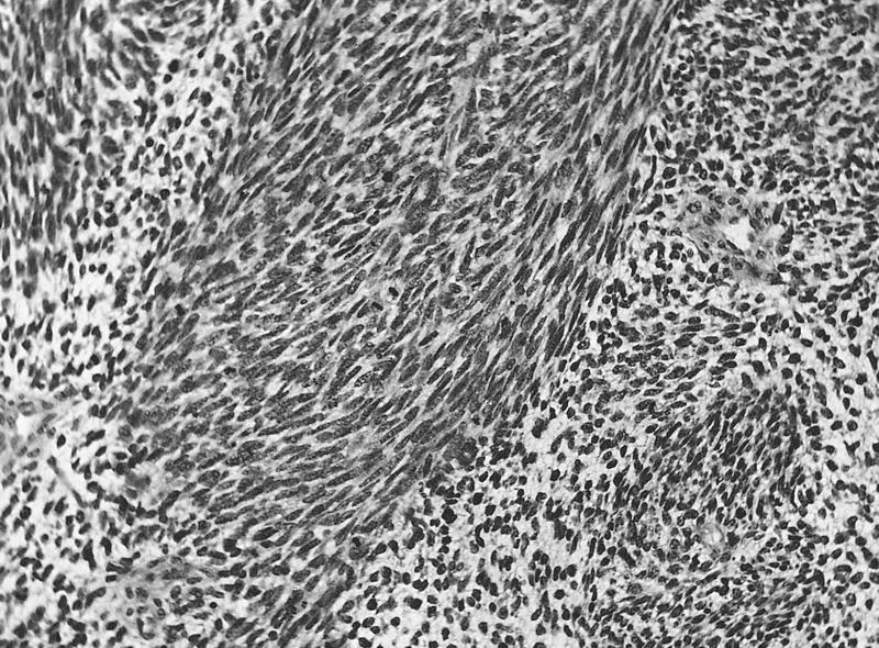
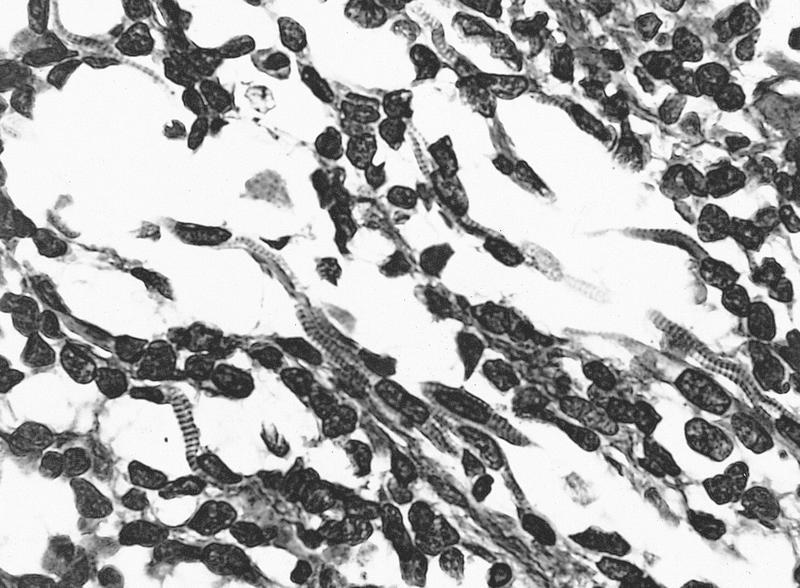
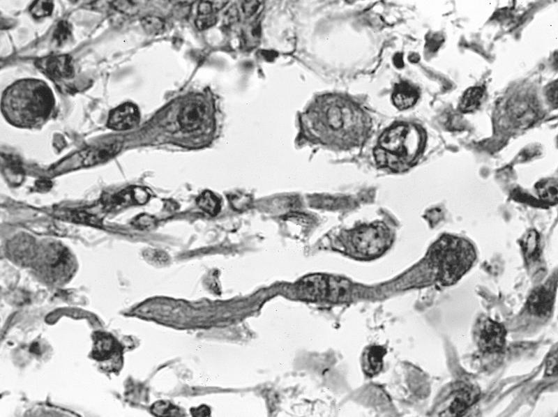
- Syncytium of strap cells with abundant eosinophilic cytoplasm
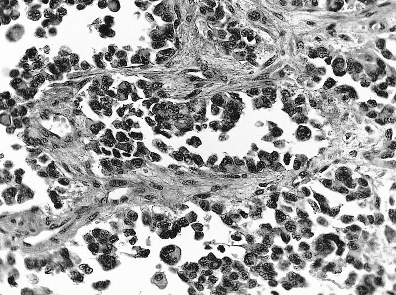
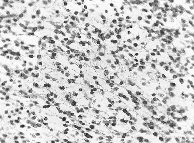
- Also closely packed small round cells with scanty cytoplasm, coarse nuclear chromatin and increased mitotic activity
- Many have minimal rhabdomyoblastic differentiation except in occasional mature strap cells with cross striations
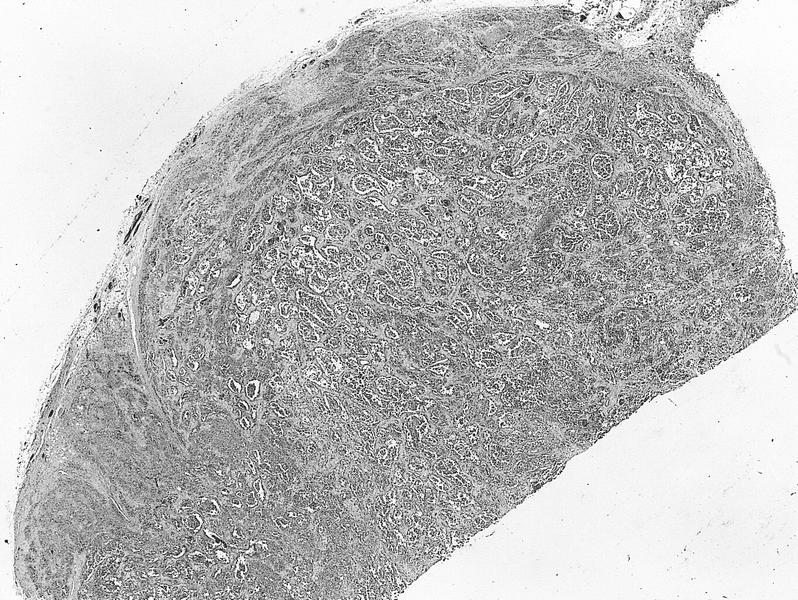
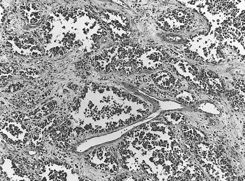
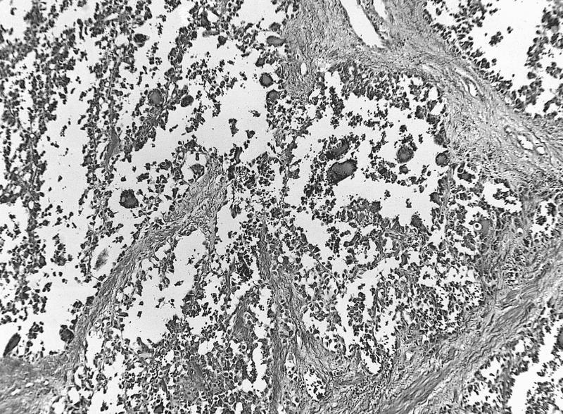
- Alveolar pattern has fibrovascular septa resembling lung alveoli
Microscopic (histologic) images
AFIP images
Cytology images
Positive stains
- Myogenin, desmin, muscle specific actin (HHF35), vimentin and neurofilament


