
Eye
Eyelid
Tumors
Eyelid cysts
Author: J. Stephen Nix, M.D.
Editorial Board Member: Ruta Gupta, M.D.
Deputy Editor-in-Chief: Kelly Magliocca, D.D.S., M.P.H.

Last author update: 18 November 2022
Last staff update: 18 November 2022
Copyright: 2004-2025, PathologyOutlines.com, Inc.
PubMed Search: Cysts eyelid

Table of Contents
Definition / general | Essential features | Terminology | ICD coding | Epidemiology | Etiology | Clinical features | Diagnosis | Case reports | Treatment | Clinical images | Gross description | Microscopic (histologic) description | Microscopic (histologic) images | Virtual slides | Videos | Sample pathology report | Differential diagnosis | Additional references | Board review style question #1 | Board review style answer #1 | Board review style question #2 | Board review style answer #2Cite this page: Nix JS. Eyelid cysts. PathologyOutlines.com website. https://www.pathologyoutlines.com/topic/eyeeyelidcysts.html. Accessed March 28th, 2025.
Definition / general
- Benign cystic lesions of the eyelid, most of which are caused by blockages of ducts (hidrocystomas) or hair follicles (epidermal cyst) or are consequence of entrapped benign, embryologic tissue (dermoid cyst)
- Includes epidermal cyst, dermoid cyst, eccrine hidrocystoma and apocrine hidrocytoma, all of which are generally cured by excision
Essential features
- Epidermal cysts feature a lining of benign keratinizing stratified squamous epithelium with a granular layer (in the absence of adnexal structures) and contain keratinaceous debris
- Dermoid cysts are embryologically derived lesions histologically identified by the presence of adnexal structures in conjunction with benign, stratified squamous epithelial cyst lining
- Eccrine hidrocystomas are caused by obstruction of eccrine sweat glands and are histologically identified by a dual layer of bland, cuboidal epithelial cells
- Apocrine hidrocystomas are caused by obstruction of apocrine sweat glands and are histologically identified by the presence of an inner layer of elongated, epithelial cells with apical snouts and decapitation secretions
Terminology
- Epidermal (infundibular) cyst, epidermal inclusion cyst, follicular cyst, infundibular type, epidermoid cyst, clinical sebaceous cyst
- Hidrocystoma, sudoriferous cyst, sweat ductal cyst
ICD coding
- ICD-10:
- H02.82 - cysts of eyelid
- H02.821 - cysts of right upper eyelid
- H02.822 - cysts of right lower eyelid
- H02.823 - cysts of right eye, unspecified eyelid
- H02.824 - cysts of left upper eyelid
- H02.825 - cysts of left lower eyelid
- H02.826 - cysts of left eye, unspecified eyelid
- H02.829 - cysts of unspecified eye, unspecified eyelid
- ICD-11:
Epidemiology
- Epidermal cysts are common, sporadic lesions that typically affect adults with a slight male predominance (StatPearls: Epidermoid Cyst [Accessed 18 August 2022])
- Periocular dermoid cysts are most often encountered in pediatric patients, with many cases arising in the superotemporal region within the first year of life (Ophthalmic Epidemiol 2019;26:117)
- Eccrine hidrocytomas generally arise along the medial or lateral eyelid in middle aged to older adults (Eye (Lond) 2005;19:77)
- Apocrine hidrocystomas often occur near the canthus in middle aged and older adults but may rarely be seen in the pediatric population (MedGenMed 2006;8:57)
Etiology
- Most epidermal cysts are thought to arise secondary to occlusion of hair follicles (Arch Ophthalmol 1988;106:270)
- Dermoid cysts are caused by entrapment of embryologic epithelium (Otolaryngol Head Neck Surg 2005;132:938)
- Eccrine hidrocystomas occur secondary to blockages of eccrine sweat glands (Eye (Lond) 2005;19:77)
- Apocrine hidrocystomas result from blockages of apocrine sweat glands (MedGenMed 2006;8:57)
Clinical features
- Epidermal cysts typically present as solitary, subcutaneous nodules and keratinaceous contents may appear cheese-like (StatPearls: Epidermal Inclusion Cyst [Accessed 18 August 2022])
- Dermoid cysts present as subcutaneous nodules, often superotemporally in the vicinity of the zygomaticofrontal suture (Int Ophthalmol 2011;31:93)
- Most hidrocystomas appear as single, dome shaped lesions with varying tints (including transparent, skin colored, brown, blue and blue-black hues) though examples of multiple hidrocystomas do occur (MedGenMed 2006;8:57)
Diagnosis
- Histopathologic examination of excised tissue
Case reports
- 8 and 11 month old boys with upper eyelid lesions (Ophthalmic Plast Reconstr Surg 2009;25:146)
- 15 month old boy with a right upper eyelid mass (Saudi J Ophthalmol 2015;29:312)
- 9 year old girl with drooping of the right upper eyelid (Indian J Ophthalmol 2017;65:884)
- 35 year old man with a large right upper eyelid swelling over a period of 10 years (Indian J Ophthalmol 2012;60:211)
- 70 year old man with a large, cystic swelling of the forehead (Indian J Otolaryngol Head Neck Surg 2019;71:59)
Treatment
- Excision is usually curative
Clinical images
Images hosted on other servers:

Epidermal cyst


Dermoid cyst


Hidrocystoma
Gross description
- Benign eyelid cysts are often received as small excisions with nodules, papules or grossly identifiable cysts
- Keratinaceous cyst contents in epidermal and dermoid cysts may appear yellow to white, cheese-like or chalky (StatPearls: Epidermal Inclusion Cyst [Accessed 18 August 2022])
- Hidrocystomas are thin walled and usually contain clear cyst contents (MedGenMed 2006;8:57)
Microscopic (histologic) description
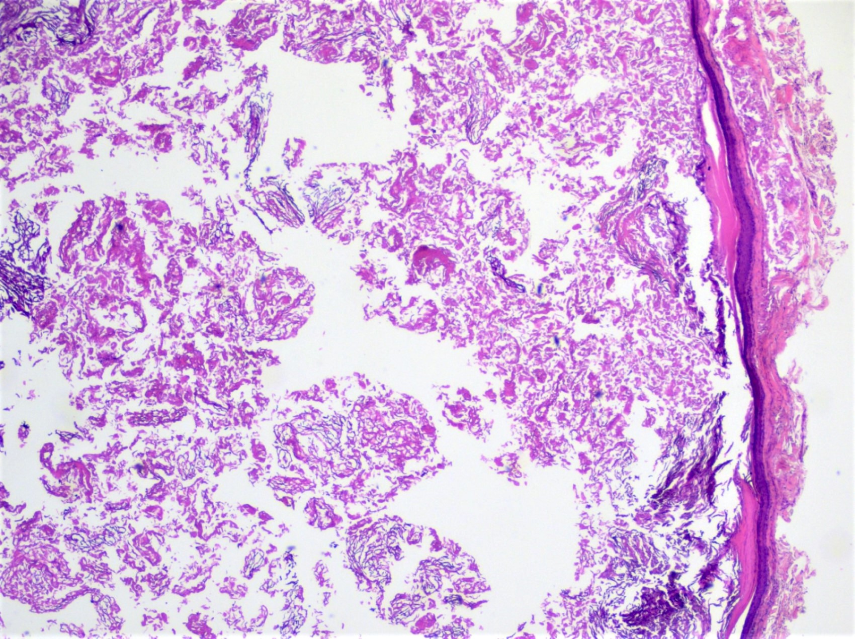
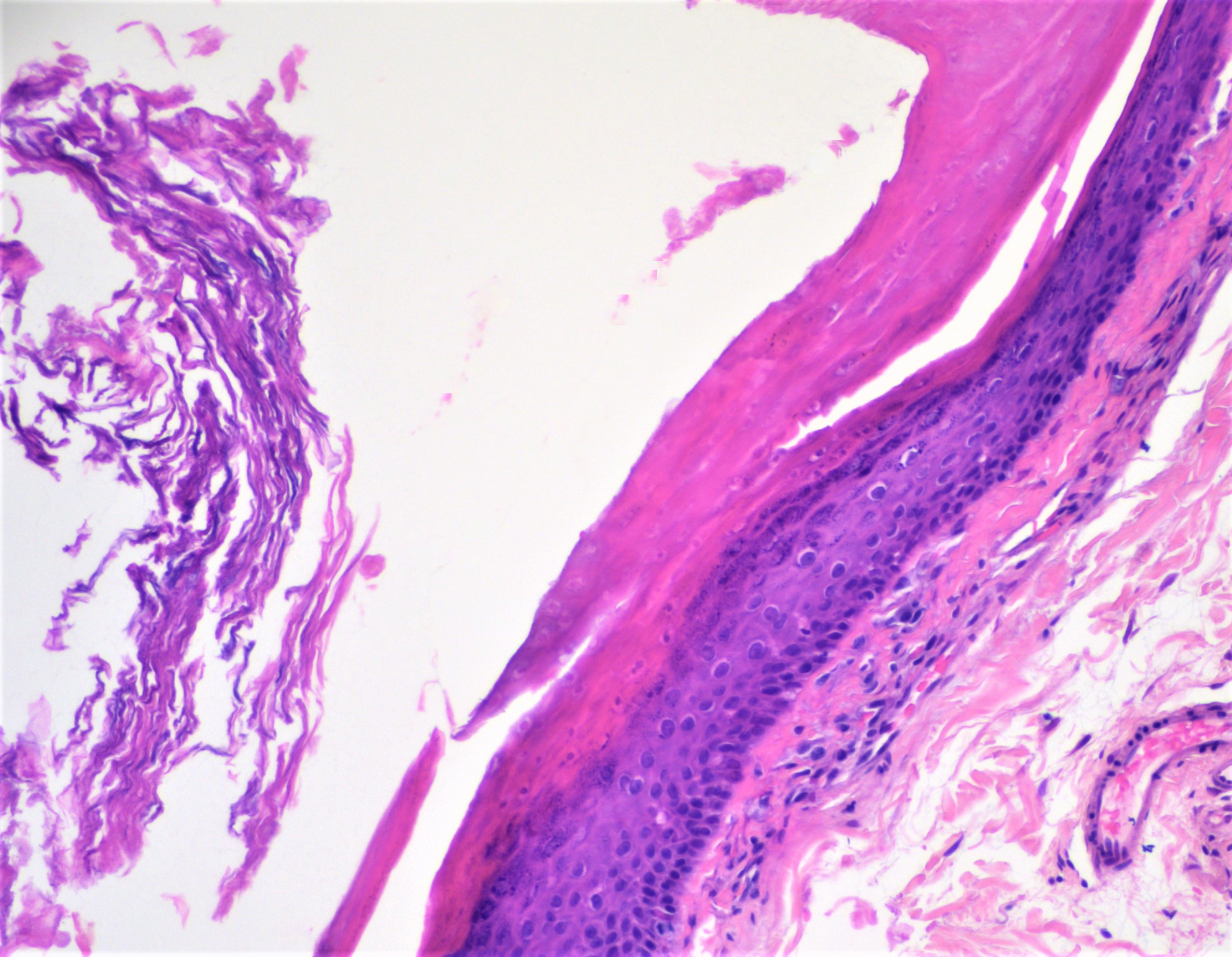
- Epidermal cyst:
- Bland, keratinizing stratified squamous epithelial cyst lining with a granular layer of keratohyaline granules in the absence of adnexal structures
- Keratinaceous debris cyst contents
- Rupture of cyst may induce an inflammatory response, including a foreign body giant cell reaction
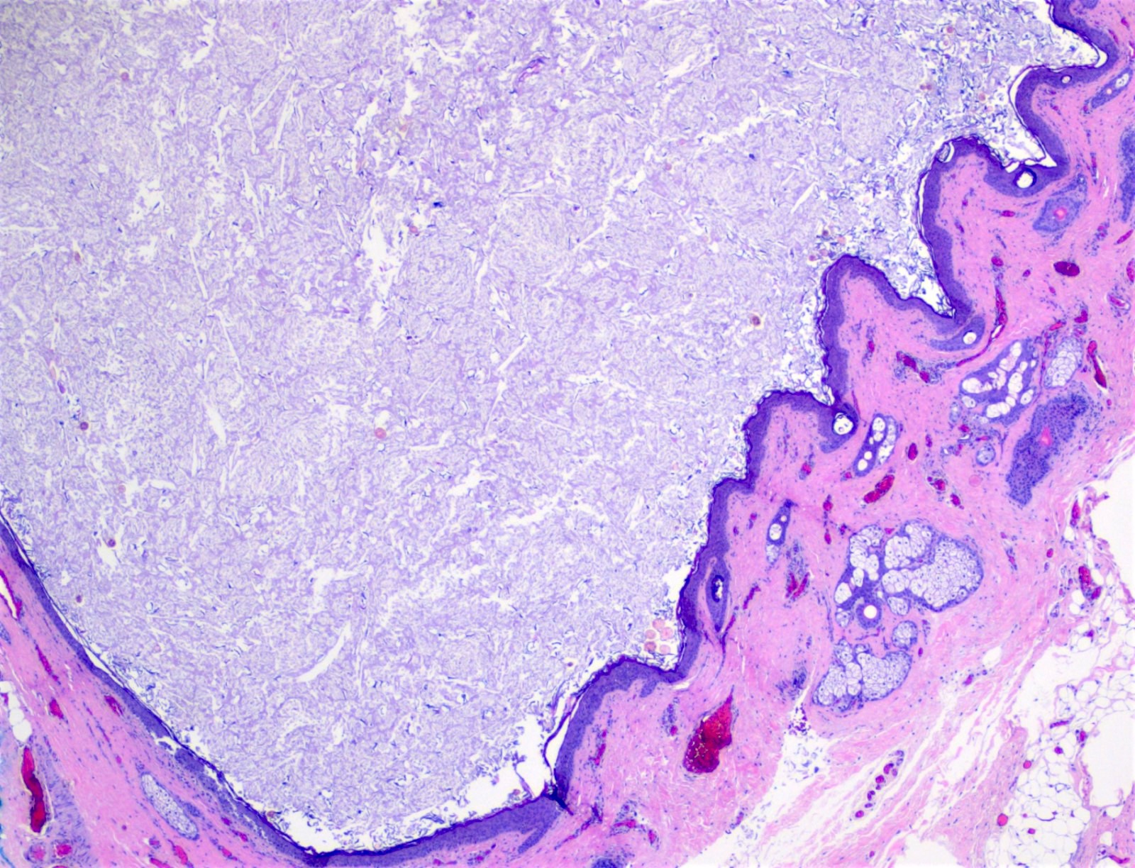
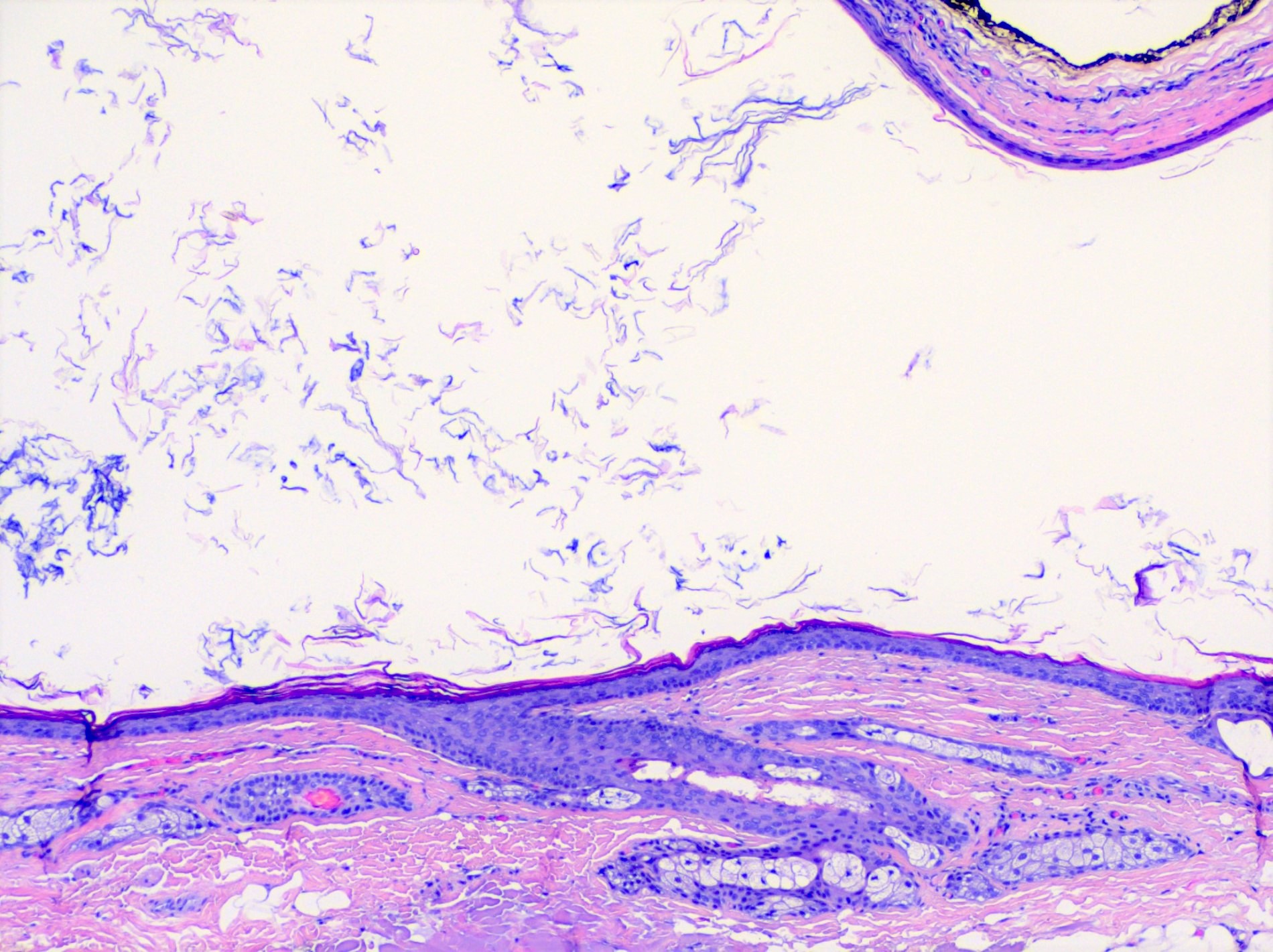
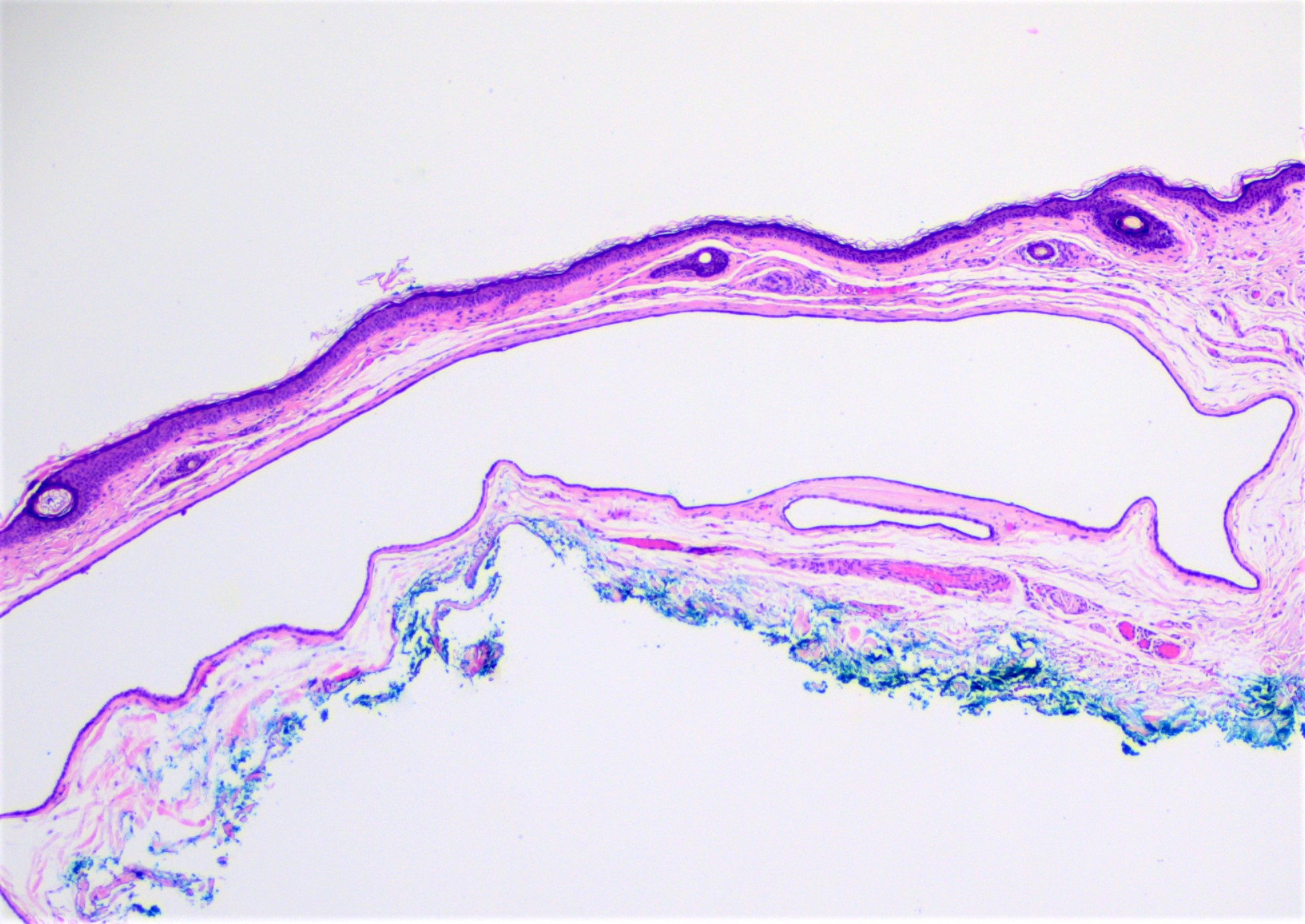
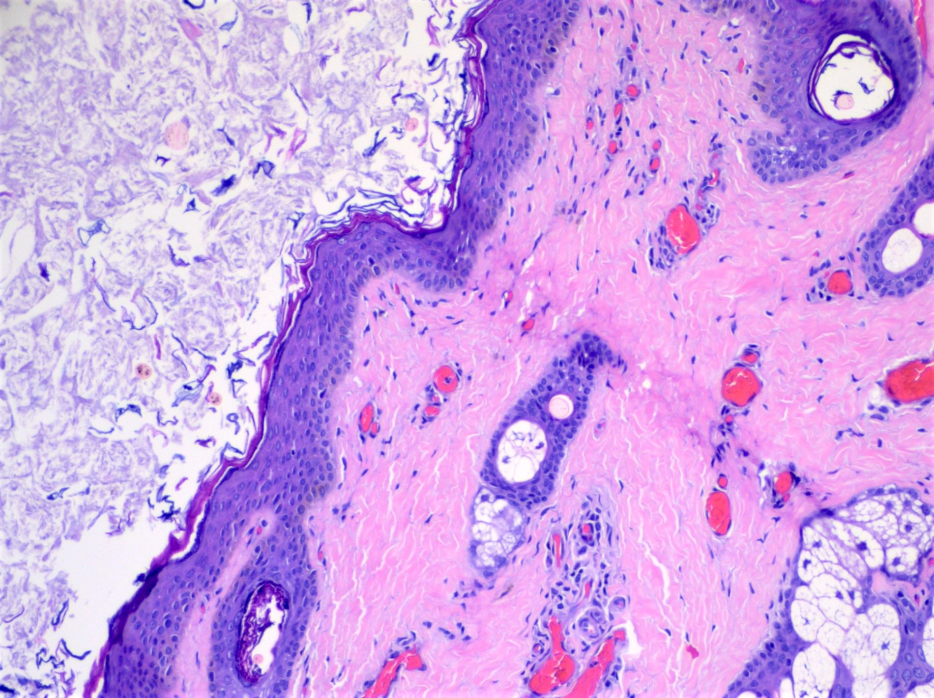
- Dermoid cyst:
- Features adnexal structures, such as hair follicles and sebaceous glands, in conjunction with bland, stratified squamous epithelial cyst lining
- Keratinaceous debris in cyst contents
- Like epidermal cysts, rupture may lead to an inflammatory response and a foreign body giant cell reaction
- Conjunctivoid dermoid cysts with nonkeratinizing squamous epithelium and goblet cells may rarely be encountered in addition to other rare features, such as oncocytic and basaloid differentiation (Ophthalmic Plast Reconstr Surg 2008;24:69, J Eur Acad Dermatol Venereol 2001;15:268)
- Eccrine hidrocystoma:
- Subepithelial cyst with dual lining of variably attenuated, cuboidal cells
- Unilocular or multilocular
- Cyst cavity may appear clear / empty or contain eosinophilic, proteinaceous material
- Apocrine hidrocystoma:
- Subepithelial cyst with an inner epithelial lining of elongated (columnar), eosinophilic cells with apical snouts and decapitation secretions
- Unilocular or multilocular
- Like eccrine hidrocystomas, the cyst cavity may appear clear / empty or contain eosinophilic, proteinaceous material
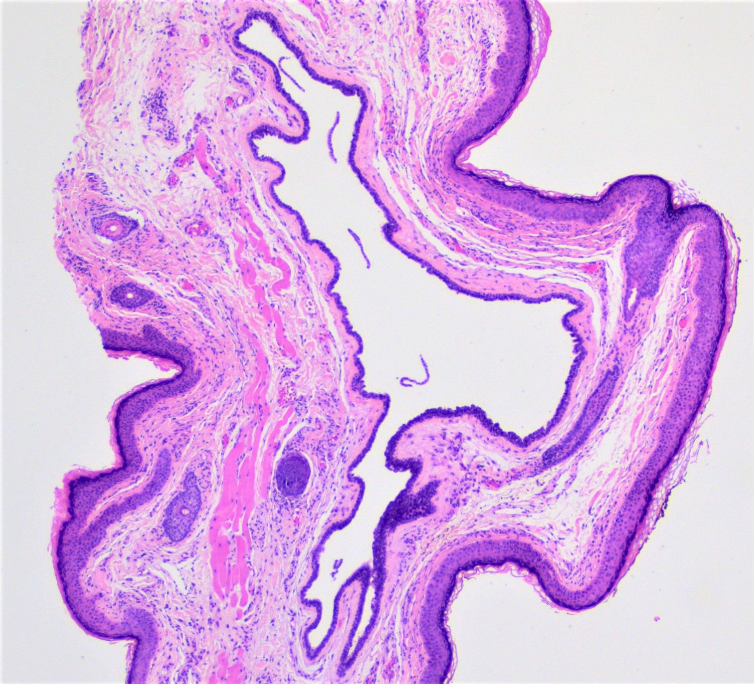
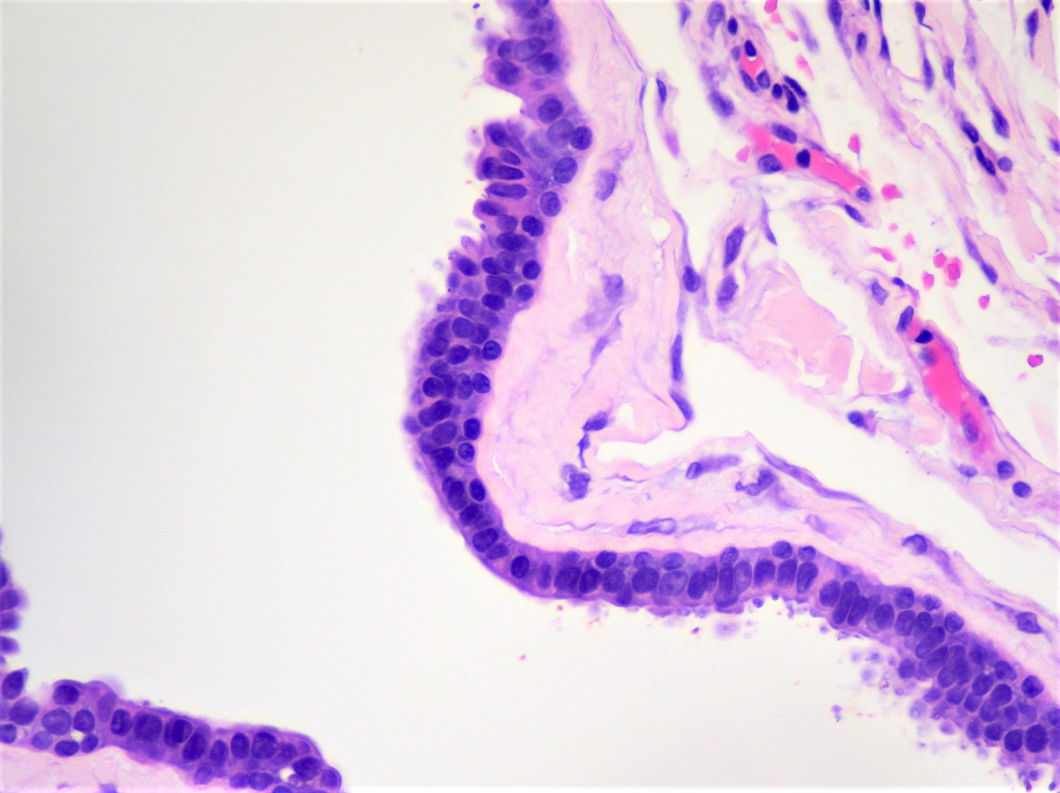
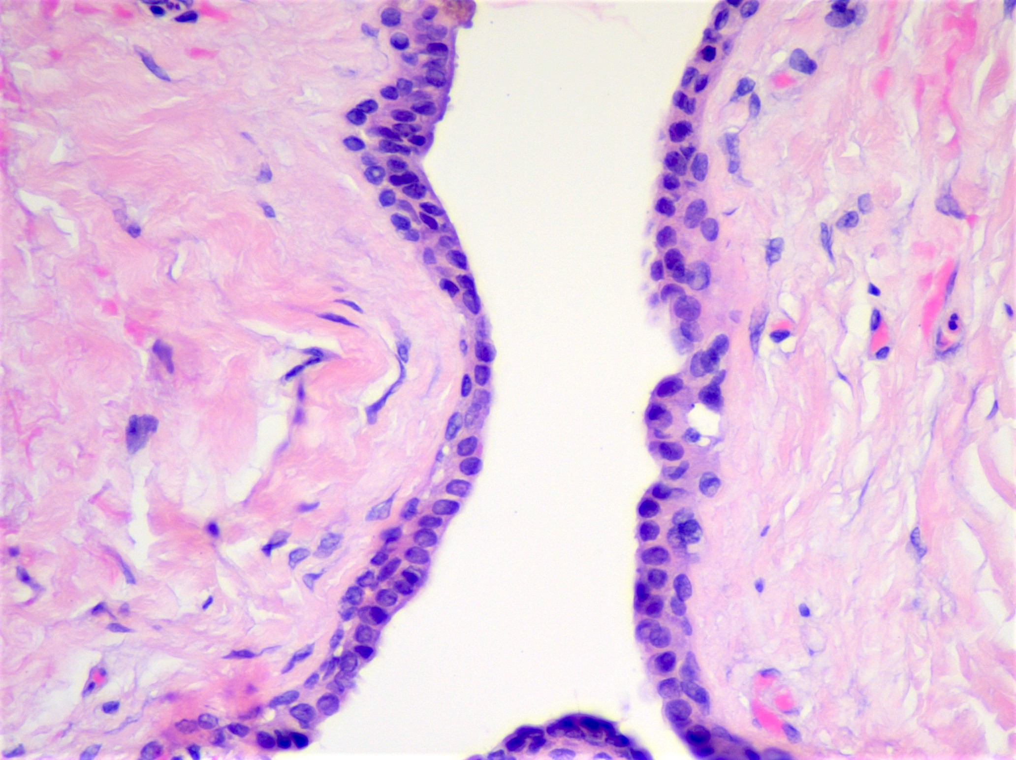
Microscopic (histologic) images
Contributed by J. Stephen Nix, M.D.
Epidermal cyst, keratinaceous contents
Epidermal cyst, granular layer
Dermoid cyst, keratinaceous contents
Dermoid cyst, adnexal structures
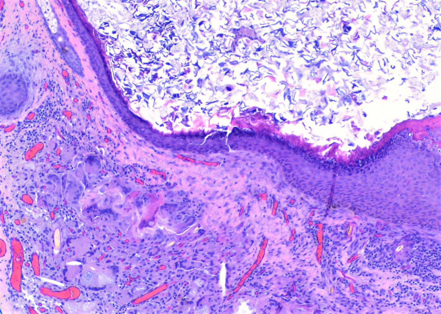
Dermoid cyst with rupture
Eccrine hidrocystoma
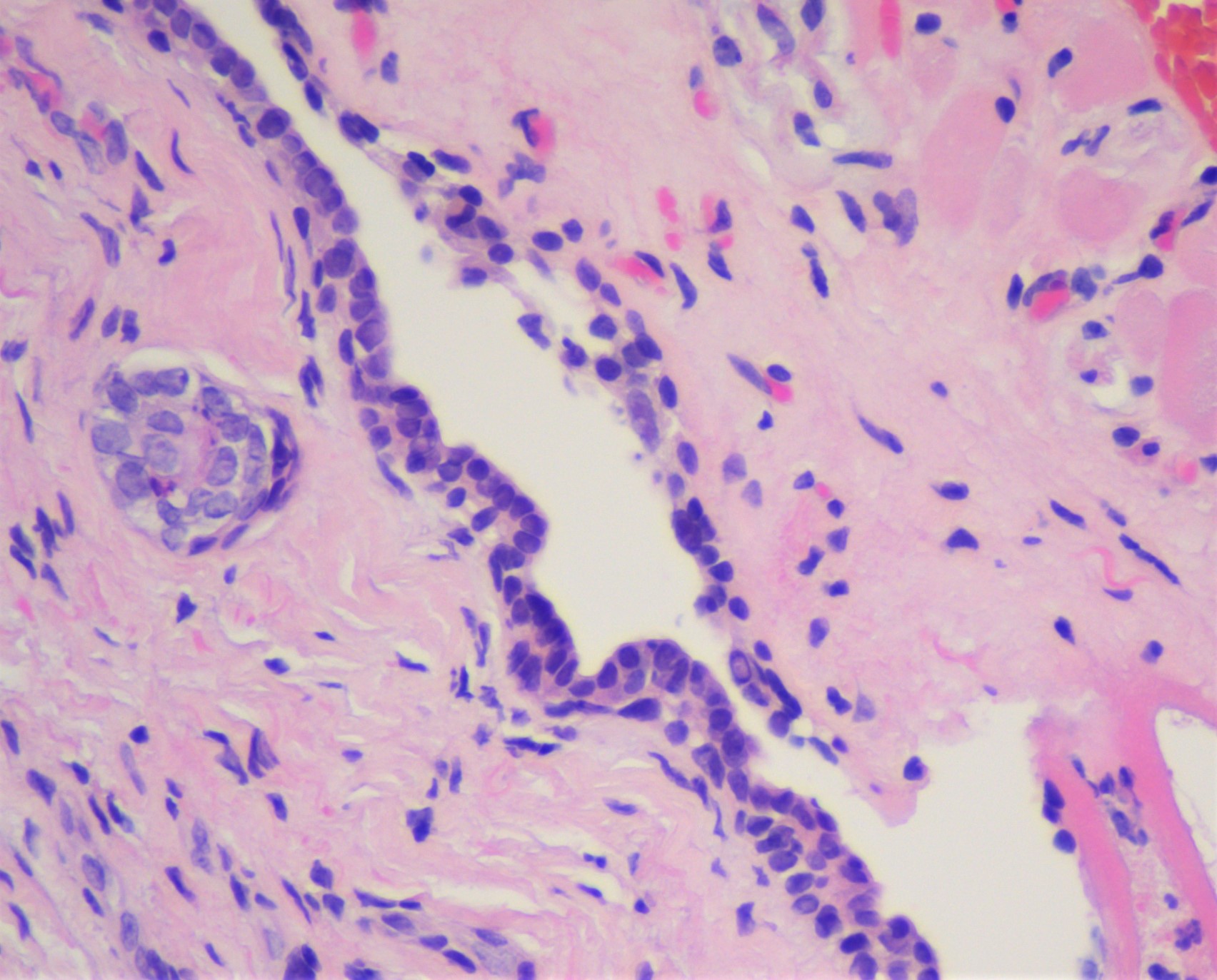
Eccrine hidrocystoma, cuboidal bilayer
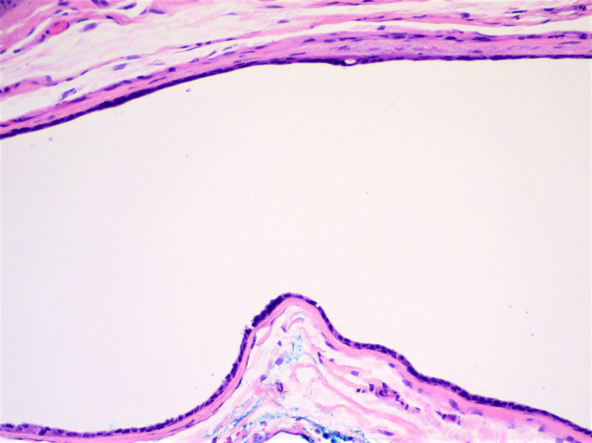
Eccrine hidrocystoma, attenuated lining
Apocrine hidrocystoma
Apocrine hidrocystoma, inner cyst lining
Virtual slides
Images hosted on other servers:

Apocrine hidrocystoma of skin
Videos
Hidrocystoma: 5 minute pathology pearls by Dr. Jared Gardner
Dermoid cyst: 5 minute pathology pearls by Dr. Jared Gardner
Sample pathology report
- Eyelid, left upper, excision:
- Epidermal cyst (see comment)
- Comment: Histologic examination shows a cyst with a bland keratinizing squamous epithelial lining with a granular layer and keratinaceous cyst contents in the absence of adnexal structures.
- Periocular nodule, excision:
- Dermoid cyst (see comment)
- Comment: Histologic examination shows a cyst with a bland keratinizing squamous epithelial lining and adjoining adnexal structures.
- Eyelid, right upper, excision:
- Apocrine hidrocystoma (see comment)
- Comment: Histologic examination shows a subepithelial cyst that features an inner layer of elongated, eosinophilic cells with apical snouts and decapitation secretions.
- Eyelid, left lower, excision:
- Eccrine hidrocystoma (see comment)
- Comment: Histologic examination shows a subepithelial cyst with an epithelial lining composed of a dual layer of flattened, cuboidal cells.
Differential diagnosis
- Chalazion:
- Lipogranulomatous inflammation that lacks an epithelial cyst lining
- Typically presents as a tender eyelid nodule
Additional references
Board review style question #1
A 68 year old man presents with a skin colored nodule of the lateral left lower eyelid. Based on the histologic image, what is the most likely etiology of the lesion?
- Blockage of an apocrine sweat gland
- Blockage of an eccrine sweat gland
- Entrapped embryologic tissue
- Obstruction of a pilosebaceous unit
Board review style answer #1
B. Blockage of an eccrine sweat gland. The histologic image shows an epithelial lining composed of a dual layer of cuboidal cells, consistent with the diagnosis of eccrine hidrocystoma. Eccrine hidrocystomas are caused by blockages of eccrine sweat glands. Apocrine hidrocystomas result from blockages of apocrine sweat glands (A). Entrapped embryonal tissue is encountered in a dermoid cyst (C) and obstruction of a pilosebaceous unit may result in an epidermal cyst (D).
Comment Here
Reference: Eyelid cysts
Comment Here
Reference: Eyelid cysts
Board review style question #2
A 1 year old girl undergoes excision of a superotemporal periocular nodule and the histologic findings are provided in the associated image. What is the diagnosis?
- Apocrine hidrocystoma
- Dermoid cyst
- Eccrine hidrocystoma
- Epidermal cyst
Board review style answer #2
B. Dermoid cyst. Periocular dermoid cysts most often occur in pediatric patients and are diagnosed histologically by the presence of adnexal structures in association with a bland stratified squamous epithelial cyst lining. Apocrine hidrocystomas demonstrate a cyst lining with an inner layer of elongated, eosinophilic cells with apical snouts and decapitation secretions (A). Eccrine hidrocystomas feature a cyst lining composed of a dual layer of cuboidal cells (C). Epidermal cysts have a cyst lining of bland keratinizing stratified squamous epithelium with a granular layer in the absence of adnexal structures (D).
Comment Here
Reference: Eyelid cysts
Comment Here
Reference: Eyelid cysts
