Table of Contents
Definition / general | Essential features | Terminology | ICD coding | Epidemiology | Sites | Pathophysiology | Etiology | Diagrams / tables | Clinical features | Diagnosis | Prognostic factors | Case reports | Treatment | Clinical images | Gross description | Microscopic (histologic) description | Microscopic (histologic) images | Sample pathology report | Differential diagnosis | Additional references | Board review style question #1 | Board review style answer #1 | Board review style question #2 | Board review style answer #2Cite this page: Hasby Saad EA. Keratoconus. PathologyOutlines.com website. https://www.pathologyoutlines.com/topic/eyecorneakeratoconus.html. Accessed April 2nd, 2025.
Definition / general
- Complex, genetically heterogeneous multifactorial degenerative disorder
- Prevalence ranges between 1/2,000 and 1/50,000 in the general population
- Characterized by corneal ectasia and thinning
- Visual acuity decreases due to progressive myopia, irregular astigmatism and often, corneal apical opacification
Essential features
- Bilateral, progressive ectatic disease of the cornea that becomes cone shaped
- Leading indicator for corneal transplantation
- Complex disorder with both genetic and environmental factors
- Disease progression of keratoconus affects the epithelium, Bowman layer, stroma and Descemet membrane of the cornea but not the corneal endothelium
- Reference: StatPearls: Keratoconus [Accessed 30 September 2021]
Terminology
- Conical cornea
- KC (keratoconus)
- KCN (keratoconus)
ICD coding
Epidemiology
- Black and Latino > Caucasian
- M:F = 6:1
- Presents at puberty and progresses until the third to fourth decades of life but can occur or progress at any age
- Progresses more rapidly in young patients and stabilizes approximately 20 years after initial onset
- Reference: eMedicine: Keratoconus [Accessed 24 September 2021]
Sites
- Often affects both eyes
- Can lead to very different vision between the eyes
- Reference: American Academy of Ophthalmology: Keratoconus Symptoms [Accessed 24 September 2021]
Pathophysiology
- All layers of the cornea are believed to be affected
- Characteristic structural changes include:
- Epithelial basement membrane fragmentation and scarring
- Breaks in the anterior limiting lamina (i.e. Bowman membrane)
- Axial stromal thinning and scarring
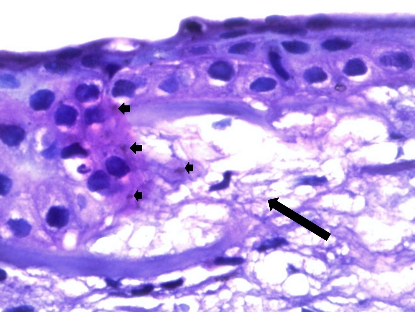
- Deposition of iron in the basal epithelial cells forms Fleischer rings
- Breaks and folds close to the Descemet membrane form commonly seen striae and rarely, acute hydrops when aqueous humor enters corneal stroma
- Keratoconic corneas have been shown to have:
- Altered antioxidant enzymes, accumulations of cytotoxic reactive oxygen / nitrogen species, activated caspase pathways and mitochondrial DNA damage
- Abnormal oxidative stress related properties that can induce activation of degradative enzymes and degradation of tissue inhibitors of metalloproteinases
- Genomic deletion in the superoxide dismutase 1 (SOD1) gene has also been associated with the disease
- Some data suggests an inflammatory component (Eye (Lond) 2014;28:189)
Etiology
- Family history: increased chance of getting keratoconus; patient's children's eyes should be checked for signs starting around age 10
- Age: starts in teenage years; might show earlier in childhood and can less commonly affect people 40 and older
- Certain disorders: Down syndrome, Ehlers-Danlos syndrome, osteogenesis imperfecta and retinitis pigmentosa
- Inflammation: allergies, asthma or atopic eye disease can break down the tissue of the cornea
- Hard eye rubbing over time can break down the cornea; it can also make keratoconus progress faster if already present
- Race: people who are black or Latino are ~50% more likely to get it than people who are white
- Reference: WebMD: What Is Keratoconus? [Accessed 24 September 2021]
Clinical features
- Blurred or distorted vision, classical perception of multiple ghost images known as monocular polyopia
- Increased sensitivity to bright light and glare, which can cause problems with night driving
- Need for frequent changes in eyeglass prescriptions
- Sudden worsening or clouding of vision
- Reference: Clin Ophthalmol 2013;7:2019
Diagnosis
- Physical examination
- Medical history
- Eye examination with measuring corneal curvature using a manual keratometer
- Slit lamp examination:
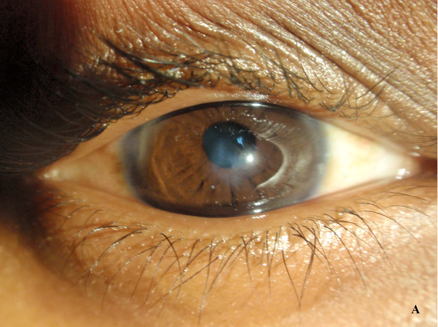
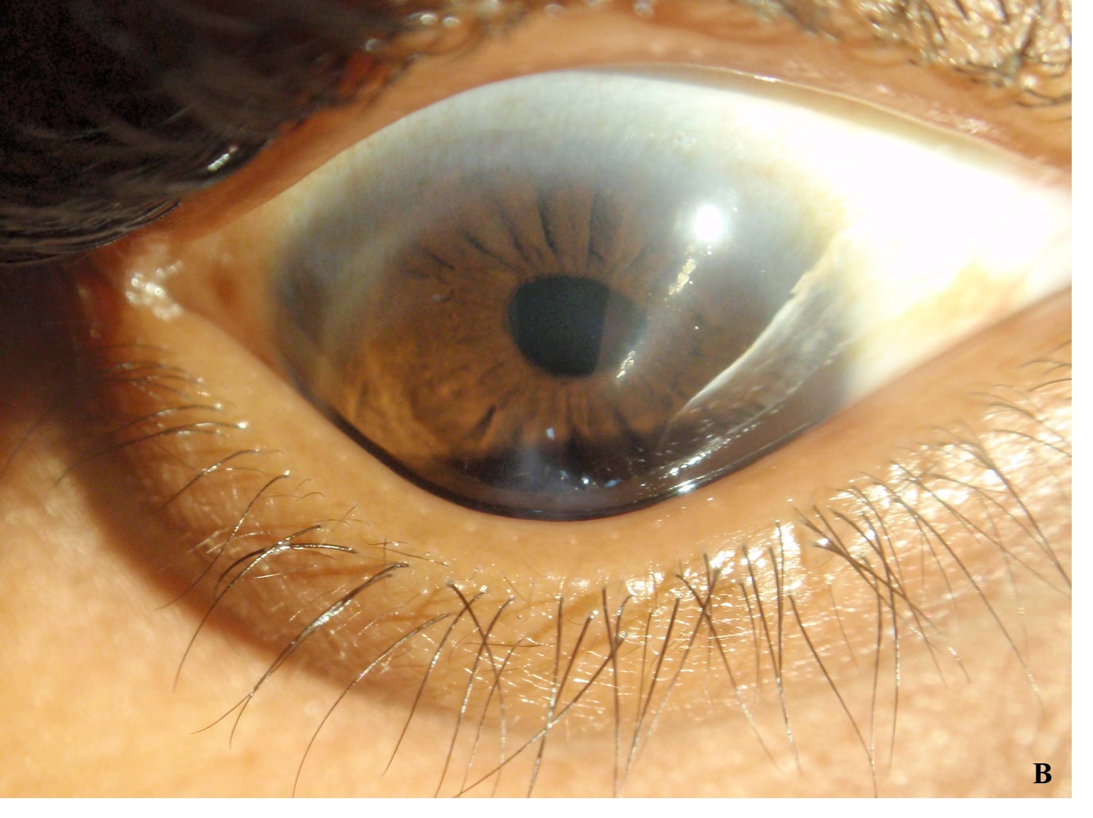
- Fleischer ring: a ring of yellow-brown to olive-green pigmentation
- Vogt striae: fine stress lines within the cornea caused by stretching and thinning
- Munson sign: V shaped indentation in the lower eyelid when the person's gaze is directed downwards is highly pronounced
- Handheld keratoscope: for noninvasive visualization of the surface of the cornea by projecting series of concentric rings of light onto the cornea
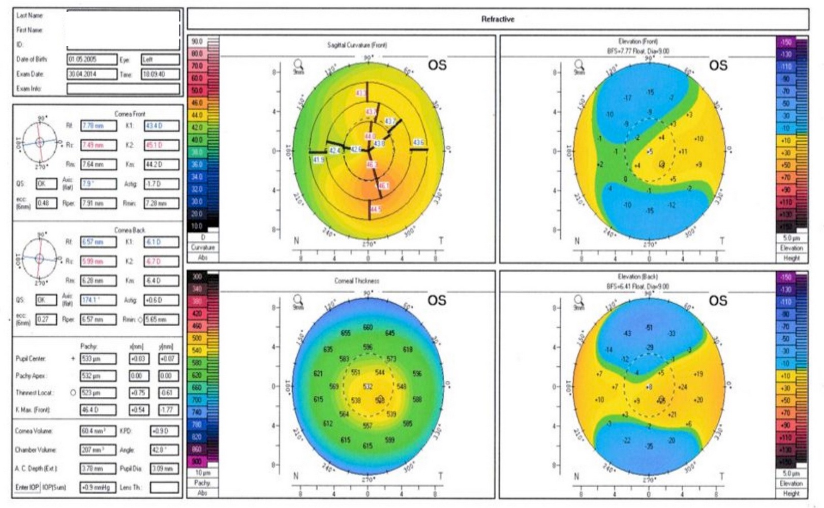
- Corneal topography provides map indicating any distortions or scarring in the cornea, including steepening of curvature that is usually below the centerline of the eye; helps in detection at early stages
- Reference: Wikipedia: Keratoconus [Accessed 24 September 2021]
Prognostic factors
- Severe vision threatening disorder that commonly indicates corneal transplantation
Case reports
- 17 year old girl, 21 year old man and 22 year old man with rare complications of stromal thinning up to Descemet membrane 3 - 6 years post corneal collagen crosslinking (CXL) (Indian J Ophthalmol 2020;68:224)
- 19 year old man presenting with an unusual case of hydrops in keratoconus (Indian J Ophthalmol 2018;66:309)
- 25 year old man with epithelial remodeling masquerading as keratoconus progression (Indian J Ophthalmol 2020;68:3053)
Treatment
- In the early stages, eyeglasses or soft contact lenses
- As the disorder progresses and the cornea continues to thin and change shape, rigid gas permeable (RGP) contact lenses
- Corneal crosslinking
- Corneal transplantation
- In severe cases, due to scarring, extreme thinning or contact lens intolerance
- Keratoconus cornea is replaced with healthy donor tissue
- Reference: StatPearls: Keratoconus [Accessed 30 September 2021]
Clinical images
Gross description
- Wrinkled corneal button after transplantation
- Cornea is cone shaped
- Often has Fleischer ring (brown, stainable intraepithelial iron arc surrounds conical portion of cornea)
- Reference: StatPearls: Keratoconus [Accessed 30 September 2021]
Microscopic (histologic) description
- In vitro study of fixed, processed corneas with light microscopy shows 2 microscopic patterns:
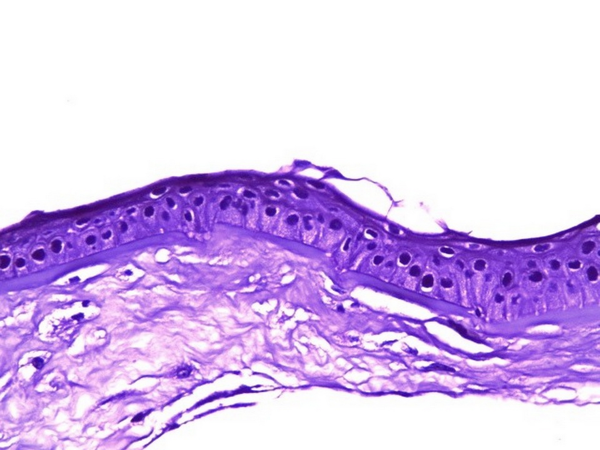
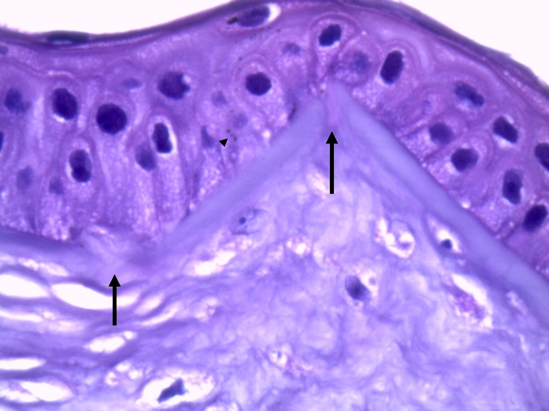
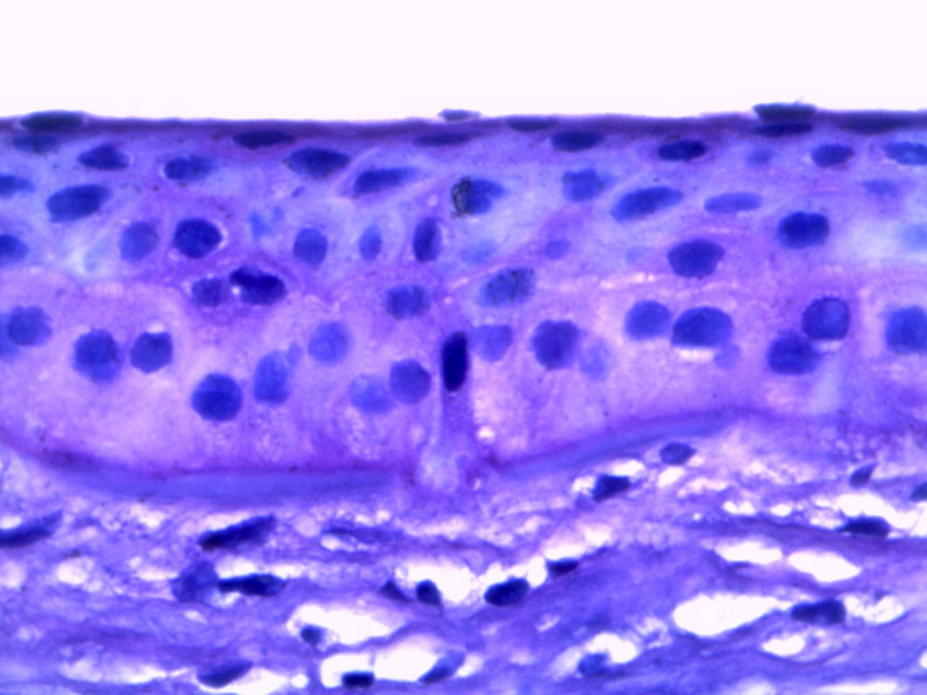
- Typical pattern (identified in > 80%): both stromal and central epithelial thinning with multiple Bowman layer breaks
- Atypical pattern: lacks breaks in Bowman layer and has less thinning of the central epithelium
- Alterations in different layers of cornea are:
- Epithelium: cellular enlargement with irregular arrangement and apoptosis
- Stroma: loss in collagenous lamella, reduction in keratocyte density with appearance of nonkeratocyte cells
- Descemet membrane: morphological folds and irregularities; its rupture with entering of aqueous humor into corneal epithelium and stroma is a serious complication for keratoconus known as acute corneal hydrops
- Endothelium: does not exhibit any changes during keratoconus progression
- Using optical coherence tomography (OCT) for in vivo examining cornea, Sandali et al. proposed a classification system for keratoconus with 5 distinct stages (Ophthalmology 2013;120:2403):
- Stage 1: thinner corneal epithelium and stroma at the conus than control
- Stage 2: hyperreflective anomalies in Bowman layer are noticed with thickening epithelium and opaque stroma
- Stage 3: increased epithelial thickening and stromal thinning with disruptions in Bowman layer
- Stage 4: shows panstromal scarring
- Stage 5: considered as the acute form of keratoconus (hydrops) with Descemet membrane rupture and total corneal scar
- Reference: Biomed Res Int 2017;2017:7803029
Microscopic (histologic) images
Sample pathology report
- Cornea, keratoplasty:
- Keratoconus (see comment)
- Comment: Sections in keratoconus cornea show stromal and central epithelial thinning with multiple Bowman layer breaks. Brown iron deposits are seen within basal layer of corneal epithelium.
Differential diagnosis
- Pellucid marginal degeneration:
- Often considered a variant of keratoconus
- Corneal thinning occurs about 1 mm above the inferior limbus, resulting in advanced against the rule corneal astigmatism that may be observed on corneal topography or tomography
- Terrien marginal corneal degeneration:
- Slowly progressive noninflammatory, unilateral or asymmetrically bilateral peripheral corneal thinning
- Associated with corneal neovascularization, opacification and lipid deposition
- Keratoglobus:
- Extremely rare corneal disease in which the entire cornea thins from limbus to limbus, sometimes to the point where spontaneous perforation becomes possible
- Contact lens induced corneal warpage:
- Corneas swell (rather than thin) from hypoxia
- Stromal striations similar to Vogt striae occur
- Corneal ectasia postrefractive laser treatment
Additional references
Board review style question #1
A 25 year old woman complaining of blurred vision and polyopia comes to the ophthalmology department. On examination, a bilateral V shaped indentation in the lower eyelids is apparent when she looks downwards and corneal topography shows steeping of corneal curvature. She undergoes keratoplasty and the removed cornea is sent for pathological examination. What is the most likely diagnosis?
- Calcific band keratopathy
- Corneal dystrophy
- Keratitis
- Keratoconus
Board review style answer #1
Board review style question #2
Board review style answer #2


