
Eye
General
Anatomy & histology-conjunctiva
Authors: Sena Zengin, M.D., J. Stephen Nix, M.D.
Editorial Board Member: Ruta Gupta, M.D.
Deputy Editor-in-Chief: Kelly Magliocca, D.D.S., M.P.H.


Last author update: 10 August 2023
Last staff update: 10 August 2023
Copyright: 2004-2025, PathologyOutlines.com, Inc.
PubMed Search: Anatomy histology conjunctiva

Table of Contents
Definition / general | Essential features | Terminology | Embryology and physiology | Diagrams / tables | Clinical features | Gross description | Microscopic (histologic) description | Microscopic (histologic) images | Positive stains | Videos | Additional references | Board review style question #1 | Board review style answer #1 | Board review style question #2 | Board review style answer #2Cite this page: Zengin S, Nix JS. Anatomy & histology-conjunctiva. PathologyOutlines.com website. https://www.pathologyoutlines.com/topic/eyeconjunctivahistology.html. Accessed March 28th, 2025.
Definition / general
- Mucous membrane that covers, protects and lubricates the posterior surface of the eyelids (palpebral, also known as tarsal, conjunctiva) and anterior surface of the globe (bulbar conjunctiva) as well as provides redundant folds (forniceal conjunctiva) to aid eye movement
Essential features
- Nonkeratinizing squamous epithelium with goblet cells overlying loose connective tissue
- Aids in protecting and lubricating the eye
- Bulbar conjunctiva covers the anterior globe
- Palpebral conjunctiva covers the posterior surface of the eyelids
- Forniceal conjunctiva forms redundant folds to aid eye movement
Terminology
- Palpebral (tarsal) conjunctiva covers the posterior surface of the eyelid and is associated with the tarsal plate and sebaceous (Meibomian) glands
- Tarsal plate is the thick fibroconnective tissue that provides structural support to the eyelid and houses sebaceous (Meibomian) glands (J Anat 2005;206:37)
- Palpebral accessory lacrimal glands of Wolfring are present in the palpebral conjunctiva (Invest Ophthalmol Vis Sci 2012;53:6738)
- Meibomian glands are sebaceous glands of the tarsal plate that secrete meibum through holocrine secretion (Ophthalmology 2017;124:S20)
- Bulbar conjunctiva covers the anterior globe (excluding the cornea) and can be further divided into scleral conjunctiva, limbal conjunctiva and plica semilunaris
- Scleral conjunctiva covers the sclera, the dense, fibrous connective tissue of the eye
- Limbal conjunctiva forms an annular rim around the cornea
- Plica semilunaris is a fold of conjunctiva lateral to the caruncle
- Limbus includes the border between the corneal and conjunctival epithelium and is an important location for multipotent stem cells (Eye (Lond) 1989;3:101)
- Characterized by fibrovascular ridges that exist in a radial orientation, known as the palisades of Vogt, which are interspaced by limbal epithelial crypts that house stem cells that mediate corneal epithelial regeneration and superficial wound healing (Trans Am Ophthalmol Soc 1982;80:155, Br J Ophthalmol 2005;89:529)
- Caruncle is the fleshy pink prominence at the medial aspect of the eye; it contains conjunctiva, skin, hair follicles and other adnexal structures (Eye (Lond) 2009;23:1004)
- Forniceal conjunctiva forms redundant folds in the cul de sac of the upper and lower fornices (the areas where the bulbar and palpebral conjunctivas meet) and allows for movement of the globe and eyelids (StatPearls: Mucous Membrane Graft [Accessed 28 June 2023])
- Pseudoglands of Henle are infoldings, invaginations or tangentially sectioned conjunctival epithelium that resemble glands, often in the context of chronic inflammation (Ophthalmology 2001;108:135)
- Conjunctival vascular supply
- Conjunctival blood supply is primarily derived from the ophthalmic artery and its branches, such as the anterior ciliary arteries (Eye (Lond) 1988;2:533)
- Conjunctival lymphatic drainage
- Conjunctiva drains to the preauricular and submandibular lymph nodes (Plast Reconstr Surg 2017;139:628e)
- Conjunctival innervation
- Sensory innervation of the conjunctiva is derived from the infratrochlear, infraorbital, lacrimal and long ciliary nerves (StatPearls: Anatomy, Head and Neck, Eye Conjunctiva [Accessed 28 June 2023])
Embryology and physiology
- Conjunctiva arises from the surface ectoderm, while the tarsal plate originates from mesenchyme (Ophthalmic Plast Reconstr Surg 2016;32:407)
- Tarsal plate originates from mesenchyme and neural crest cells (Ophthalmic Plast Reconstr Surg 2016;32:407)
- Additional mesenchyme and neural crest derivatives include connective tissue and pericytes (Ophthalmic Plast Reconstr Surg 2016;32:407)
- Sebaceous (Meibomian) glands and hair follicles are of mixed origin with contributions from the surface ectoderm (Ophthalmic Plast Reconstr Surg 2016;32:407)
- Key functions of the conjunctiva include production of tear constituents and the maintenance of ocular surface homeostasis (Acta Biomater 2018;67:259)
Diagrams / tables
Contributed by Sena Zengin, M.D. and J. Stephen Nix, M.D.
Conjunctiva
Clinical features
- Pterygium appears as a wing-like lesion of encroaching bulbar conjunctiva over the cornea (see Pterygium) (Am J Pathol 2011;178:817)
- Pinguecula appears as a yellow to brown nodular lesion of the bulbar conjunctiva near the limbus without extension over the cornea (see Pinguecula) (Graefes Arch Clin Exp Ophthalmol 2006;244:104)
- Inflammation of the conjunctiva (conjunctivitis) manifests as eye redness with watery discharge associated with viral and allergic etiologies and purulent discharge, a sign of bacterial infection (see Chronic conjunctivitis, Infectious conjunctivitis, Conjunctivitis-general) (StatPearls: Conjunctivitis (Nursing) [Accessed 31 July 2023])
Gross description
- Routine surgical specimens of benign conjunctiva are typically small, unoriented fragments of tan-pink tissue
Microscopic (histologic) description
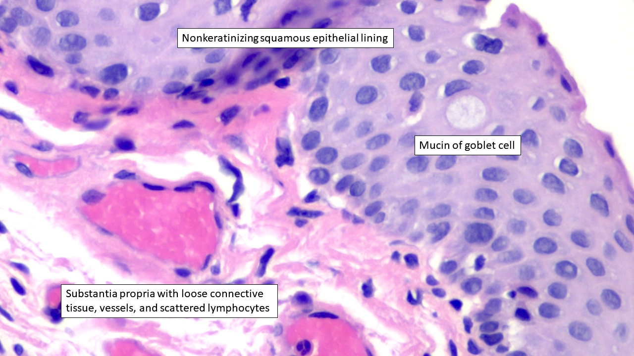
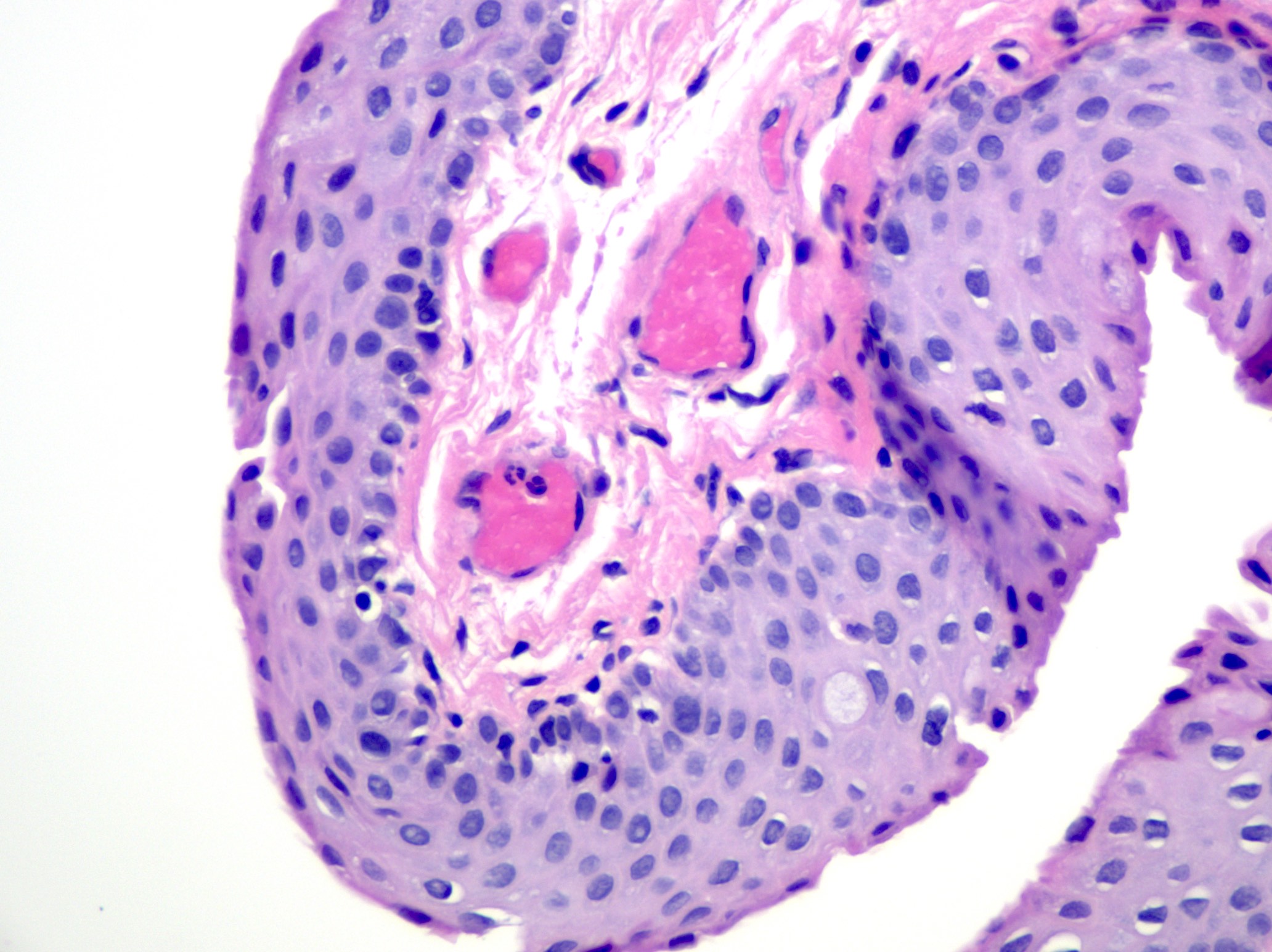
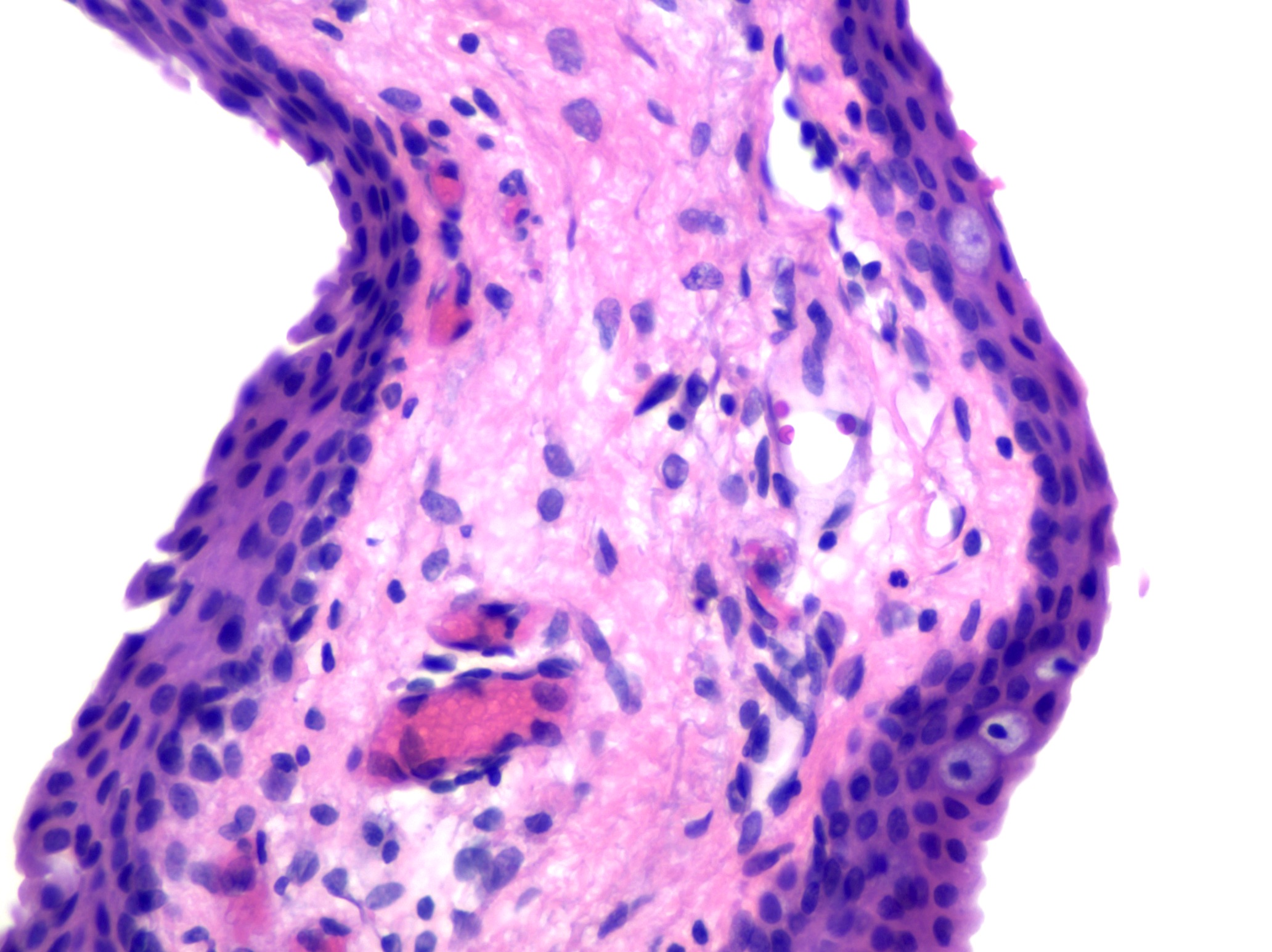
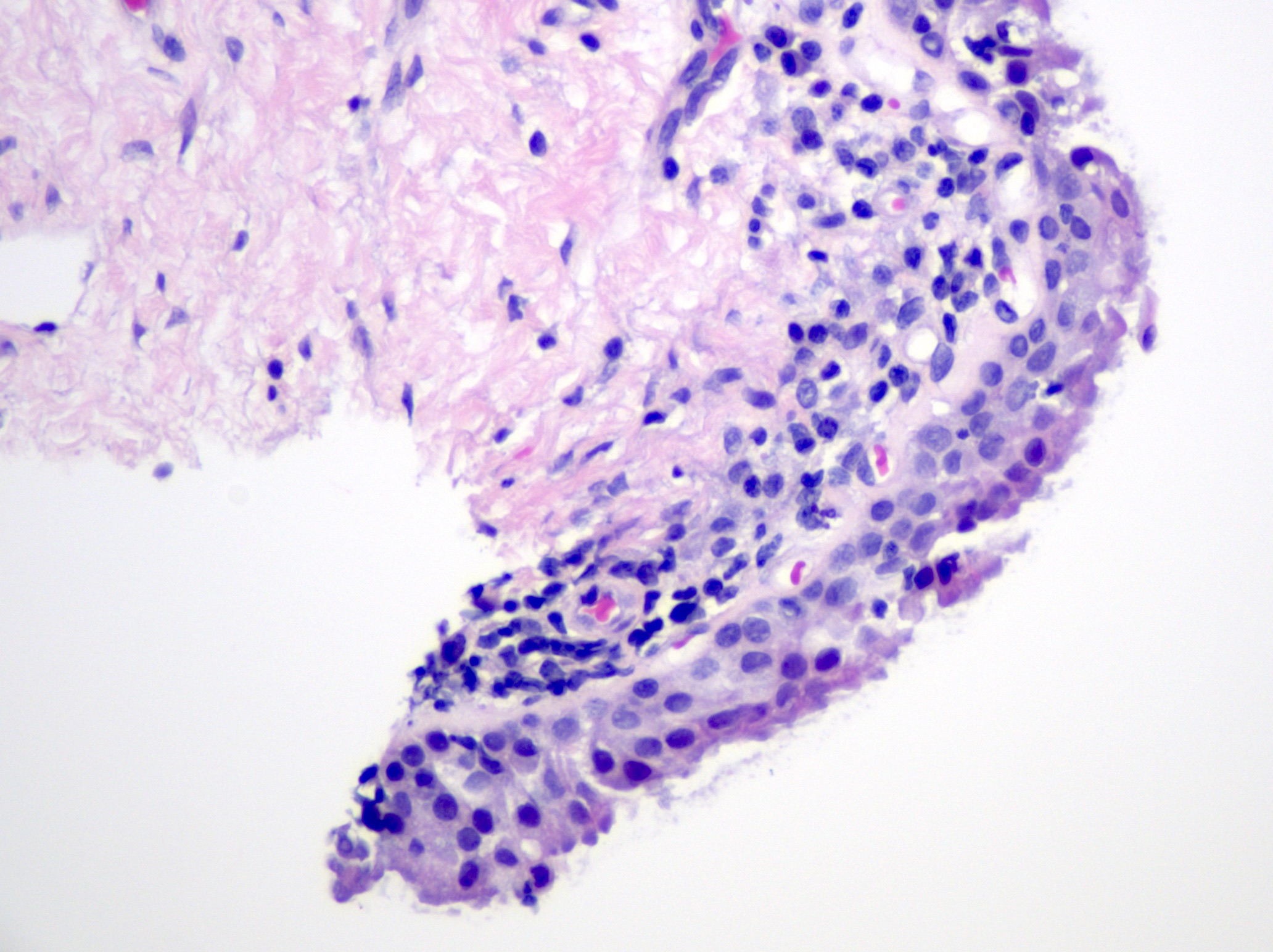
- Nonkeratinizing squamous epithelium with scattered goblet cells overlying loose connective tissue of the substantia propria with limited chronic inflammation (including lymphocytes and plasma cells) and subepithelial glands, such as sebaceous (Meibomian) glands in the vicinity of the tarsal plate (Exp Eye Res 2014;129:172, Invest Ophthalmol Vis Sci 2011;52:1938)
- Tarsal plate is identified by dense fibroconnective tissue in association with sebaceous (Meibomian) glands (J Anat 2005;206:37)
- Infoldings of conjunctival epithelium, particularly in the context of chronic conjunctivitis, may mimic glandular tissue, a histologic appearance known as pseudoglands of Henle (Ophthalmology 2001;108:135)
- Caruncle is identified by the presence of adnexal structures in addition to conjunctiva
- Degenerative changes related to the conjunctiva include pterygium and pinguecula; both are caused by ultraviolet light damage
- Pterygium histologically appears as benign conjunctival epithelium with corneal Bowman membrane, underlying elastotic (solar) degeneration and neovascularization (see Pterygium) (Am J Pathol 2011;178:817)
- Pinguecula histologically demonstrates solar elastotic degeneration with variably present, hyalinized and amorphous material (see Pinguecula) (Graefes Arch Clin Exp Ophthalmol 2006;244:104)
- Inflammatory changes may also be encountered
- Follicular conjunctivitis is comprised of lymphocytic nodules in the substantia propria and causes a bulge of the overlying epithelium; etiologies include infections, drug reaction, manifestations of systemic diseases and idiopathic causes
- Papillary conjunctivitis is a polygonal distortion of the epithelium, often affecting tarsal conjunctiva and may be caused by chronic mechanical irritation, allergy, topical drugs and as a complication of contact lenses (Ocul Surf 2020;18:396)
- Granulomatous conjunctivitis may be caused by foreign material, infection, systemic diseases or sarcoidosis (see Granulomatous conjunctivitis)
Microscopic (histologic) images
Contributed by J. Stephen Nix, M.D.
Nonkeratinizing squamous conjunctiva
Cuboidal and columnar appearance
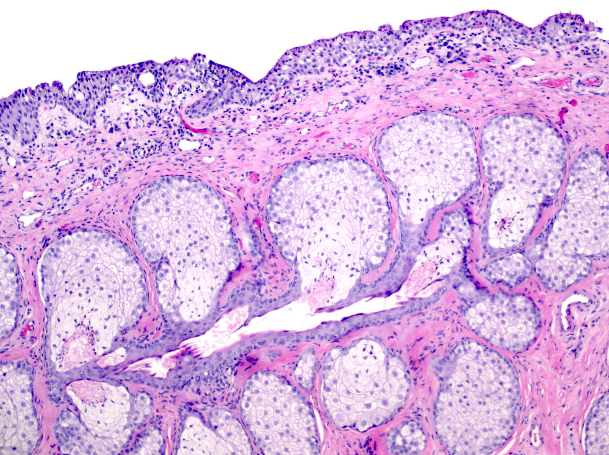
Tarsal plate, Meibomian glands

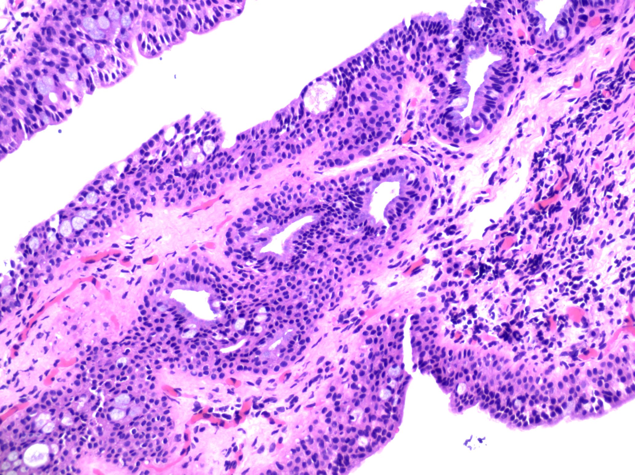
Caruncle, chronic conjunctivitis
Pseudoglands of Henle
Positive stains
- Though stains are not routinely used in practice, the normal conjunctiva is positive for pancytokeratins (AE1 / AE3) and CK7 (Invest Ophthalmol Vis Sci 1997;38:142)
Videos
Pathology lecture: conjunctiva
Additional references
Board review style question #1

Based on the histologic features shown above, from what location is the biopsy taken?
- Bulbar conjunctiva
- Caruncle
- Cornea
- Forniceal conjunctiva
- Palpebral conjunctiva
Board review style answer #1
E. Palpebral conjunctiva. The presence of dense fibroconnective tissue constituting the tarsal plate and sebaceous (Meibomian) glands is helpful for identifying the palpebral (tarsal) conjunctiva. Answer B is incorrect because the caruncle is identified by the presence of adnexal structures, such as pilosebaceous units. Answer C is incorrect because the cornea features corneal epithelium, Bowman membrane, corneal stroma, Descemet membrane and endothelium. Answers A and D are incorrect because the bulbar and forniceal conjunctivas are not seen in association with the tarsal plate.
Comment Here
Reference: Anatomy & histology-conjunctiva
Comment Here
Reference: Anatomy & histology-conjunctiva
Board review style question #2
Based on the histologic features shown, from what location is the following biopsy with increased chronic inflammation and reactive changes taken?
- Bulbar conjunctiva
- Caruncle
- Cornea
- Forniceal conjunctiva
- Palpebral conjunctiva
Board review style answer #2
B. Caruncle. The caruncle is identified by the presence of adnexal structures, such as the pilosebaceous unit seen in this histologic image. Answer E is incorrect because thick fibroconnective tissue of the tarsal plate and sebaceous (Meibomian) glands would be helpful for identifying the palpebral conjunctiva. Answer C is incorrect because the cornea features corneal epithelium, Bowman membrane, corneal stroma, Descemet membrane and endothelium. Answers A and D are incorrect because the bulbar and forniceal conjunctivas would not feature an associated pilosebaceous unit.
Comment Here
Reference: Anatomy & histology-conjunctiva
Comment Here
Reference: Anatomy & histology-conjunctiva
