


Eye
General
Anatomy & histology-uvea
Author: Nat Pernick, M.D.

Last author update: 1 February 2014
Last staff update: 10 May 2024
Copyright: 2004-2024, PathologyOutlines.com, Inc.
PubMed Search: Uvea anatomy

Cite this page: Pernick N. Anatomy & histology-uvea. PathologyOutlines.com website. https://www.pathologyoutlines.com/topic/eyeanatomyuvea.html. Accessed July 15th, 2024.
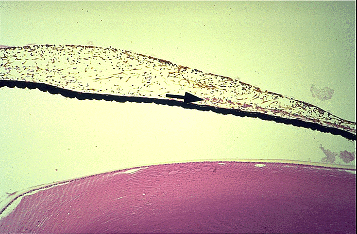
Choroid
- Middle layer of globe between outer sclera and inner retina
- Highly vascular but no lymphatics
- Extends from ciliary body to optic nerve
- Inner aspect is adherent to retinal pigment epithelium; outer surface is loosely attached to overlying sclera
- Stroma contains abundant pigmented melanocytes
- Bruch membrane: separates choroid from overlying retinal pigment epithelium, is 2 - 4 microns thick, has 5 distinct layers (basal lamina of overlying retinal pigment epithelium, collagenous layer, elastic fiber rich layer, collagenous layer and basal lamina of endothelial cells of choriocapillaris), thickens with age, has focal excrescences known as drusen
- Choriocapillaris: in innermost choroidal stroma adjacent to Bruch membrane, connects with arterial and venous channels from vessels in outer choroidal stroma to nourish outer retinal layers
Iris
- Thin diaphragm of tissue with central opening (pupil)
- Forms boundary of anterior and posterior chamber
- Highly textured with folds and crypts
- Part of middle layer of eye (also ciliary body and choroid)
- Normally rests gently upon lens and bulges slightly forward
- Consists of stroma and posterior epithelial lining (2 closely apposed epithelial layers, with numerous melanosomes); contains sphincter muscle within stroma that controls pupil
- Anterior iris lacks a cellular lining
- Color is due to number of stromal melanocytes; blue irises have few stromal melanocytes; brown irises have numerous melanocytes
- Blood vessels are usually surrounded by a thick collar of collagen fibers, resembling arteriolosclerosis
- Fewer melanosomes and melanocytes in patients with ocular and oculocutaneous albinism
- Regulates amount of light reaching pupil; muscles of iris dilate or constrict pupil in response to parasympathetic or sympathetic nerve impulses; normal diameter of pupil is 1 - 8 mm
- Iridectomy: excision of small segment of iris; place on filter paper to avoid folding
- Ectropion uveae: fibrovascular tissue on anterior surface of iris everts the papillary margin and pulls pigmented epithelia onto anterior surface of iris
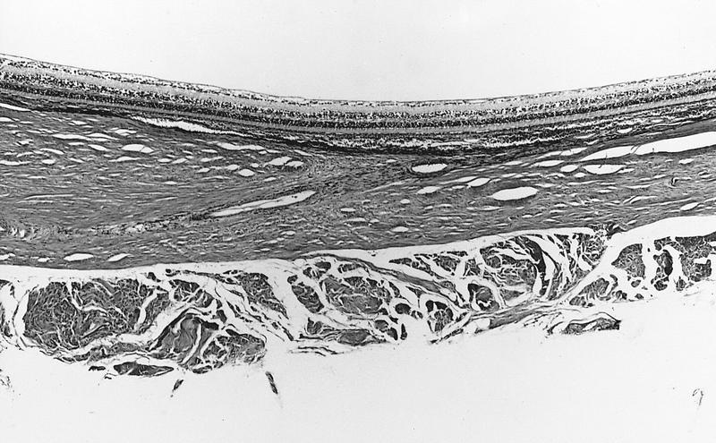
Sclera
- 80% of surface area of eye
- Begins at periphery of cornea, extends posteriorly to optic nerve
- Relatively rigid; protects eye from trauma and maintains intraocular pressure
- Visible anteriorly under transparent conjunctiva; white in adults
- Thickness varies; 0.3 mm at insertion of rectus muscles, 0.8 mm at limbus and 1.0 mm at insertion of optic nerve
- Weakly attached to underlying choroid by thin collagen fibers
- Heals poorly due to few blood vessels or fibroblasts
- Aging related changes include calcification between collagen fibers, senile scleral plaques
- Composed of episclera, stroma and lamina fusca
- Episclera: most superficial part of sclera, located between fibrous structure that envelopes the globe (Tenon capsule) and scleral stroma; composed of loose collagen fibers and fibroblasts with numerous vessels, occasional melanocytes and mononuclear white blood cells
- Stroma: largest component of sclera, randomly arranged bands of collagen with occasional elastic fibers and fibroblasts; minimal blood vessels except in perforating emissiary canals, accompanied by nerves and scattered melanocytes; rarely contains a prominent nerve (nerve loop of Axenfeld) in an emissiarial canal near limbus, which may mimic a neurofibroma; anterior ciliary arteries perforate sclera near insertion of rectus muscles, posterior ciliary arteries pass through sclera near optic nerve; vortex veins exit sclera posterior to equator of eye
- Lamina fusca: innermost layer of sclera with loose collagen fibers, fibroblasts and scattered melanocytes
Drawings
Images hosted on other servers:

Choroid and iris
Images hosted on other servers:

Iris: front view
Microscopic (histologic) images
AFIP images
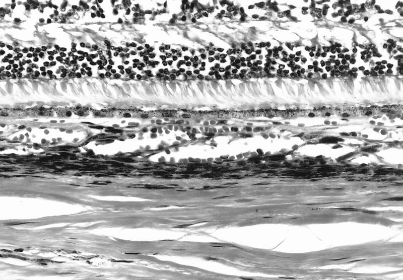
Vascularized pigmented stroma
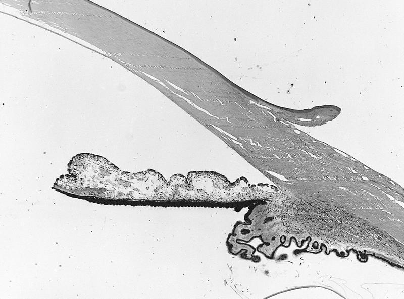
Uveal tissue extends into sclera
Iris and ciliary body with open anterior chamber angle
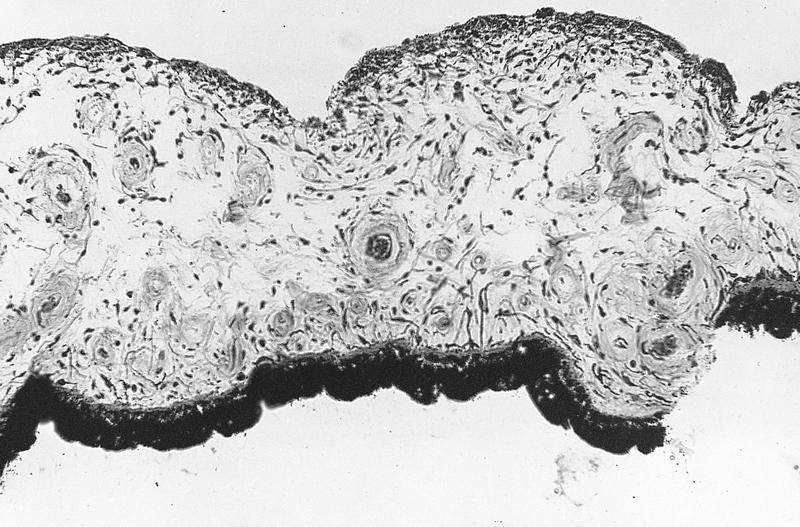
Normal iris
Images hosted on other servers:
Iris and lens
