
Esophagus
Other tumors
Esophageal sarcoma-overview
Authors: Feriyl Bhaijee, M.D., Israh Akhtar, M.D.


Last author update: 2 December 2013
Last staff update: 20 March 2025 (update in progress)
Copyright: 2003-2025, PathologyOutlines.com, Inc.
PubMed Search: Sarcoma [title] esophagus


Table of Contents
Definition / general | Epidemiology | Sites | Clinical features | Diagnosis | Radiology description | Radiology images | Prognostic factors | Treatment | Clinical images | Gross description | Gross images | Microscopic (histologic) description | Microscopic (histologic) images | Positive stains | Flow cytometry description | Electron microscopy description | Molecular / cytogenetics description | Differential diagnosis | Additional referencesCite this page: Bhaijee F, Akhtar I. Esophageal sarcoma-overview. PathologyOutlines.com website. https://www.pathologyoutlines.com/topic/esophagussarcomagen.html. Accessed March 31st, 2025.
Definition / general
- Malignant mesenchymal tumors of the esophagus with variable degrees of differentiation, biologic behavior and prognosis
- Examples
- Angiosarcoma: high grade malignancy of endothelial cells
- Ewing sarcoma: highly malignant small round blue cell tumor characterized by recurrent chromosomal translocations [t(11;22) EWSR1-FLI1 or t(21;22) EWSR1-ERG] and membranous MIC2 / CD99 overexpression
- Fibrosarcoma: spindle cell neoplasm of low grade to intermediate grade malignancy
- Gastrointestinal stromal tumor (GIST): mesenchymal tumor of digestive tract, likely originating from multipotential progenitors of interstitial cells of Cajal
- Hemangiopericytoma (solitary fibrous tumor): ubiquitous mesenchymal tumor showing uncertain line of differentiation (not true microvascular pericytes)
- Kaposi sarcoma: uncommon, low grade, vascular malignancy caused by Kaposi sarcoma herpesvirus / human herpesvirus 8 (KSHV / HHV8) infection
- Leiomyosarcoma: malignant smooth muscle tumor
- Liposarcoma: malignant adipocytic / lipomatous tumor
- Malignant peripheral nerve sheath tumor (MPNST): malignant tumor arising from peripheral nerve or a neurofibroma or in extraneural soft tissue and showing nerve sheath differentiation
- Osteosarcoma: mesenchymal malignancy characterized by neoplastic cells that produce osteoid matrix
- Synovial sarcoma (SS): malignant mesenchymal tumor showing epithelial differentiation, either overtly (biphasic SS) or by IHC alone (monophasic SS)
- Undifferentiated pleomorphic sarcoma: highly malignant pleomorphic neoplasm lacking any specific line of differentiation
Epidemiology
- Extremely rare
Sites
- Likely arise from submucosal or intramural elements
Clinical features
- Asymptomatic mass
- Obstructive features: dysphagia, odynophagia, nausea, vomiting
Diagnosis
- Biopsy or resection with immunohistochemical or molecular ancillary studies
Radiology description
- Highly variable
- Destructive mass with variable hemorrhage or necrosis
- Strictures or obstruction
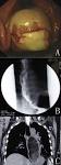
Radiology images
Images hosted on other servers:

Dilated thoracic esophagus

Gastrographin esophagography
Prognostic factors
- Poor prognostic factors
- High histologic grade
- Cytologic atypia, nuclear pleomorphism
- Necrosis
- Mitotic activity
- High histologic grade
- Poor degree of differentiation
Treatment
- Treatment options depend on type of sarcoma and clinical stage
- Treatment modalities include surgery, chemotherapy, radiotherapy
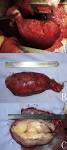
Clinical images
Images hosted on other servers:

GIST

Leiomyosarcoma
Liposarcoma
Gross description
- Often large tumor with infiltrative margins and variable hemorrhage, necrosis
Gross images
AFIP images
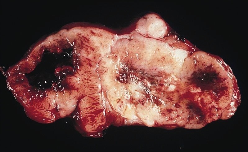
Sarcoma
Images hosted on other servers:

GIST
Liposarcoma
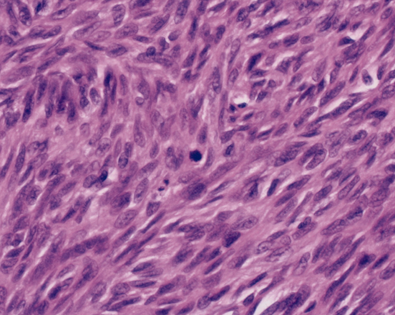
Microscopic (histologic) description
- Highly variable
- Round blue cell tumors: Ewing sarcoma, small cell variant of osteosarcoma
- Spindle cell tumors: angiosarcoma, fibrosarcoma, GIST, hemangiopericytoma, Kaposi sarcoma, leiomyosarcoma, MPNST, synovial sarcoma
- Adipocytic differentiation: liposarcoma
- Osteoid production: osteosarcoma
- Undifferentiated: pleomorphic sarcoma
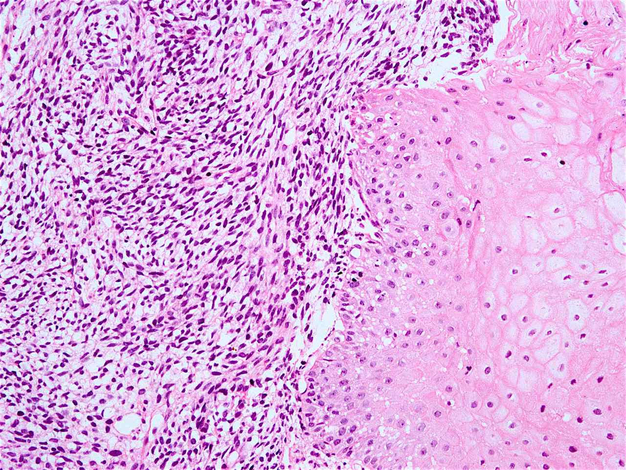
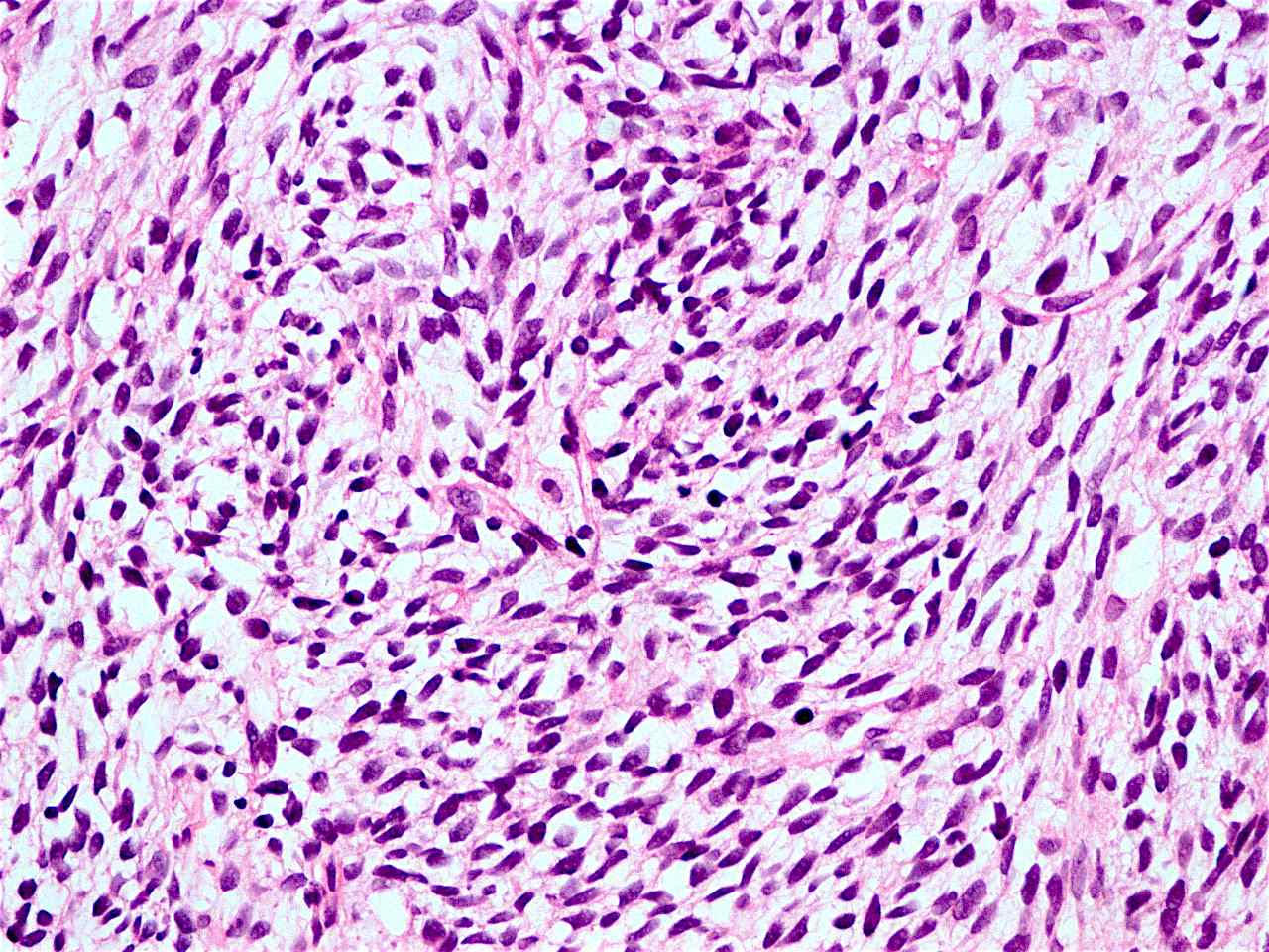
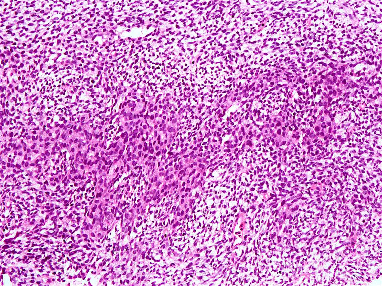
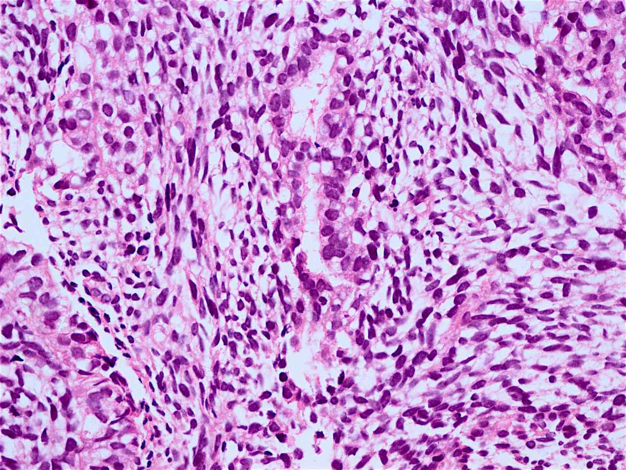
Microscopic (histologic) images
Contributed by AFIP and Dr. Zafar Ali
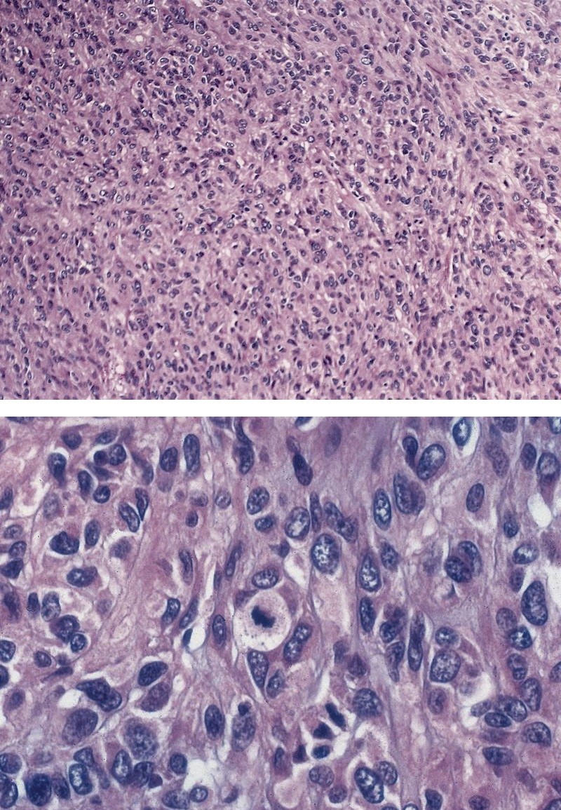
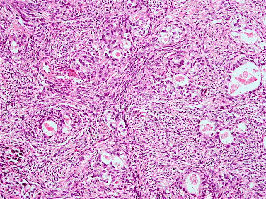
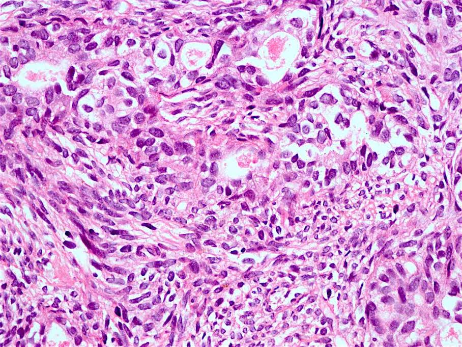
Sarcoma, not otherwise specified
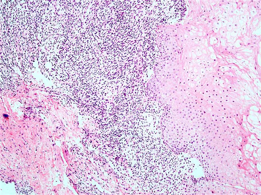
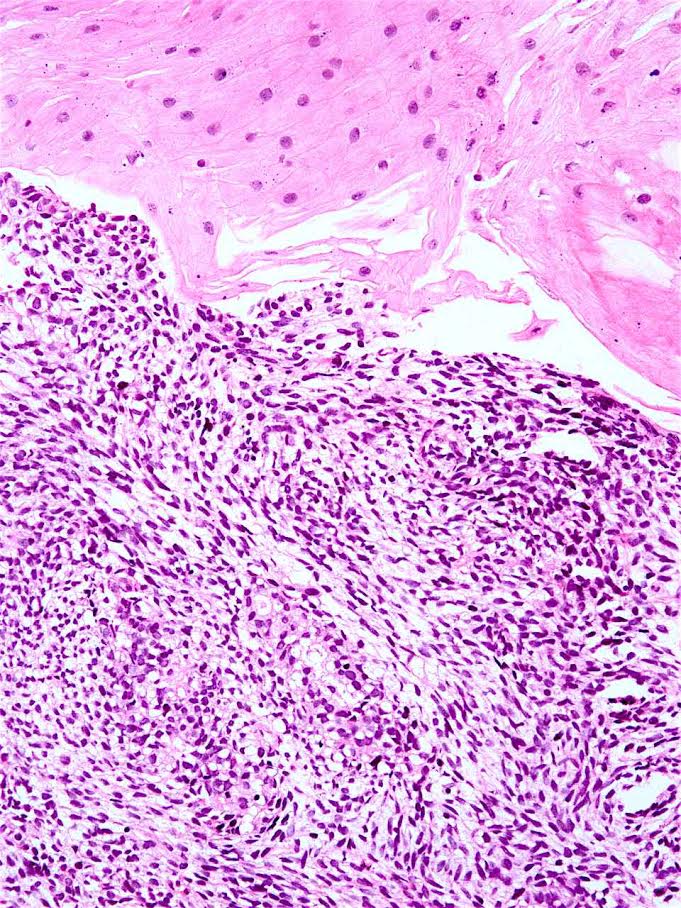
36 year old man with mass just below upper esophageal sphincter, synovial sarcoma (biphasic)

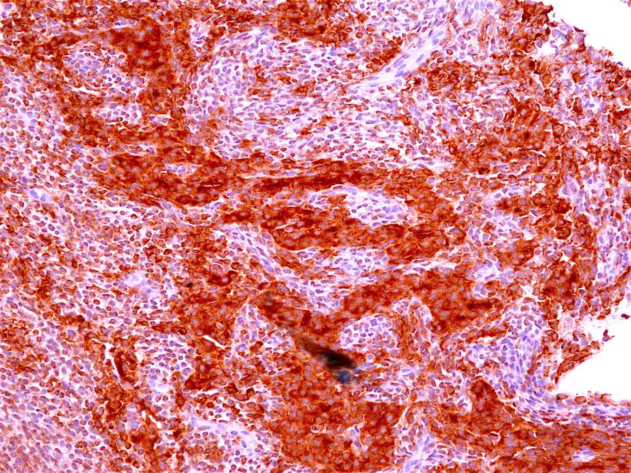
CD99
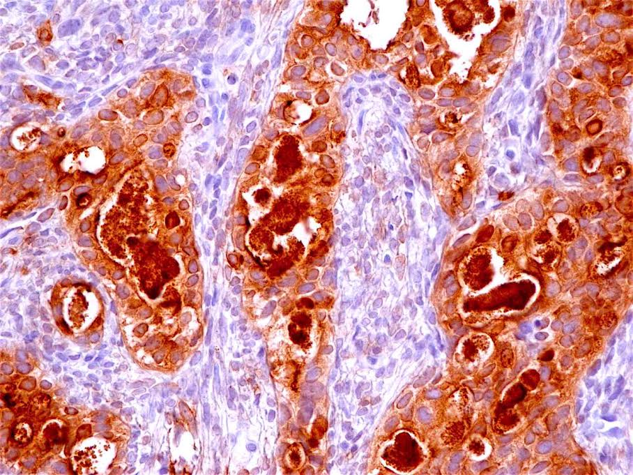
EMA
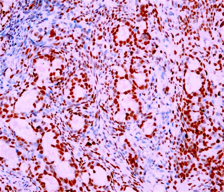

TLE1
Images hosted on other servers:
Leiomyosarcoma

Liposarcoma

Osteosarcoma
Positive stains
- Angiosarcoma: endothelial markers (factor VIII related antigen, CD31, CD34)
- Ewing sarcoma: MIC2 / CD99 (100% sensitive but not specific), FLI1 gene product (90%), neuron specific enolase, vimentin, p53
- GIST: CD117 / c-kit, DOG1, CD34, focal SMA, focal desmin, focal S100
- Hemangiopericytoma: CD34 (95%), CD99 (70%), CD57 (50%), focal nuclear beta catenin
- Kaposi sarcoma: HHV8, factor VIII related antigen, CD34, CD31, FLI1, D2-40, VEGFR3, BCL2
- Leiomyosarcoma: smooth muscle markers (SMA, desmin, caldesmon, calponin)
- Liposarcoma: S100 (lipogenic zones); MDM2, CDK4 (well differentiated tumors)
- MPNST: S100 (weak / focal in spindled MPNST, diffuse / strong in epithelioid MPNST)
- Synovial sarcoma: focal high molecular weight cytokeratin
- Undifferentiated pleomorphic sarcoma: CD68
Flow cytometry description
- Useful to exclude hematopoietic malignancies
Electron microscopy description
- Angiosarcoma: Weibel-Palade bodies
- Ewing sarcoma: extensive cytoplasmic glycogen deposits
- Hemangiopericytoma: undifferentiated spindle cell or fibroblastic features
- Leiomyosarcoma: features of smooth muscle differentiation (thin filaments, pinocytic vesicles, attachment plaques, interrupted external lamina)
- Liposarcoma: nonmembrane bound intracytoplasmic lipid droplets of varying sizes / densities
- MPNST: tumor cells with external laminae, indicating schwannian differentiation
- Osteosarcoma: osteoid matrix comprises nonperiodic fibrils, scattered collagen fibers and hydroxyapatite calcium crystals
Molecular / cytogenetics description
- RT PCR or FISH
- Ewing sarcoma: recurrent chromosomal translocations including t(11;22)(q24;q12) EWS-FLI1, t(21;22)(q22;q12) EWS-ERG, t(7;22) EWS-ETV1, t(17;22) EWS-E1AF, t(2;22) EWS-FEV
- GIST: mutually exclusive mutations of activating KIT (95%) or platelet derived growth factor alpha (PDGFRA) receptor tyrosine kinase (5%)
- Kaposi sarcoma: HHV8 detected by PCR
- Leiomyosarcoma: no consistent genetic events reported
- Well differentiated liposarcoma: giant marker or supernumerary ring chromosomes with amplification of 12q12-15 region, including MDM2, CDK4 and other genes
- Myxoid / round cell liposarcoma: t(12;16) DDIT3-TLS or t(12;22) DDIT3-EWS balanced translocations
- Synovial sarcoma: characteristic t(X;18) SSYT-SSX1 / 2 translocations
Differential diagnosis
- Benign spindle cell proliferations, e.g.
- Inflammatory fibroid polyp: PDGFRA+, CD34+, CD117-, mixed inflammatory infiltrate (especially eosinophils)
- Leiomyoma: rare atypia, mitotic activity, or necrosis
- Ossifying fibromyxoid tumor: usually bland cytology and peripheral ossification
- Schwannoma: diffuse S100+, Verocay bodies
- Lymphoma: usually CD45 (LCA)+, flow cytometry may be diagnostic
- Melanoma: melanoma markers+ (S100, HMB45, MART, MITF, MelanA, SOX10, tyrosinase)
- Other round blue cell tumors, e.g.
- Alveolar rhabdomyosarcoma: desmin+, myogenin+, myoD1+
- Desmoplastic small round cell tumor (DSRCT): striking desmoplasia, coexpression of cytokeratin / desmin, WT1+
- Glomus tumor: round to polygonal cells with scant cytoplasm and marked cellularity uniformity; SMA+, desmin-, S100-
- Neuroendocrine (small cell) carcinoma: neuroendocrine markers+ (CD56, chromogranin, synaptophysin)
- Poorly differentiated carcinoma: focal cytokeratin+
