
Esophagus
Benign tumors
Leiomyoma
Author: Elliot Weisenberg, M.D.

Last author update: 1 January 2013
Last staff update: 14 September 2023
Copyright: 2003-2025, PathologyOutlines.com, Inc.
PubMed Search: Esophageal leiomyoma[TI] full text[sb]


Table of Contents
Definition / general | Clinical features | Case reports | Treatment | Clinical images | Gross description | Gross images | Microscopic (histologic) description | Microscopic (histologic) images | Cytology images | Positive stains | Negative stains | Differential diagnosisCite this page: Weisenberg E. Leiomyoma. PathologyOutlines.com website. https://www.pathologyoutlines.com/topic/esophagusleiomyoma.html. Accessed April 1st, 2025.
Definition / general
- Most common benign tumor of esophagus (eMedicine: Esophageal Leiomyoma [Accessed 15 February 2019], Surg Clin North Am 1983;63:625), 8% incidence in autopsy studies (Hum Pathol 1981;12:1006)
Clinical features
- Median age 35 years, 2/3 men, usually single, 24% multiple (seedling tumors)
- Usually arises from inner circular muscle; most common in distal esophagus, rarely polypoid
- Benign behavior (Ann Thorac Surg 2005;79:1122)
- Minute (1 - 2 mm "seedling") tumors are often near the gastroesophageal junction and are asymptomatic
- Multiple tumors are associated with MEN1 syndrome (Am J Pathol 2001;159:1121)
- Large tumors may cause obstructive symptoms
Case reports
- 26 year old woman with multinodular growth pattern simulating carcinoma (Dis Esophagus 2007;20:187)
- 64 year old man with coexisting early squamous cell carcinoma (Jpn J Clin Oncol 2004;34:751)
- 75 year old man with coexisting leiomyosarcoma (J Exp Clin Cancer Res 2005;24:487)
Treatment
- Excise if significant symptoms (Ann Thorac Cardiovasc Surg 2007;13:78), endoscopic enucleation for small tumors (Singapore Med J 2006;47:901), esophagectomy for large tumors (World J Gastroenterol 2005;11:4258)
Clinical images
Images hosted on other servers:

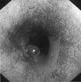
Endoscopic examination
Gross description
- Circumscribed, mural, solitary mass, 2 - 5 cm (surgical specimens), bulges into lumen, may be polypoid
- Pinkish gray white with whorled cut surface; mucosal surface is only rarely ulcerated
Gross images
AFIP images
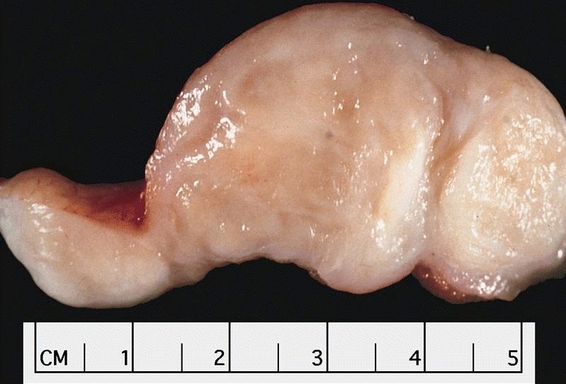
Bulging, white, whorled cut surface
Images hosted on other servers:


Submucosal tumor
With squamous cell carcinoma
Microscopic (histologic) description
- Similar to classic endometrial leiomyoma; circumscribed lesion of circular muscularis propria or muscularis mucosae composed of intersecting fascicles of bland spindle cells with abundant cytoplasm
- Variable fibrosis in center of large leiomyomas
- Occasional calcification; no / rare mitotic figures; no atypia, no cellular foci
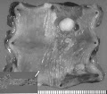
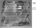
Microscopic (histologic) images
AFIP images

Cluster of seedling leiomyomas
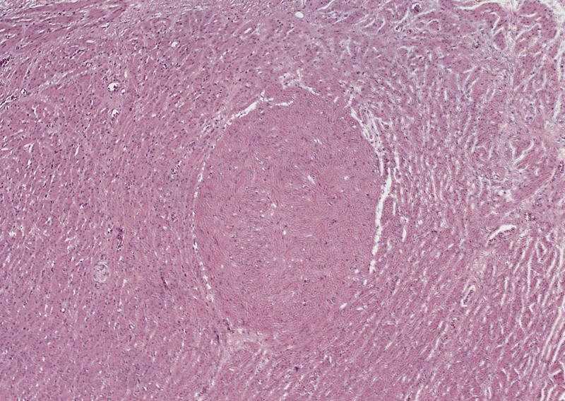
Seedling leiomyoma of muscularis propria
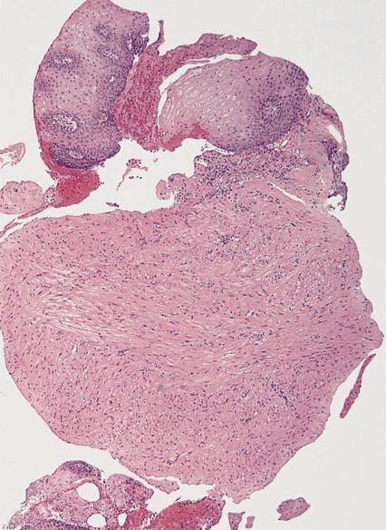
Leiomyoma with overlying squamous epithelium
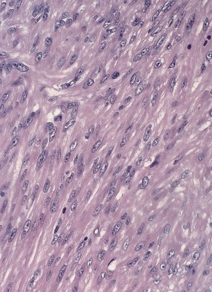
Mature, hypertrophied smooth muscle cells

Multiple seedling leiomyomas
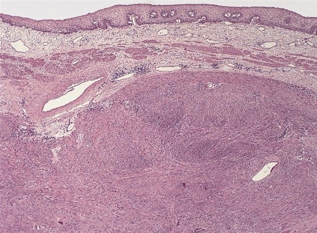
Leiomyoma of muscularis propria

Fascicular growth pattern
Images hosted on other servers:

Squamous cell carcinoma and leiomyoma
Cytology images
Images hosted on other servers:


Groups of spindled cells with low cellularity
Positive stains
Differential diagnosis
- GIST: very rare; solid, myxoid and perivascular patterns; more cellular by H&E and cytology, CD117+, CD34+, variable desmin and actin immunoreactivity (Am J Surg Pathol 2000;24:211)
