Table of Contents
Definition / general | Essential features | CPT coding | Sites | Case reports | Cytology description | Cytology images | Immunohistochemistry | Sample pathology report | Differential diagnosis | Additional references | Board review style question #1 | Board review style answer #1 | Board review style question #2 | Board review style answer #2Cite this page: Alshaikh S. Suspicious for malignancy. PathologyOutlines.com website. https://www.pathologyoutlines.com/topic/cytopathologylungsuspicious.html. Accessed April 3rd, 2025.
Definition / general
- Suspicious for malignancy is used in respiratory cytopathology when some cytopathological features are suggestive of malignancy but there are insufficient features either in number or quality to confirm a diagnosis of malignancy (Acta Cytol 2023;67:80)
- Risk of malignancy (ROM), which falls under the suspicious for malignancy category, is 82% with a range of 54.5 - 90%; in a recent study, ~5% of the cases were placed in this category (Acta Cytol 2023;67:80)
Essential features
- Category of suspicious for malignancy is used when cytomorphological features are suspicious but not diagnostic of malignancy (due to insufficient features characteristic of malignancy or insufficient number of atypical cells to be diagnosed as malignancy) so that further diagnostic management can be planned
- When a case is categorized as suspicious for malignancy, the report should include which differential diagnoses are suspected, including non-small cell carcinoma, neuroendocrine tumors, small and large cell neuroendocrine carcinomas, lymphoma, sarcoma and metastatic carcinomas
CPT coding
Sites
- Lung, bronchus
- Lung, parenchyma
Case reports
- 72 year old woman with an incidentally detected lung mass (Diagn Cytopathol 2022;50:E156)
- 75 year old man with a known history of tuberculosis (TB), shortness of breath and a dry cough (Diagn Cytopathol 2011;39:927)
- 76 year old man with a histologically confirmed KRAS mutated, thyroid transcription factor 1 (TTF1) positive, grade 1, mucinous adenocarcinoma (Diagn Cytopathol 2024;52:E172)
Cytology description
- Suspicious for malignancy category is usually applied in cases where there is significant cytopathological atypia but there are only a small number of cells that show these features or in cases where the qualitative features are not definitive for a diagnosis of malignancy, for instance
- Significant loss of cell and nuclear polarity
- Nuclear crowding
- Significant nuclear membrane irregularities
- Significant abnormalities of chromatin pattern and hyperchromasia
- Significantly increased N:C ratio
- Loss of cilia
- Mild to moderate anisonucleosis (Cytojournal 2023;20:42)
Cytology images
Immunohistochemistry
- If the quality and quantity of suspicious cells on direct smears and in cell blocks prevent further definition of the diagnosis, a suspicious for malignancy categorization and a statement regarding the type of carcinoma suspected should be included in the report
- Positive immunohistochemical stains for neuroendocrine markers on a small amount of extensively crushed material in a cell block from a lung fine needle aspiration biopsy (FNAB) may favor a neuroendocrine tumor (NET) and exclude lymphoma but to define the tumor grade, there may be insufficient tumor to do the proliferation marker Ki67
- Thyroid transcription factor 1 (TTF1) as a marker of lung adenocarcinoma and p40 as a marker of squamous differentiation are often used for further classification of non-small cell carcinomas; however, these stains do not separate benign from malignant as they are expressed in benign reactive respiratory epithelial cells
- Reference: Acta Cytol 2024;68:351
Sample pathology report
- Right lung lobe, 50 mm mass, endobronchial ultrasound guided FNA:
- FNAB right lung lobe, suspicious for malignancy; necrosis with scattered atypical squamous epithelium suspicious for squamous cell carcinoma (see report below)
- Microscopic description: There is marked necrotic material and keratinous debris as well as rare minute tissue fragments of keratinized cells with small pyknotic, hyperchromatic nuclei and a low to moderate N:C ratio.
- Comment: The features are suspicious for keratinizing squamous cell carcinoma. Correlation with clinical and imaging findings is required.
- Lung, left upper lobe, 30 mm mass, sputum sample:
- Suspicious for malignancy; atypical glandular cells (see report below)
- Microscopic description: Few small, crowded epithelial cells with large, pleomorphic, irregular nuclei, prominent nucleoli and vacuolated cytoplasm are present. These epithelial cells lack cilia.
- Comment: The above cytopathological findings are suspicious for adenocarcinoma. Correlation with imaging and clinical finding is required. Recommend further workup with bronchial brush and FNA.
Differential diagnosis
- Inflamed reactive epithelium:
- Reactive epithelial cells have cilia and regular nuclear membrane
- Prior radiation or chemotherapy (particularly for lung carcinomas):
- May result in significant atypia in a sample with low cellularity
Additional references
Board review style question #1
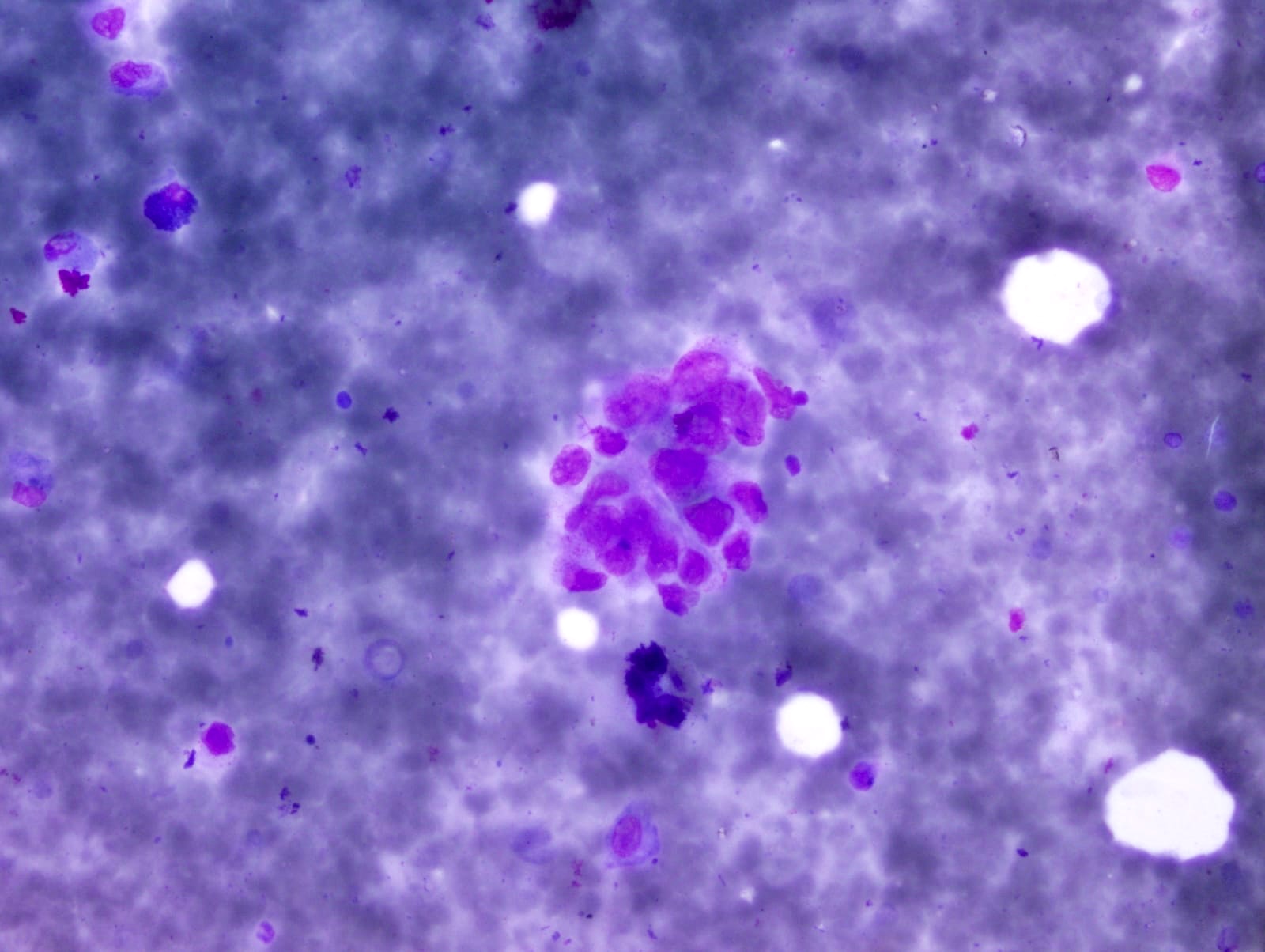
A 79 year old smoker presents with dyspnea. A computed tomography (CT) scan shows a 3 cm mass in his right lung. Endobronchial ultrasound (EBUS) guided fine needle aspiration of the mass is shown in the image above. It shows only a small number of atypical cells in a necrotic background. Which of the following is the best diagnostic category?
- Atypical
- Benign
- Insufficient / inadequate
- Malignant
- Suspicious for malignancy
Board review style answer #1
E. Suspicious for malignancy. There is significant cytopathological atypia including nuclear enlargement, nuclear pleomorphism, irregular nuclear membrane, hyperchromatic chromatin and nuclear crowding but there are only a small number of cells showing these features. Answer C is incorrect because a specimen categorized as insufficient / inadequate lacks atypical cells. Answer B is incorrect because a specimen categorized as benign should show benign cytopathological features. Answer A is incorrect because a specimen categorized as atypical demonstrates features predominantly seen in benign lesions and minimal features that may raise the possibility of a malignant lesion. Answer D is incorrect because a specimen categorized as malignant should show unequivocal cytopathological features of malignancy.
Comment Here
Reference: Suspicious for malignancy
Comment Here
Reference: Suspicious for malignancy
Board review style question #2
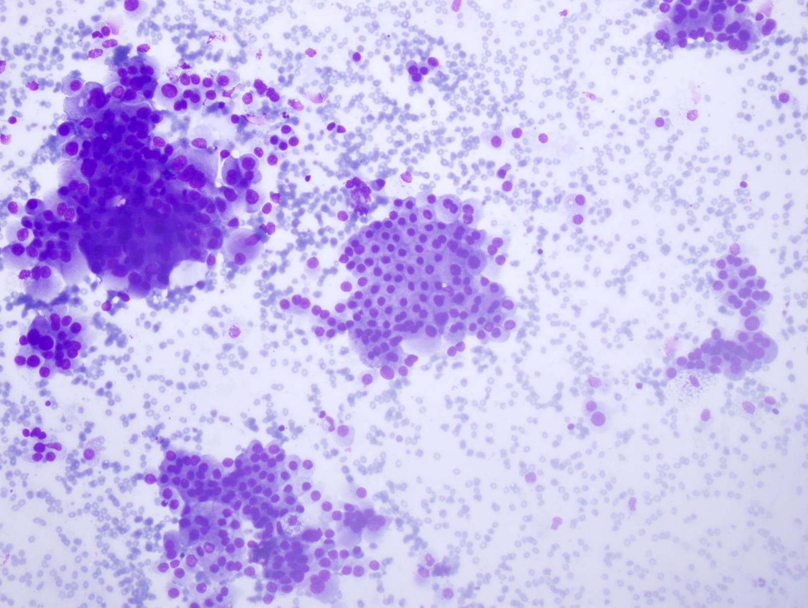
A 55 year old man presented with a centrally located 3.5 cm mass. An FNA was performed and the Diff-Quik smear (image shown above) shows a rare cluster of medium sized pleomorphic atypical cells, with high N:C ratio, irregular nuclear membranes and hyperchromatic nuclei. Which of the following is the most likely diagnosis?
- Adenocarcinoma
- Atypical
- Insufficient / inadequate
- Small cell carcinoma
- Suspicious for malignancy
Board review style answer #2
E. Suspicious for malignancy. There is one cluster that shows marked cytopathological atypia including nuclear enlargement, nuclear pleomorphism, irregular nuclear membrane, hyperchromatic chromatin and nuclear crowding but there are only a small number of cells showing these features. Answer C is incorrect because a specimen categorized as insufficient / inadequate lacks atypical cells. Answer B is incorrect because a specimen categorized as atypical should show features predominantly seen in benign lesions and minimal features that may raise the possibility of a malignant lesion. Answer D is incorrect because a specimen categorized as small cell carcinoma should show sufficient cellularity and cytopathological features of malignancy, including oval to elongated nuclei, absent nucleoli, scant cytoplasm, molding, necrosis and apoptosis. Answer A is incorrect because a specimen categorized as adenocarcinoma should show sufficient clusters of cohesive cells, with foamy vacuolated cytoplasm, fine chromatin and prominent nucleoli.
Comment Here
Reference: Suspicious for malignancy
Comment Here
Reference: Suspicious for malignancy


