Table of Contents
Definition / general | Essential features | Terminology | Sites | Clinical features | Treatment | Gross description | Microscopic (histologic) description | Microscopic (histologic) images | Positive stains | Negative stains | Electron microscopy description | Molecular / cytogenetics description | Sample pathology report | Differential diagnosis | Board review style question #1 | Board review style answer #1Cite this page: Gonzalez RS. Perineurioma. PathologyOutlines.com website. https://www.pathologyoutlines.com/topic/colontumorperineurioma.html. Accessed April 1st, 2025.
Definition / general
- Benign polypoid lesion occurring in the colonic mucosa or submucosa
- Peripheral nerve sheath tumor, often with overlying serrated / hyperplastic epithelium
Essential features
- Uncommon mucosal epithelial-mesenchymal lesion of the colon
- Usually occurs in middle aged women
- Benign, does not recur
Terminology
- Also known as benign fibroblastic polyp of the colon (Am J Surg Pathol 2004;28:374, Am J Surg Pathol 2008;32:1088)
Sites
- Usually in distal colon; rarely in small intestine
Clinical features
- F > M, with a median age of 51 (Am J Surg Pathol 2005;29:859)
Treatment
- Excision (benign behavior)
Gross description
- Small sessile polyp (median size 0.4 cm)
Microscopic (histologic) description
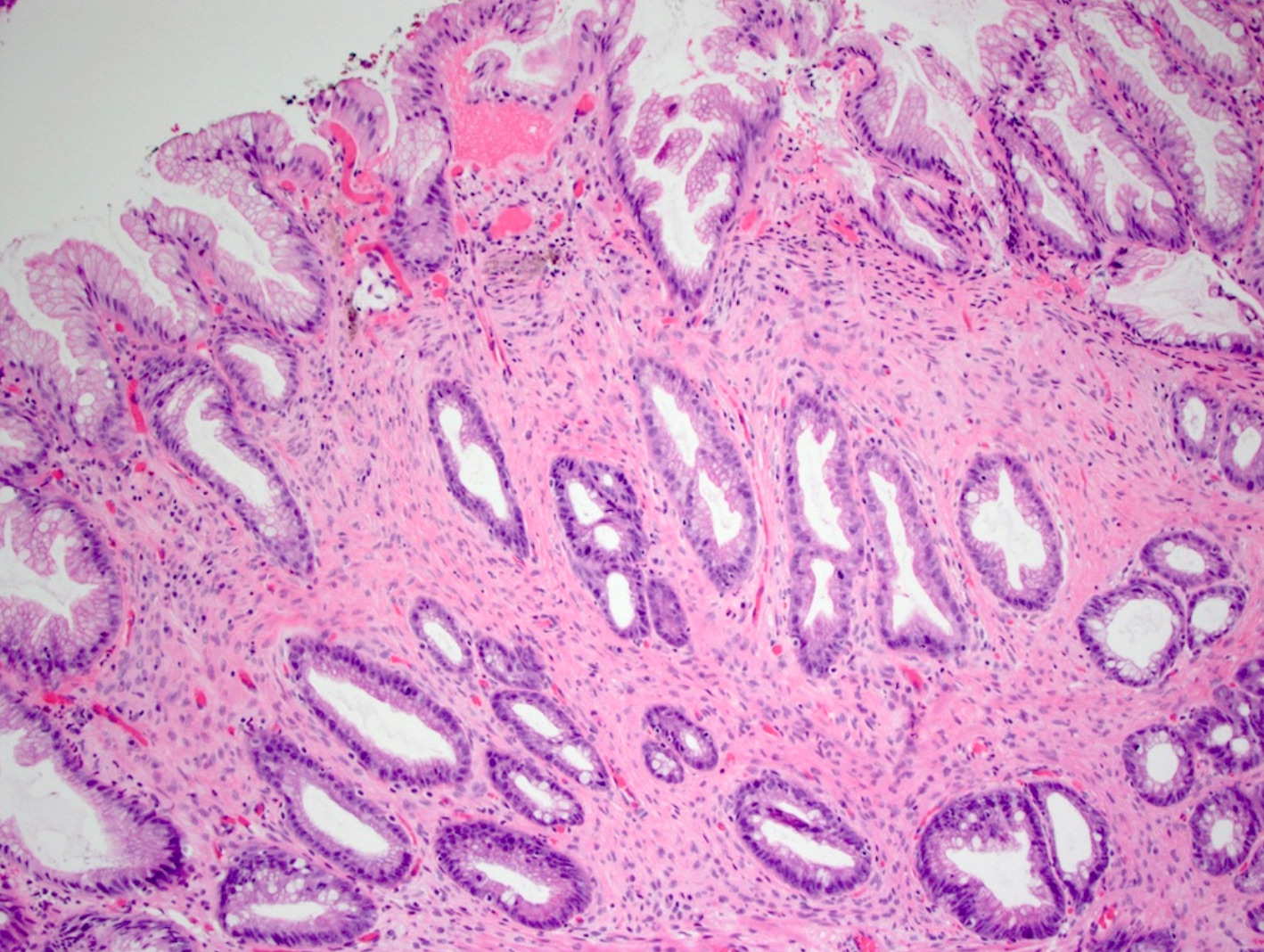
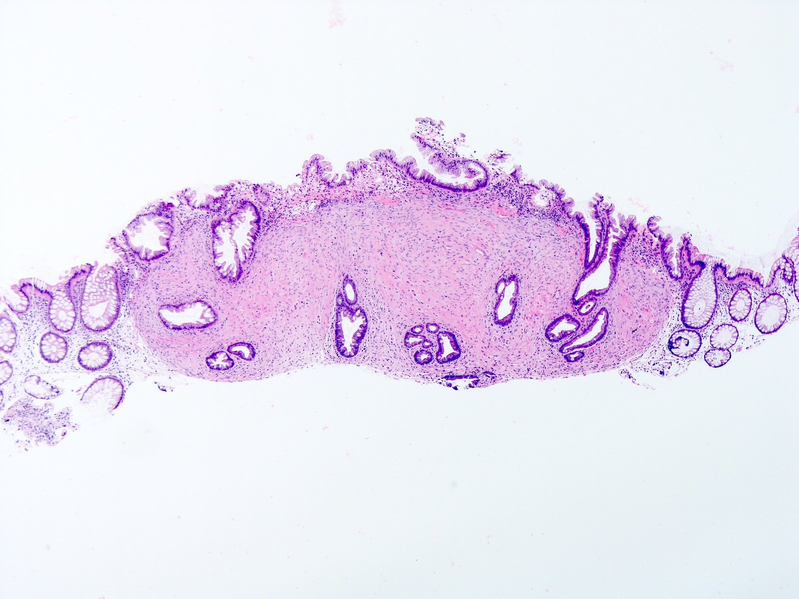
- Poorly circumscribed spindle cell proliferation, usually confined to the mucosa, with ovoid nuclei and pale indistinct cytoplasm, in a background of fine collagenous stroma
- No atypia, no pleomorphism, no mitotic figures
- Entrapped / overlying colonic crypts appear hyperplastic or serrated in the majority of cases
- Perineurial proliferation sometimes focally present in otherwise typical sessile serrated polyp / adenoma (Am J Surg Pathol 2011;35:1373)
Microscopic (histologic) images
Negative stains
Electron microscopy description
- Long bipolar cytoplasmic processes, prominent pinocytotic vesicles
Molecular / cytogenetics description
- BRAF mutation often occurs in overlying serrated epithelium, if present (Am J Surg Pathol 2013;37:745)
Sample pathology report
- Ascending colon, polypectomy:
- Perineurioma (see comment)
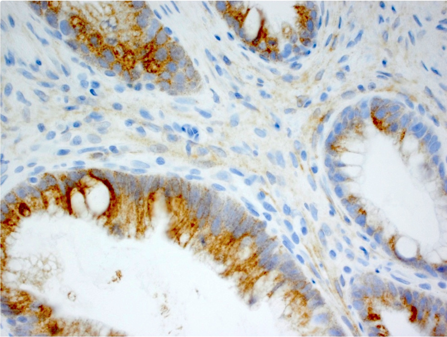
- Comment: Immunohistochemical stains show the lesion is positive for EMA (focal) and negative for S100.
Differential diagnosis
- Mucosal Schwann cell hamartoma:
- Ganglioneuroma:
- Positive for S100
- Ganglion cells present
- Inflammatory fibroid tumor:
- Perivascular spindling of cells
- Prominent eosinophils
- Strong CD34 positivity
- Neurofibroma:
- Unlikely to involve mucosa
- Benign epithelioid peripheral nerve sheath tumor:
- Cells are epithelioid
- Lesion extends into superficial submucosa (Am J Surg Pathol 2005;29:1310)
- Tactile corpuscle-like body:
- Usually small, incidental finding in mucosal biopsy (Am J Surg Pathol 2015;39:1668)
Board review style question #1
Board review style answer #1


