Table of Contents
Definition / general | Epidemiology | Clinical features | Grading | Radiology description | Radiology images | Prognostic factors | Case reports | Treatment | Gross description | Microscopic (histologic) description | Microscopic (histologic) images | Positive stains | Negative stains | Electron microscopy description | Molecular / cytogenetics description | Differential diagnosisCite this page: Abdelzaher E. Astroblastoma, MN1 altered. PathologyOutlines.com website. https://www.pathologyoutlines.com/topic/cnstumorastroblastoma.html. Accessed April 2nd, 2025.
Definition / general
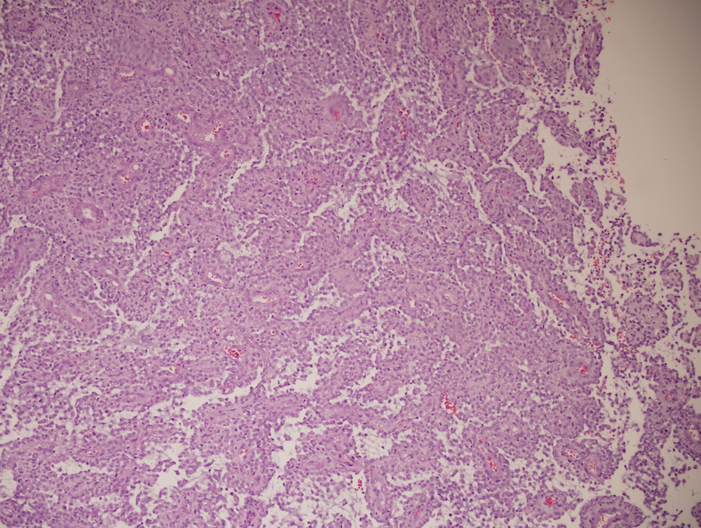
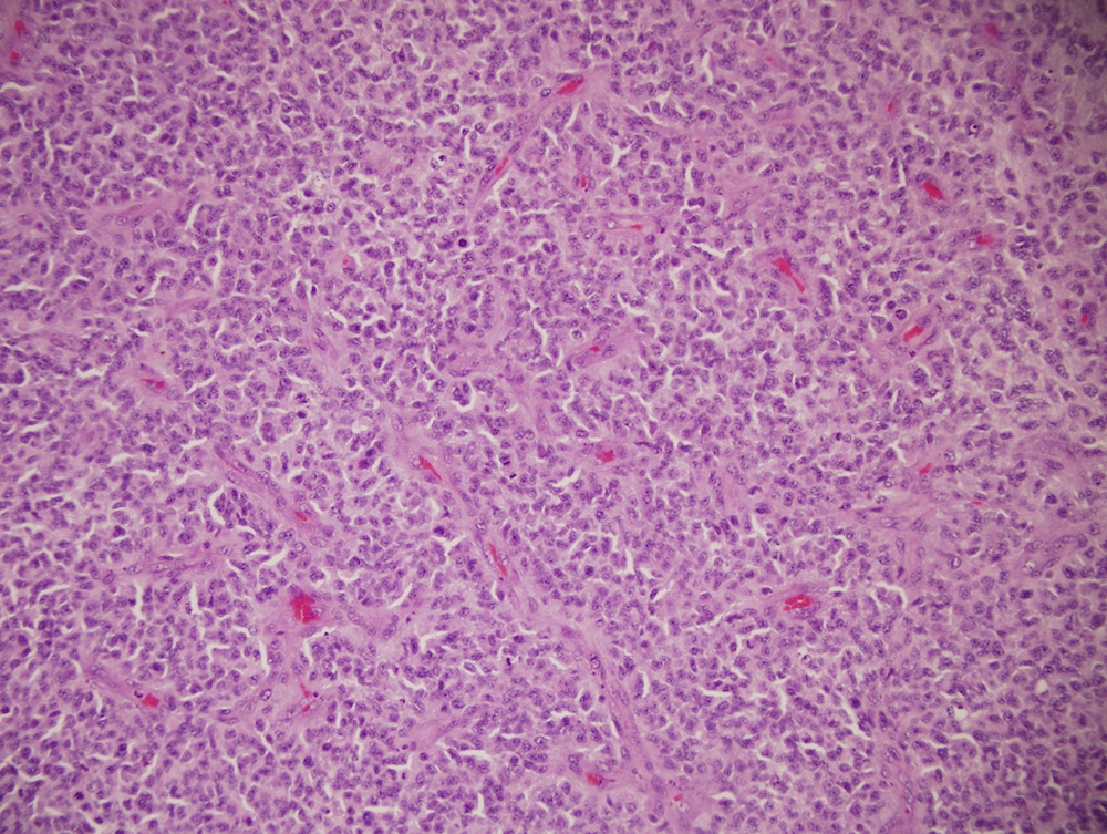
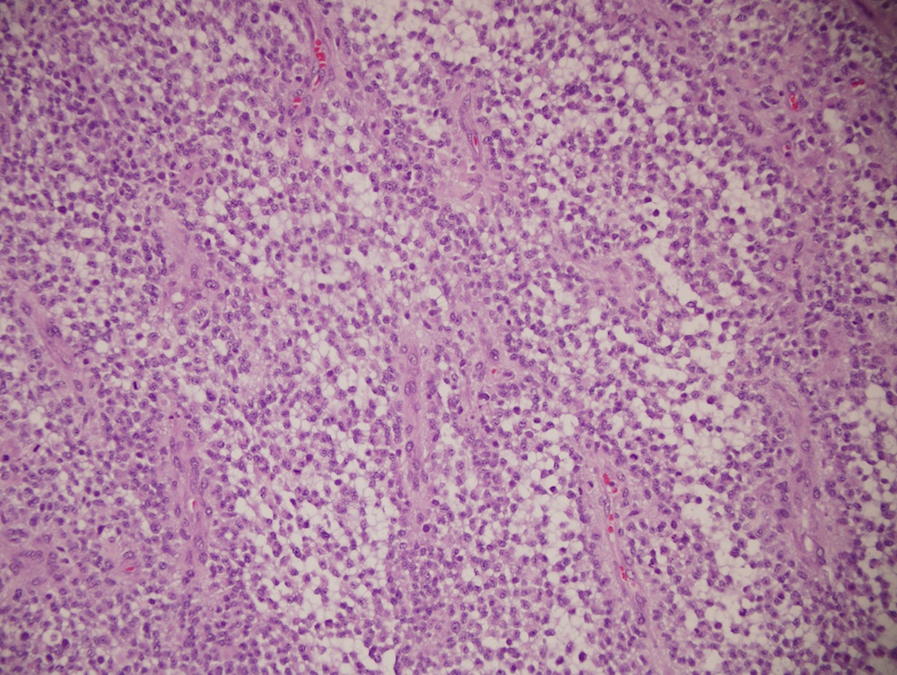
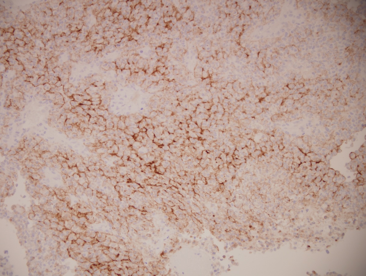
- Rare ( < 3% of primary brain gliomas), compact glial neoplasm with perivascular pseudorosettes formed of GFAP+ cells arranged around central, often sclerotic, blood vessels
- Controversial entity - relationship with ependymoma is not clear
Epidemiology
- Usually children and young adults, median age 11 years, range 1 - 58 years
Clinical features
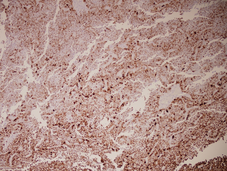
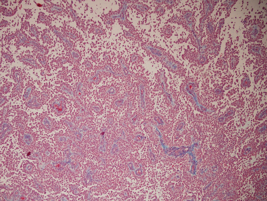
- Features of astrocytoma and ependymoma; expresses nonfibrillar form of GFAP (so PTAH-)
Grading
- No WHO grade assigned for astroblastoma or anaplastic astroblastoma
Radiology description
- Discrete supratentorial cerebral, often superficial, contrast enhancing mass
- Cystic change is common
Prognostic factors
- Anaplastic histology predicts poor prognosis (J Neurooncol 1998;40:59)
Case reports
- Newborn boy with large cerebral mass (Case #312)
- 6 year old girl with intraventricular astroblastoma (J Neurosurg Pediatr 2008;1:152)
- 8 year old girl with tumor in frontoparietal lobe (Neuropathology 2006;26:72)
- 10 year old girl with recurrent low grade astroblastoma with signet ring-like cells and high proliferative index (Fetal Pediatr Pathol 2013;32:284)
- 15 year old girl with headache and diplopia (J Korean Med Sci 2004;19:772)
Treatment
- Resection (adequate for well differentiated tumors), more aggressive treatment needed for malignant tumors
Gross description
- Well circumscribed, peripheral, cerebral hemispheric masses
- Firm, often cystic
Microscopic (histologic) description
- Well circumscribed with discrete pushing borders, occasionally infiltrative in high grade lesions
- Perivascular pseudorosettes resembling ependymoma but with thick processes from cell body to adventitia of vessel
- Also vascular hyalinization, little fibrillar background
- Limit diagnosis to tumors in which these features predominate (other tumors have these features focally)
- High grade astroblastomas have hypercellular and mitotically active regions, often with vascular proliferation or necrosis with pseudopalisading; rare features are signet ring cells (Neuropathology 2002;22:200)
Negative stains
- PTAH, synaptophysin, cytokeratin
Electron microscopy description
- Abundant intermediate filaments forming bundles in tumor cytoplasm, membrane junctions and external lamina when cells are in contact with collagen fibers (Surg Neurol 1991;35:116)
Molecular / cytogenetics description
- +20q, +19 (Brain Pathol 2000;10:342)
Differential diagnosis
- Ependymoma: more fibrillar, nuclei smaller and less pleomorphic, true rosettes, less sclerosis (AJNR Am J Neuroradiol 2002;23:243, Childs Nerv Syst 2005;21:211)
- Pilocytic astrocytoma: biphasic piloid areas with Rosenthal fibers alternating with spongy microcystic areas with eosinophilic granular bodies
- Pleomorphic xanthoastrocytoma: fascicular pattern, pleomorphic cells, lipidized cells, eosinophilic granular bodies, perivascular lymphocytes


