Table of Contents
Definition / general | Case reports | Microscopic (histologic) description | Microscopic (histologic) images | Positive stains | Negative stains | Molecular / cytogenetics description | Electron microscopy images | Differential diagnosis | Additional referencesCite this page: Perunovic B. Large cell neuroendocrine carcinoma. PathologyOutlines.com website. https://www.pathologyoutlines.com/topic/cervixlargecellneuroendocrine.html. Accessed April 2nd, 2025.
Definition / general
- Rare ( < 1% of cervical carcinomas)
- Mean age 34 years, range 21 to 62 years
- Presents with abnormal Pap smear or vaginal bleeding
- Aggressive behavior, similar to lung counterpart, with early metastases to regional lymph nodes and liver, lung, bone and brain (Int J Gynecol Pathol 2003;22:226)
- Median survival < 2 years
Case reports
- 27 year old woman with 6 cm cervical mass (Case #327)
- 35 year old woman with small cell component (Gynecol Oncol 1998;68:69)
- 39 year old woman with carcinomatous meningitis (J Postgrad Med 2004;50:311)
- Patient with HSIL (Pathology 1999;31:158)
Microscopic (histologic) description
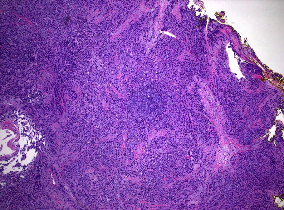
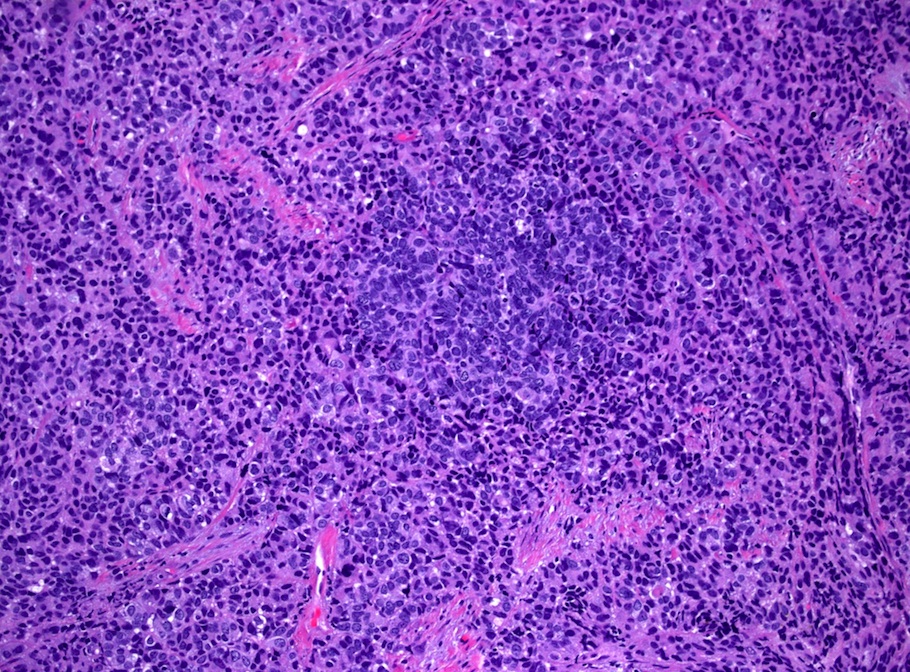
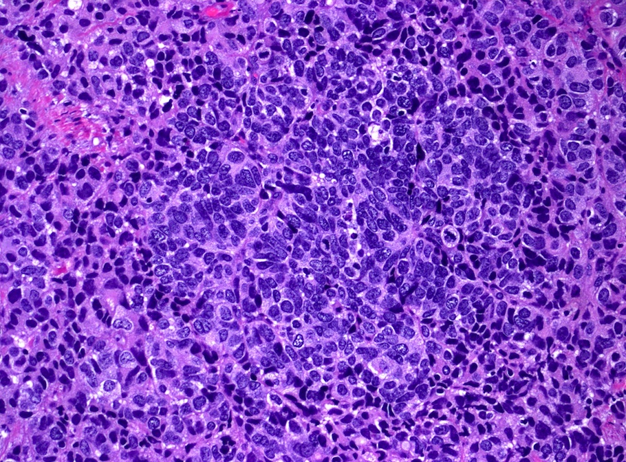
- Defined as moderate to severe nuclear atypia, neuroendocrine differentiation with cells larger than typical small cell carcinoma
- Insular, trabecular, glandular and solid growth patterns
- Usually eosinophilic cytoplasmic granules, > 10 MF/10 HPF and extensive necrosis
- Angiolymphatic invasion
- Often with adjacent adenocarcinoma in situ
Positive stains
- Keratin (MNF116) in paranuclear dot-like pattern
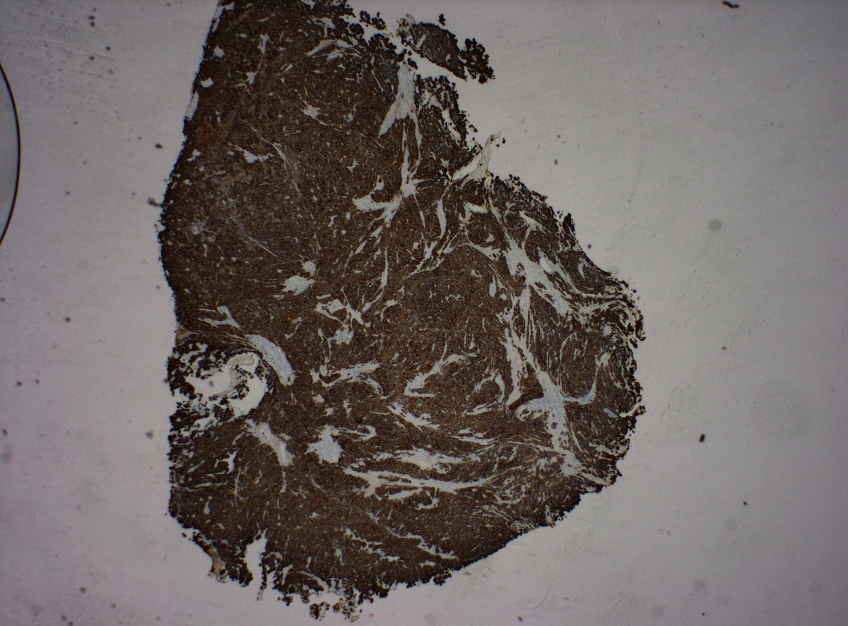
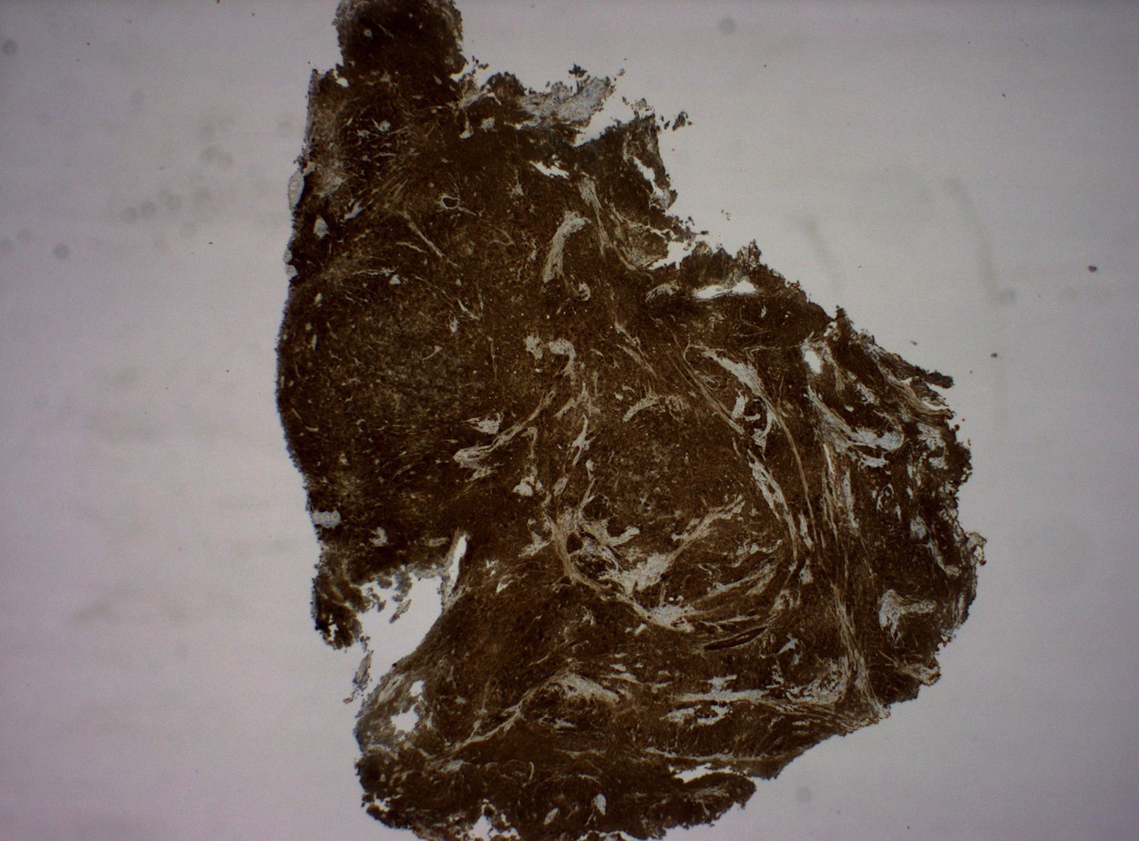
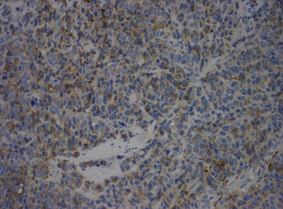
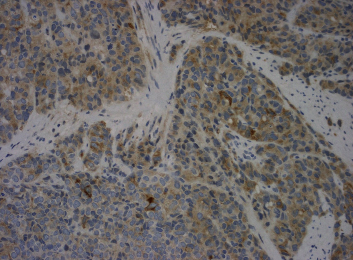
- Chromogranin or synaptophysin, vascular endothelial growth factor (Int J Gynecol Cancer 2005;15:646)
- HepPar1 (J Clin Pathol 2004;57:48)
- Alpha fetoprotein (Acta Cytol 2003;47:799)
Molecular / cytogenetics description
- HPV16 and HPV18 are usually present (J Clin Pathol 2002;55:108)
Differential diagnosis
- Atypical carcinoid tumor
- Poorly differentiated carcinoma
Additional references


