Table of Contents
Definition / general | Epidemiology | Pathophysiology | Etiology | Clinical features | Prognostic factors | Case reports | Treatment | Gross description | Gross images | Microscopic (histologic) description | Microscopic (histologic) images | Cytology description | Cytology images | Positive stains | Negative stains | Electron microscopy description | Differential diagnosis | Additional referencesCite this page: Perunovic B., Sunassee A, Askeland R. Adenosquamous carcinoma. PathologyOutlines.com website. https://www.pathologyoutlines.com/topic/cervixadenosquamous.html. Accessed December 24th, 2024.
Definition / general
- Carcinoma containing a mixture of glandular and squamous components
- ~10% of cervical carcinomas (Virchows Arch 2009;455:253)
- Glassy cell carcinoma is a rare variant of poorly differentiated adenosquamous carcinoma; aggressive subtype with rapid growth, early metastases and poor prognosis (Cytojournal 2013;10:17, Eur J Obstet Gynecol Reprod Biol 2014;179:232)
Epidemiology
- Glassy cell carcinoma:
- Younger age group (mean 41 years), associated with pregnancy, HPV 16 and 18 in tumor cells (Cytojournal 2013;10:17)
- Peak incidence third to fourth decades (Cytojournal 2013;10:17)
- Some studies have noted an association with pregnancy
Pathophysiology
- Glassy cell carcinoma:
- The presence of HPV 18 might stimulate biphasic squamous and glandular differentiation (Int J Gynecol Pathol 2002;21:134)
- Cytokeratin expression is similar to that of reserve cells or immature squamous cells of cervix (Int J Gynecol Pathol 2002;21:134)
Etiology
- May arise from subcolumnar reserve cells in basal layer of endocervix
- More common during pregnancy
- Associated with HPV18 (Virchows Arch 2009;455:253, Int J Gynecol Pathol 2012;31:294)
- Frequent loss of ARID1A protein expression (Int J Gynecol Cancer 2012;22:208)
Clinical features
- Same prognosis as other cervical carcinomas when stratified by grade and stage but most cases are high grade
- Glassy cell carcinoma:
- 1 - 2% of cervical carcinomas
- Historically considered more aggressive with poorer prognosis than ordinary adenosquamous carcinoma or adenocarcinoma, although recent studies show less or no difference (APMIS Suppl 1991;23:119, Am J Obstet Gynecol 2004;190:67)
- May have peripheral blood eosinophilia
Prognostic factors
- Glassy cell carcinoma has poor prognostic factors
- Angiolymphatic invasion, deep stromal invasion, large tumor size
- HER2 overexpression may correlate with more aggressive behavior and worse clinical outcome (Acta Cytol 2006;50:418)
Case reports
- 27 year old woman with stage IB2 tumor diagnosed at 19 weeks gestation (Aust N Z J Obstet Gynaecol 2015;55:94)
- 29 year old woman with signet ring cell carcinoma (Pathol Int 2004;54:787)
- 33 year old woman with history of post coital bleeding and vaginal discharge (UPMC Case #100)
- 44 year old and 67 year old women (Ceska Gynekol 1999;64:279, Ginekol Pol 2011;82:936)
- 72 year old woman with endometrial extension of cervical tumor (Int J Gynecol Cancer 2004;14:625)
- Metastasis to infusion port site (Gynecol Oncol 1999;74:130)
- Patient with myometrial recurrence during pregnancy (Gynecol Oncol 2000;76:409)
- Patient with metastasis to port site (Gynecol Oncol 1999;74:130)
Treatment
- Radical hysterectomy and chemoradiation
- Cisplatin based chemoradiation: overall survival now comparable to squamous cell carcinoma of cervix, although historically poorer prognosis
- Poorer prognosis with radiation alone (Gynecol Oncol 2014;135:208, Gynecol Oncol 2014;135:462)
Gross description
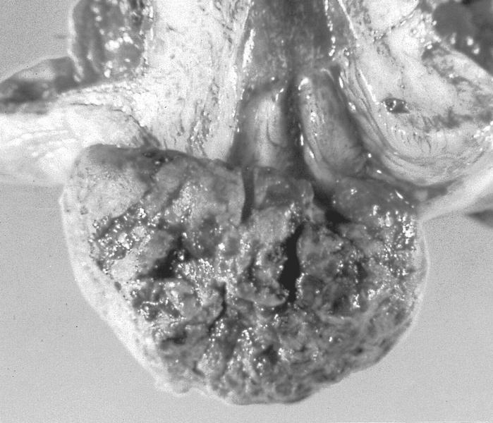
- Glassy cell carcinoma: bulky exophytic mass with barrel shape cervix (Cytojournal 2013;10:17)
Microscopic (histologic) description
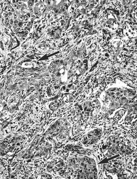
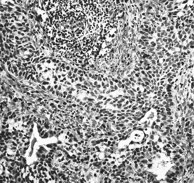
- Usually defined as biphasic pattern of well defined malignant glandular and squamous components clearly identifiable without special stains
- Glandular component usually endocervical and poorly differentiated with cytoplasmic vacuoles or luminal mucin
- Squamous component also is poorly differentiated
- If endometrioid call endometrioid carcinoma with squamous differentiation
- Glassy cell carcinoma:
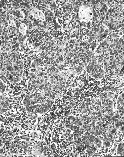
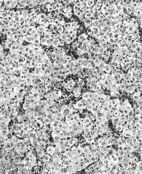
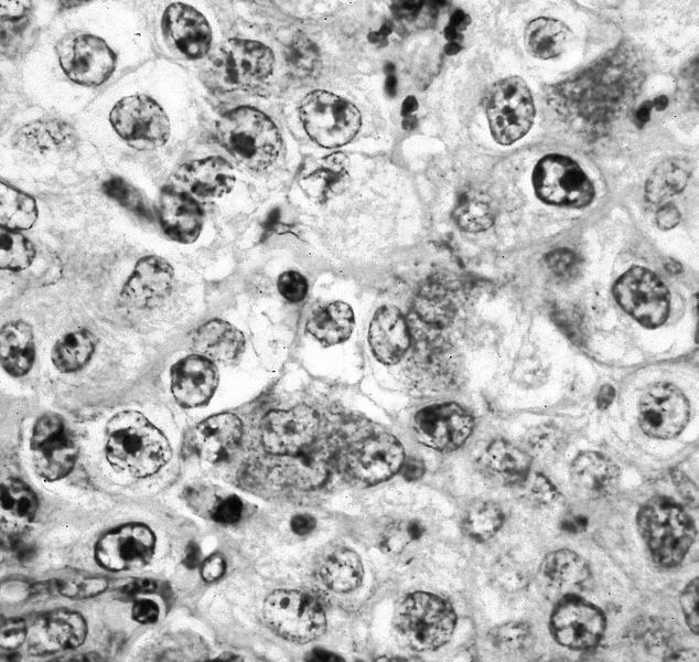
- Solid nests of markedly pleomorphic, polygonal tumor cells with prominent cell membrane, glassy and eosinophilic cytoplasm, large eosinophilic nuclei, prominent nucleoli, surrounded by heavy inflammatory infiltrate containing eosinophils
- Frequent mitotic figures
- Pure cases have no histologic evidence of glandular or squamous differentiation (i.e. no intracellular bridges, no dyskeratosis, no intracellular glycogen), which is detectable only by electron microscopy
- Often less invasion than is suspected
Microscopic (histologic) images
Cytology description
- Often not diagnosed on pap smear (Cancer Cytopathology 2004;102:210)
- Papillary subtype (thin layer cytology):
- High cellularity
- Multiple small, papillary clusters of basaloid to columnar cells
- Discernible fibrovascular cores
- Background of loosely dispersed bland looking columnar cells and high grade squamous intraepithelial lesion
- Scattered adenocarcinoma cells containing intracytoplasmic vacuoles (Acta Cytol 2003;47:649)
- Glassy cell carcinoma:
- Tumor cells arranged in sheets or clusters
- Distinct cell borders with moderate to abundant finely granular (ground glass-like) cytoplasm
- Large round/oval vesicular nuclei with one or more prominent nucleoli
- Chromatin varies from finely dispersed to coarse and irregular (Acta Cytol 2004;48:99, Zhonghua Bing Li Xue Za Zhi 2011;40:523)
- Cytoplasmic vacuolization and bizarre cells with multinucleation may be seen (Acta Cytol 2001;45:407)
- Mitotic figures frequently seen
- Background inflammatory infiltrate including frequent eosinophils, neutrophils, plasma cells, lymphocytes and necrotic debris
- Focal abortive keratin production; squamous or glandular differentiation may be present
- Focal clear cell differentiation may be present
Positive stains
- p63 (squamous component), CK7
- Glassy cell carcinoma:
- MUC1 / EMA, MUC2, CEA (focal), CAM5.2, p63 (Zhonghua Bing Li Xue Za Zhi 2011;40:523)
- PAS+ cell wall
- Vimentin
- Focal mucin
Electron microscopy description
- Glandular features include mucous secretory vacuoles, true lumen formation and scattered glycogen
- Tonofilaments and secretory products
- Note: most undifferentiated cervical carcinomas have ultrastructural features of squamous or glandular differentiation
- Glassy cell carcinoma:
- Glassy features may be due to cytoplasmic polyribosomes, abundant tonofilaments and abundant dilated rough endoplasmic reticulum (Am J Clin Pathol 1991;96:520)
- Adenosquamous features include well developed desmosomal complexes and microvilli
- Occasional intracellular lumina (Cancer 1983;51:2255)
Differential diagnosis
- Adenocarcinoma with coexisting SIL:
- Usually no mixing of tumor elements
- Adenoid basal carcinoma (Pathol Int 2005;55:445)
- Extension of endometrial adenocarcinoma:
- Bulk of tumor is in endometrium
- Squamous cell carcinoma with focal mucin droplets
- Large cell nonkeratinizing squamous cell carcinoma:
- Cell membrane is less well defined, cytoplasm is less finely granular, coarser chromatin distributed along nuclear membrane
- Also poor staining or fixation makes it resemble glassy cell carcinoma
- Lymphoepithelioma-like carcinoma (Acta Cytol 1999;43:285)
Additional references


