Table of Contents
Definition / general | Essential features | Terminology | Pathophysiology | Clinical features | Interpretation | Uses by pathologists | Prognostic factors | Microscopic (histologic) description | Microscopic (histologic) images | Positive staining - normal | Positive staining - disease | Negative staining | Sample pathology report | Additional references | Board review style question #1 | Board review style answer #1Cite this page: Bhattarai N, Malhotra S, Crane GM. CD79a. PathologyOutlines.com website. https://www.pathologyoutlines.com/topic/cdmarkerscd79a.html. Accessed March 30th, 2025.
Definition / general
- CD79a, identified as the alpha chain of the B cell antigen receptor complex, is a transmembrane protein synthesized by the CD79A gene located on chromosome 19
- Expressed over full range of B cell development from early B cell precursors, preceding immunoglobulin heavy chain gene rearrangement and persisting through B cell maturation (Blood 1995;86:1453, Appl Immunohistochem Mol Morphol 2001;9:97)
Essential features
- CD79a / CD79b are membrane bound immunoglobulins that form the B cell receptor (BCR) complex
- Expressed from early B cell precursors until advanced plasma cell stages (Blood 1995;86:1453)
- Membranous and cytoplasmic expression useful for confirming B cell lineage or following therapy with monoclonal antibodies against other B cell antigens
Terminology
- Also called MB1 membrane glycoprotein, IgMα and Igα
Pathophysiology
- CD79a, a 226 amino acid membrane protein, is the product of the human mb1 gene situated on chromosome 19, specifically at band q13.2 (OMIM: CD79A Antigen; CD79A [Accessed 23 January 2024])
- Has single extracellular immunoglobulin domain, a transmembrane domain and an intracytoplasmic signaling domain with an immunoreceptor tyrosine based activation motif (ITAM)
- Heterodimerizes with CD79b (B29) and constitutes an integral component of the B cell receptor (BCR) complex
- CD79a's cytoplasmic domain enables signal transduction via antigen binding to the BCR, causing rapid protein tyrosine phosphorylation and activation of pathways through the immunoreceptor tyrosine based activation motif (ITAM) (Front Immunol 2018;9:665)
- CD79a and CD79b are required for surface IgM expression in human B cells (J Immunol 2022;209:2042)
Clinical features
- Deletions or mutations in the genes encoding CD79a lead to developmental arrest at the pre-B cell stage and can be a cause of agammaglobulinemia (J Clin Invest 1999;104:1115)
- Plays an important functional role in maintaining the immature, immune suppressive phenotype of myeloid derived suppressor cells (MDSC) and in inducing the secretion of protumorigenic cytokines, thus, may be a novel target for cancer therapy (PLoS One 2013;8:e76115)
- Impairment of the glycosylation of μ and CD79a chains is associated with lower levels of expression of IgM in chronic lymphocytic leukemia (Blood 2005;105:2933)
- Synovial infiltration of CD79a positive B cells correlates with joint destruction in rheumatoid arthritis (J Rheumatol 2011;38:2301)
- Aberrant staining in megakaryocytes in postinduction acute lymphoblastic leukemia (ALL) cases (cytoplasmic and granular) (Hum Pathol Case Rep 2021;23:200467)
- Aberrant CD79a positivity in erythroid precursors postchemotherapy in lymphoma patients can complicate minimal residual disease detection after rituximab treatment (Haematologica 2007;92:855)
Interpretation
- Membranous and cytoplasmic expression is considered positive; pro-B cells may only show cytoplasmic expression (J Immunol 1991;147:2474)
Uses by pathologists
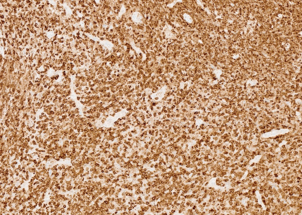
- Used as a pan-B cell marker in immunohistochemistry
- Used to differentiate ALL from other pediatric small blue round cell tumors (Appl Immunohistochem Mol Morphol 2001;9:97)
- Used to confirm B cell lineage in rituximab treated B cell neoplasms (Blood Res 2022;57:55)
- Used as part of a panel to differentiate classic Hodgkin lymphoma from nodular lymphocyte predominant Hodgkin lymphoma and aggressive B cell lymphomas that may show Hodgkin-like features (Appl Immunohistochem Mol Morphol 2016;24:535)
Prognostic factors
- Some studies report that cytoplasmic CD79a expression is associated with lower survival odds than classical acute myeloid leukemia (AML) with t(8;21)(q22;q22) (Korean J Lab Med 2007;27:388)
- Cytoplasmic CD79a can be used in monitoring of relapse in B ALL post-CD19 chimeric antigen receptor (CAR) T cell therapy (Leuk Lymphoma 2022;63:426)
- CD79a may have independent prognostic relevance for CNS involvement and CNS relapse in pediatric B cell precursor ALL (Commun Biol 2021;4:73)
- CD79a positivity may be associated with worse prognosis in classic Hodgkin lymphoma compared to CD79a negative classic Hodgkin lymphoma (J Clin Exp Hematop 2020;60:78)
Microscopic (histologic) description
Microscopic (histologic) images
Positive staining - normal
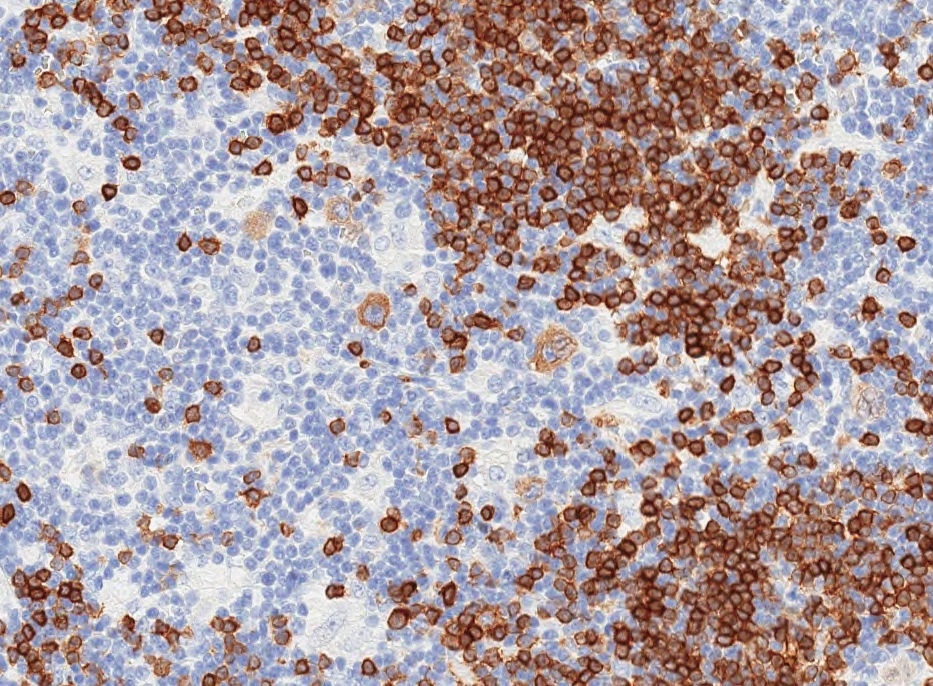
- B lymphocytes (mature and immature) and plasma cells (J Immunol 1991;147:2474)
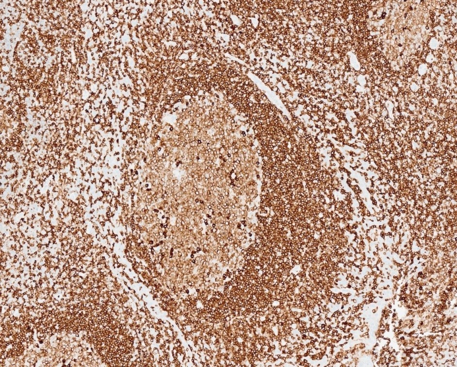
- Follicular mantle zone more strongly labeled than germinal center B cells; in spleen, marginal zones are strongly labeled (Appl Immunohistochem Mol Morphol 2001;9:97)
- Expressed weakly and transiently in early T cell differentiation (J Pathol 2002;197:341)
Positive staining - disease
- B cell lymphoid neoplasms
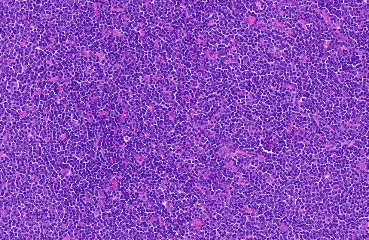
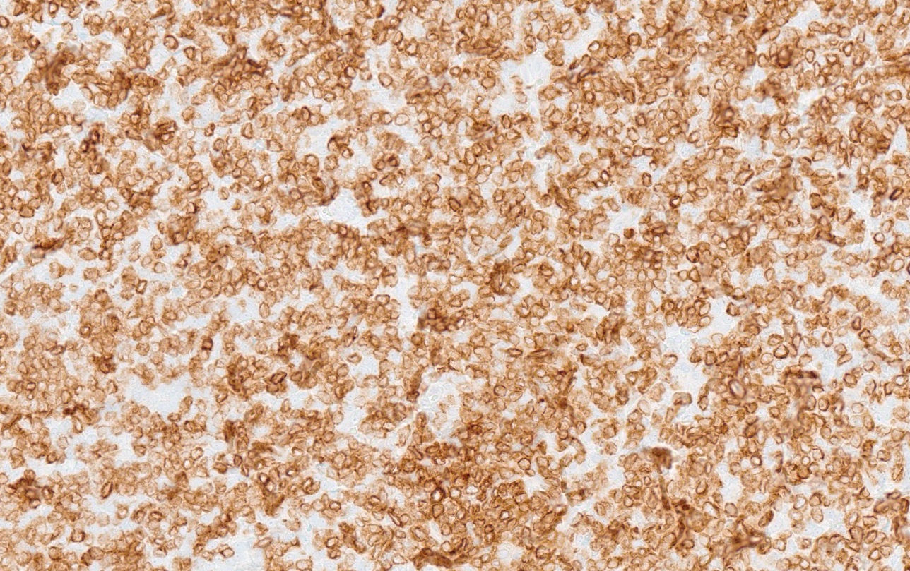
- Most low grade B cell lymphomas (membranous and cytoplasmic often with perinuclear accentuation) (Blood 1995;86:1453)
- Weak expression may be seen in follicular lymphoma (Am J Surg Pathol 2002;26:1458)
- B ALL (90% of cases, cytoplasmic, may be weak and focal) (Blood 1993;82:853)
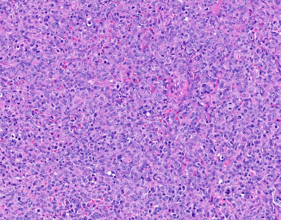
- Diffuse large B cell lymphomas (Appl Immunohistochem Mol Morphol 2001;9:97)
- Primary mediastinal large B cell lymphomas (heterogenous pattern); discordant mb1+ / Ig- expression occurs commonly in mediastinal large B cell lymphomas (Appl Immunohistochem Mol Morphol 2001;9:97, Am J Clin Pathol 2021;156:497)
- Nodular lymphocyte predominant Hodgkin lymphoma (faint) (Am J Clin Pathol 2003;119:192)
- T cell histiocyte rich large B cell lymphomas (60% of cases, weak expression) (Am J Surg Pathol 2002;26:1458)
- Myeloma / plasmacytoma (50% of cases positive) (Appl Immunohistochem Mol Morphol 2001;9:97)
- Can be used together with other markers to identify mixed phenotype acute leukemia (Arch Pathol Lab Med 2017;141:1462)
Negative staining
- Anaplastic large cell lymphoma (Hum Pathol 2004;35:455)
- T cell neoplasms
- Few T ALL may be positive (10 - 40% of cases, weak to moderate cytoplasmic positivity, 30 - 100% of leukemic cells) (J Pathol 1998;186:140, Am J Clin Pathol 2000;113:823, Cancer Res 2004;64:7399)
- Some extranodal NK / T cell lymphomas are positive (Mod Pathol 2000;13:766)
- Classic Hodgkin lymphoma (18% of cases are positive) (Mod Pathol 2004;17:1531)
- Acute myeloid leukemias; however, aberrant CD79a expression can be seen in AML cases with RUNX1 mutations and t(8; 21) (Am J Clin Pathol 2000;113:823, Pathobiology 2019;86:162)
- Myeloid sarcoma (Int J Lab Hematol 2011;33:555)
Sample pathology report
- Cervical lymph node, excisional biopsy:
- Nodular lymphocyte predominant Hodgkin (B cell) lymphoma (see comment)
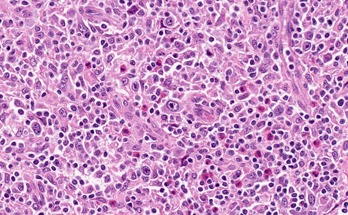
- Comment: Histologic sections show lymphoid infiltrate in diffuse and vaguely nodular patterns. The lymphoma cells are large with scant to moderate cytoplasm and large nuclei with fine chromatin (LP cells). These cells are in a background of numerous small, mature lymphocytes and epithelioid histiocytes. Immunohistochemical stains show the large, abnormal cells are positive for CD20, CD45, CD79a, BCL6 and PAX5. They are negative for CD3, CD4, CD5, CD8, CD10, CD15, CD30, ALK1, BCL2 and fascin. The large, abnormal cells overlap networks of CD21 positive follicular dendritic cells. Chromogenic in situ hybridization studies with probes for Epstein-Barr virus (EBV) encoded small RNA (EBER) are negative. PD-1 is positive on small, mature T cells surrounding the LP cells.
Additional references
Board review style question #1
Board review style answer #1
A. Complete absence of expression. Despite originating from B cells, Hodgkin and Reed-Sternberg (HRS) cells in classic Hodgkin lymphoma exhibit a disrupted B cell program, resulting in the loss or downregulation of numerous B cell markers. These include pan-B cell antigens, such as CD19, CD20, CD22 and CD79a, as well as B cell transcription factors, such as OCT2, BOB.1 and BCL6. Answers B and D are incorrect because they do not represent the usual pattern of expression in Hodgkin lymphoma. Answer C is incorrect because some cases may show weak expression of CD79a but it is not the typical pattern.
Comment Here
Reference: CD79a
Comment Here
Reference: CD79a


