Table of Contents
Definition / general | Essential features | Terminology | Pathophysiology | Clinical features | Interpretation | Uses by pathologists | Prognostic factors | Microscopic (histologic) images | Positive staining - normal | Positive staining - disease | Negative staining | Sample pathology report | Board review style question #1 | Board review style answer #1Cite this page: Fenu EM, Maracaja DLV. CD45 (LCA). PathologyOutlines.com website. https://www.pathologyoutlines.com/topic/cdmarkerscd45.html. Accessed April 1st, 2025.
Definition / general
- Ubiquitous cell surface marker of nucleated hematopoietic cells involved in the regulation and activation of the immune system
- Detectable by immunohistochemistry or flow cytometry
Essential features
- Receptor tyrosine phosphatase protein expressed on the cell surface of white blood cells and involved in B and T cell activation and signaling (Immunol Lett 2018;196:22)
- Used by pathologists to highlight inflammatory cells or determine the hematopoietic nature of tumors of unknown origin with high specificity
Terminology
- Leukocyte common antigen (LCA) or Ly-5
Pathophysiology
- High molecular weight (180 - 220 kDa) transmembrane protein with intrinsic tyrosine phosphatase activity
- Involved in regulating thymic selection and B and T cell antigen receptor mediated activation (Hum Pathol 2017;62:83)
Clinical features
- Different subsets of immune cells express different isoforms of CD45 due to variable exon splicing (Immunol Lett 2018;196:22):
- Mutations in CD45 have been implicated in cases of autosomal recessive T cell negative, B cell positive, NK cell positive severe combined immunodeficiency (Immunol Lett 2018;196:22, OMIM: Severe Combined Immunodeficiency, Autosomal Recessive, T Cell Negative, B Cell Positive, Nk Cell Positive [Accessed 13 January 2022])
Interpretation
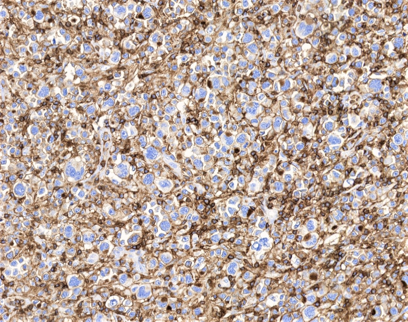
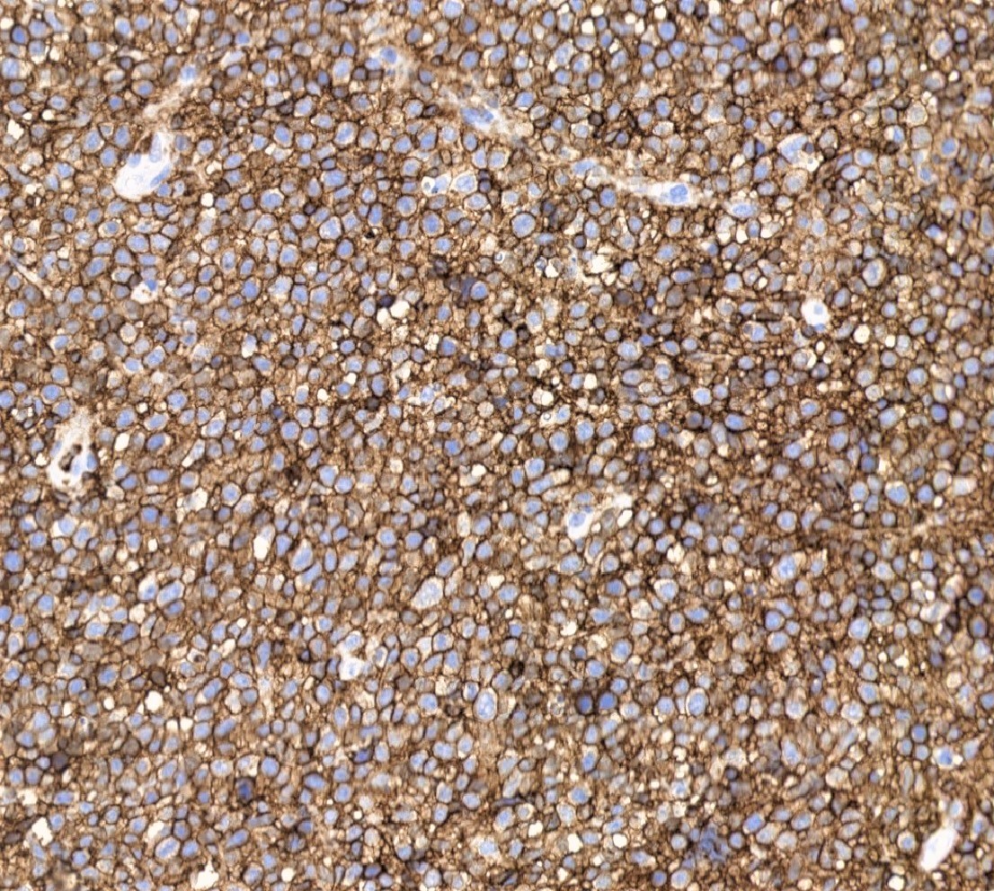
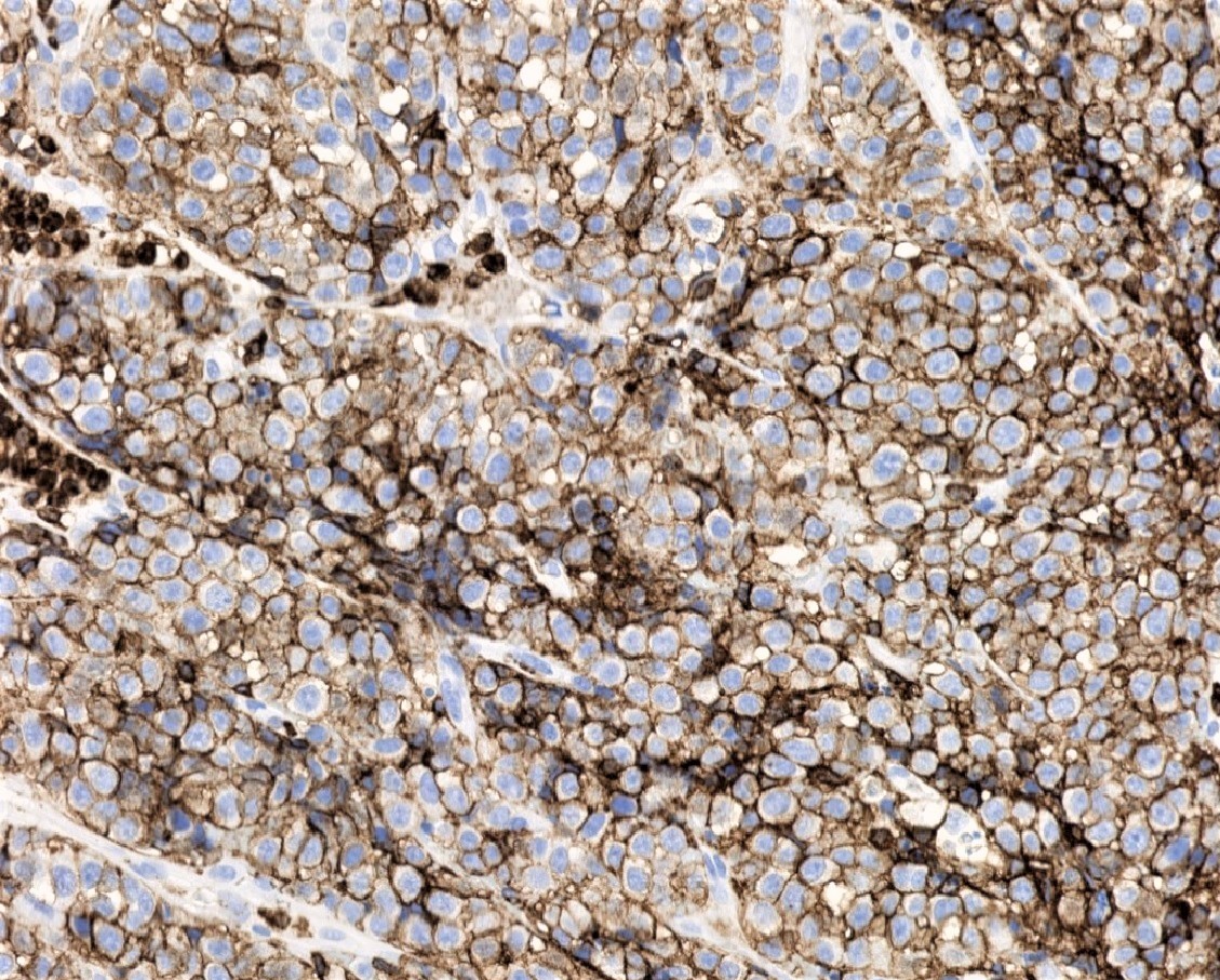
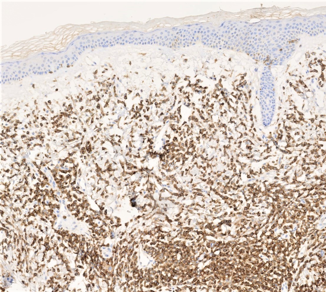
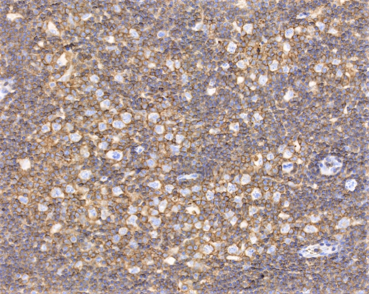
- Membranous / cytoplasmic staining
Uses by pathologists
- Confirm the hematopoietic nature of tumors
- Highlight inflammatory cells, for example in lymphocytic myocarditis or Sjögren syndrome (Hum Pathol 2017;62:83, J Oral Pathol Med 2021;50:98)
Prognostic factors
- Strong expression of CD45 may predict a worse prognosis in certain hematologic malignancies, including pediatric B cell progenitor acute lymphoblastic leukemia and multiple myeloma (Blood Cells Mol Dis 2021;89:102562, Leuk Res 2016;44:32)
- Tumor infiltrating lymphocytes, which can be highlighted by CD45, may predict a more favorable prognosis in solid tumors, like colorectal cancer or small cell carcinoma (Science 2006;313:1960, Chest 2013;143:146)
Microscopic (histologic) images
Positive staining - normal
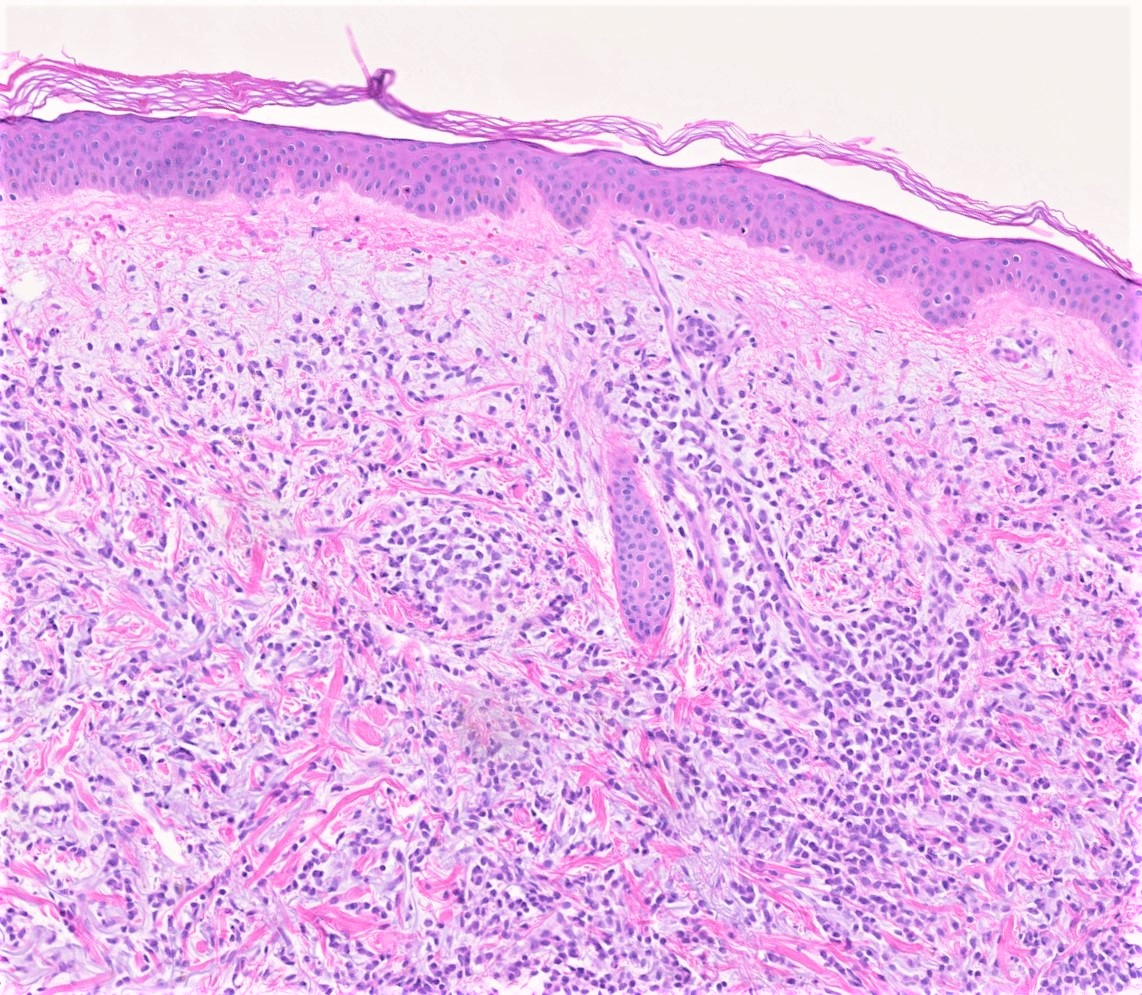
- White blood cells, including granulocytes, lymphocytes, eosinophils, basophils (dim), monocytes, macrophages / histiocytes, mast cells and plasma cells; not including red blood cells and their precursors, megakaryocytes or platelets (Immunol Lett 2018;196:22)
Positive staining - disease
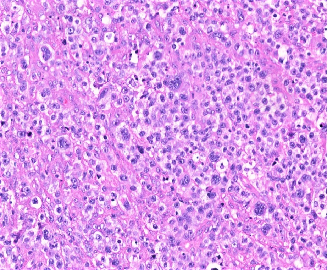
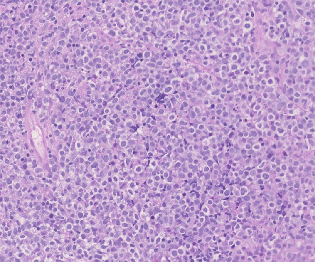
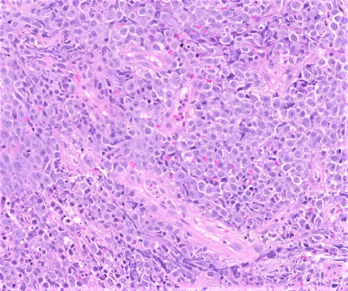
- Acute leukemias, including acute myeloid leukemia, acute lymphoblastic leukemias and leukemia cutis (Methods Mol Biol 2017;1633:33, J Hematol 2018;7:124)
- Acute myeloid leukemia blasts have a lower CD45 mean fluorescence intensity in flow cytometry than other circulating white blood cells, allowing them to be identifed as a distinct population (blast gate) (Leukemia 1997;11:1878, Am J Clin Pathol 2012;137:800)
- B lymphoblastic leukemia / lymphoma can be negative; if positive, it is usually dimly expressed and the strong expression has been described as associated with a worse prognosis in B cell progenitor acute lymphoblastic leukemia (Blood Cells Mol Dis 2021;89:102562)
- B and T cell lymphomas (most), including the lymphocyte predominant (LP) cells of nodular lymphocyte predominant Hodgkin lymphoma (Leuk Res 2008;32:263)
- Plasma cell neoplasms (variable) (Blood Cells Mol Dis 2004;32:293)
- Histiocytic tumors such as histiocytic sarcoma (Diagn Cytopathol 2018;46:790)
- Mast cell disorders, such as systemic / cutaneous mastocytosis, mastocytoma (Am J Clin Pathol 2015;143:527)
- Blastic plasmacytoid dendritic cell neoplasm (J Hematol 2018;7:124)
- Candida albicans yeast forms (Am J Clin Pathol 2000;113:59)
- Rarely carcinomas, particularly undifferentiated / neuroendocrine neoplasms or necrotic tumors (Mod Pathol 1998;11:1204, Am J Clin Pathol 1998;110:641)
Negative staining
- Red blood cells, megakaryocytes, osteoblasts and platelets (Methods Cell Biol 2011;103:221)
- Reed-Sternberg cells of classic Hodgkin lymphoma (Appl Immunohistochem Mol Morphol 2016;24:535)
- Anaplastic T cell lymphomas, plasma cell neoplasms and acute lymphoblastic leukemia / lymphoma neoplasms can occasionally have negative expression of CD45 (Appl Immunohistochem Mol Morphol 2016;24:535)
- ALK positive anaplastic large cell lymphoma may sometimes be negative for CD45 by immunohistochemistry but CD45 positive by flow cytometry (Am J Clin Pathol 2007;127:707)
- Epithelial tumors, germ cell tumors, melanoma, mesothelioma, sarcoma (note that infiltrating lymphocytes are CD45 positive and must be distinguished from tumor cells) (Appl Immunohistochem Mol Morphol 2016;24:535)
Sample pathology report
- Lymph node, right cervical excision:
- Nodular lymphocyte predominant Hodgkin lymphoma (see comment)
- Comment: There is total replacement of the nodal architecture by expansive vague nodules of small lymphocytes with sparse, relatively large tumor cells with multilobulated or round nucleus, thin nuclear membrane, finely granular chromatin and variable, small nucleoli (popcorn cells). The tumor cells are immunoreactive for CD45, CD20 and CD19 and negative for CD15 and CD30, which confirms the diagnosis.
Board review style question #1
CD45 expression is consistently seen in most hematopoietic neoplasms and it is considered a specific marker when investigating neoplasms of unknown primary origin; however, CD45 can be negative in a subset of hematopoietic neoplasms. Which of the following hematopoietic neoplasms is positive for CD45 in nearly all cases?
- Acute megakaryoblastic leukemia
- B lymphoblastic leukemia / lymphoma (B-ALL)
- Classic Hodgkin lymphoma (CHL)
- Nodular lymphocyte predominant Hodgkin lymphoma (NLPHL)
- Plasma cell neoplasm
Board review style answer #1
D. Nodular lymphocyte predominant Hodgkin lymphoma (NLPHL). CD45 is positive in nearly all cases and it is an important marker together with other B cell markers (CD20, PAX5, CD19, CD79a, CD75, OCT2 and BOB1) in differentiating classic Hodgkin lymphoma (CHL) and NLPHL. Sometimes the background staining of the hematopoietic cells can make the interpretation of CD45 difficult; however, the absence of staining in back to back Reed-Sternberg / Hodgkin cells or consistent positive staining in lymphocyte predominant (LP) cells are helpful in the differentiation of these lesions. All the other hematopoietic neoplasms listed on the question can have negative expression of CD45.
Comment Here
Reference: CD45
Comment Here
Reference: CD45


