Table of Contents
Definition / general | Essential features | Terminology | Pathophysiology | Diagrams / tables | Interpretation | Uses by pathologists | Prognostic factors | Microscopic (histologic) images | Virtual slides | Positive staining - normal | Positive staining - disease | Negative staining | Flow cytometry images | Sample pathology report | Board review style question #1 | Board review style answer #1Cite this page: Siegele B. CD10. PathologyOutlines.com website. https://www.pathologyoutlines.com/topic/cdmarkerscd10.html. Accessed January 21st, 2025.
Definition / general
- Cell membrane zinc dependent metalloendopeptidase that is widely distributed in hematopoietic cells and hematopoietic neoplasms, normal kidney tissue and renal neoplasms, as well as a wide variety of additional tissues
- Important in diagnosis, subclassification and surveillance of B lymphoblastic leukemia (B ALL) (OMIM: Membrane Metalloendopeptidase; MME [Accessed 17 January 2023])
- Useful in diagnosis of other entities but must be used with caution, as staining is nonspecific
Essential features
- Membranous marker with extensive expression in normal tissues, complicating interpretation
- Critical for use in diagnosis and subclassification of leukemias (e.g., B lymphoblastic leukemia, mixed phenotype acute leukemia, etc.) and lymphomas (e.g., follicular lymphoma, Burkitt lymphoma, diffuse large B cell lymphoma, angioimmunoblastic T cell lymphoma)
- Additional uses in diagnosis of solid tumors (e.g., renal cell carcinoma, endometrial stromal sarcoma, solid pseudopapillary neoplasm of the pancreas, etc.)
Terminology
- Cluster of differentiation 10
- Also called common acute lymphoblastic leukemia antigen (CALLA), membrane metalloendopeptidase (MME), neutral endopeptidase, membrane associated (NEP), neutral endopeptidase 24.11, neprilysin, EC 3.4.24.11, enkephalinase, atriopeptidase
Pathophysiology
- Encoded by MME gene
- 85-110 kDa cell membrane metalloendopeptidase at 3q25.1-q25.2 that inactivates bioactive peptides, including atrial natriuretic factor, bombesin, β amyloid, endothelin 1, enkephalins, gastrin and substance P and through the cytoplasmic domain, directly interacts with ezrin / radixin / moesin proteins, Lyn kinase and the phosphatase and tensin homolog (PTEN) protein (J Pediatr Hematol Oncol 2010;32:2)
- In epithelial cells, CD10 loss from methylation leads to increased cell migration, cell growth and cell survival, contributing to neoplastic development and progression (J Pediatr Hematol Oncol 2010;32:2, Biochim Biophys Acta 2005;1751:52)
- In the central nervous system, CD10 knockout mice demonstrate increased levels of Aβ peptides in the brain and exhibit amyloid-like deposits with signs of neurodegeneration in the hippocampus and behavioral deficits (J Neurosci Res 2006;84:1871, Science 2001;292:1550)
- In the peripheral nervous system, CD10 decrease or loss resulting from MME gene loss of function mutations are associated with adult onset autosomal recessive Charcot-Marie-Tooth disease (AR-CMT) (Ann Neurol 2016;79:659)
Interpretation
- Cell membrane staining: general, classic staining pattern
- Apical surface staining only: well differentiated adenocarcinoma of colon, pancreas and prostate (Am J Clin Pathol 2000;113:374)
- Apical cytoplasmic staining: microvillus inclusion disease (intense apical cytoplasmic staining in small bowel enterocytes or focal apical cytoplasmic staining in absorptive colonocytes) versus linear brush border staining of enterocytes and negative colonic staining in normal tissue) (Am J Surg Pathol 2002;26:902, Orphanet J Rare Dis 2006;1:22, Am J Surg Pathol 2010;34:970)
- Canalicular staining: hepatocellular carcinoma (versus metastatic carcinomas) (Am J Surg Pathol 2001;25:1297, Am J Surg Pathol 2002;26:978)
- Diffuse cytoplasmic or membranous / Golgi staining pattern: poorly differentiated adenocarcinoma, endometrial stromal sarcoma, melanoma, renal cell carcinoma, urothelial carcinoma (Am J Clin Pathol 2000;113:374)
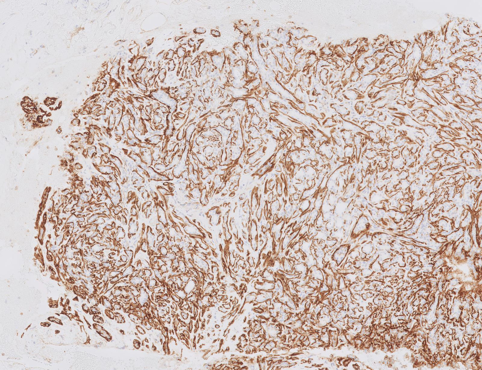
- Peritumoral stromal staining: trichoblastoma (versus epithelial staining in basal cell carcinoma and strong stromal staining in squamous cell carcinoma) (Int J Dermatol 2009;48:713, Iran J Med Sci 2013;38:100)
- Strong stromal staining: cutaneous squamous cell carcinoma (versus epithelial staining in basal cell carcinoma and peritumoral stromal staining in trichoblastoma) (Int J Dermatol 2009;48:713, Iran J Med Sci 2013;38:100)
Uses by pathologists
- B lymphoblastic leukemia (B ALL):
- One of first markers to identify leukemic cells in children, corresponding to name of common acute lymphoblastic leukemia antigen (CALLA)
- CD10 expression corresponds to the degree of differentiation of B lymphoblasts, differentiating early precursor B ALL / pro-B ALL (earliest stage) (CD10-) from common B ALL (intermediate stage) and pre-B ALL and transitional pre-B ALL (most mature stages) (CD10+)
- CD10- is associated with t(v;11q23.3); KMT2A (MLL) rearrangement, infantile (< 1 year of age) presentation and poor prognosis (Leukemia 2002;16:1233)
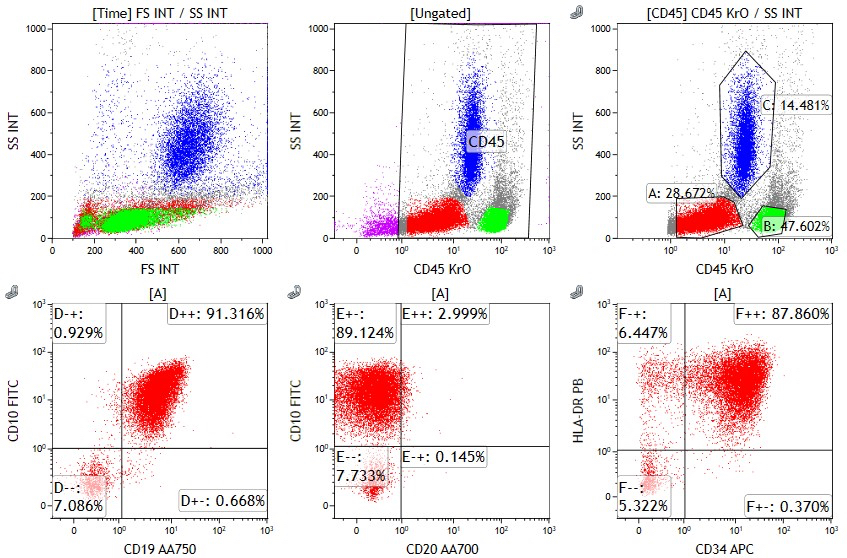
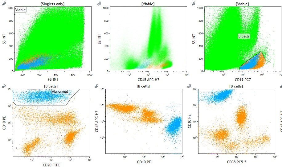
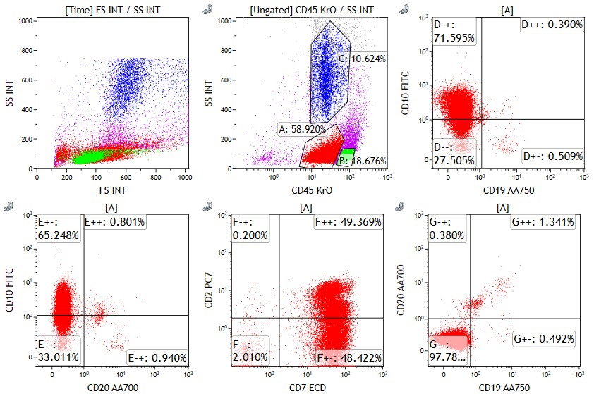
- May distinguish B lymphoblasts (aberrant overexpression or underexpression of CD10) from hematogones (CD10+) by minimal residual disease (MRD) flow cytometry (Leukemia 2001;15:1185)
- Bladder:
- Present in 40 - 50% of urothelial carcinoma and squamous cell carcinoma, with CD10 expression strongly correlated with high tumor grade and stage (Diagn Pathol 2009;4:38, Am J Clin Pathol 2005;124:371, J Environ Pathol Toxicol Oncol 2012;31:203, APMIS 2007;115:1206)
- Breast:
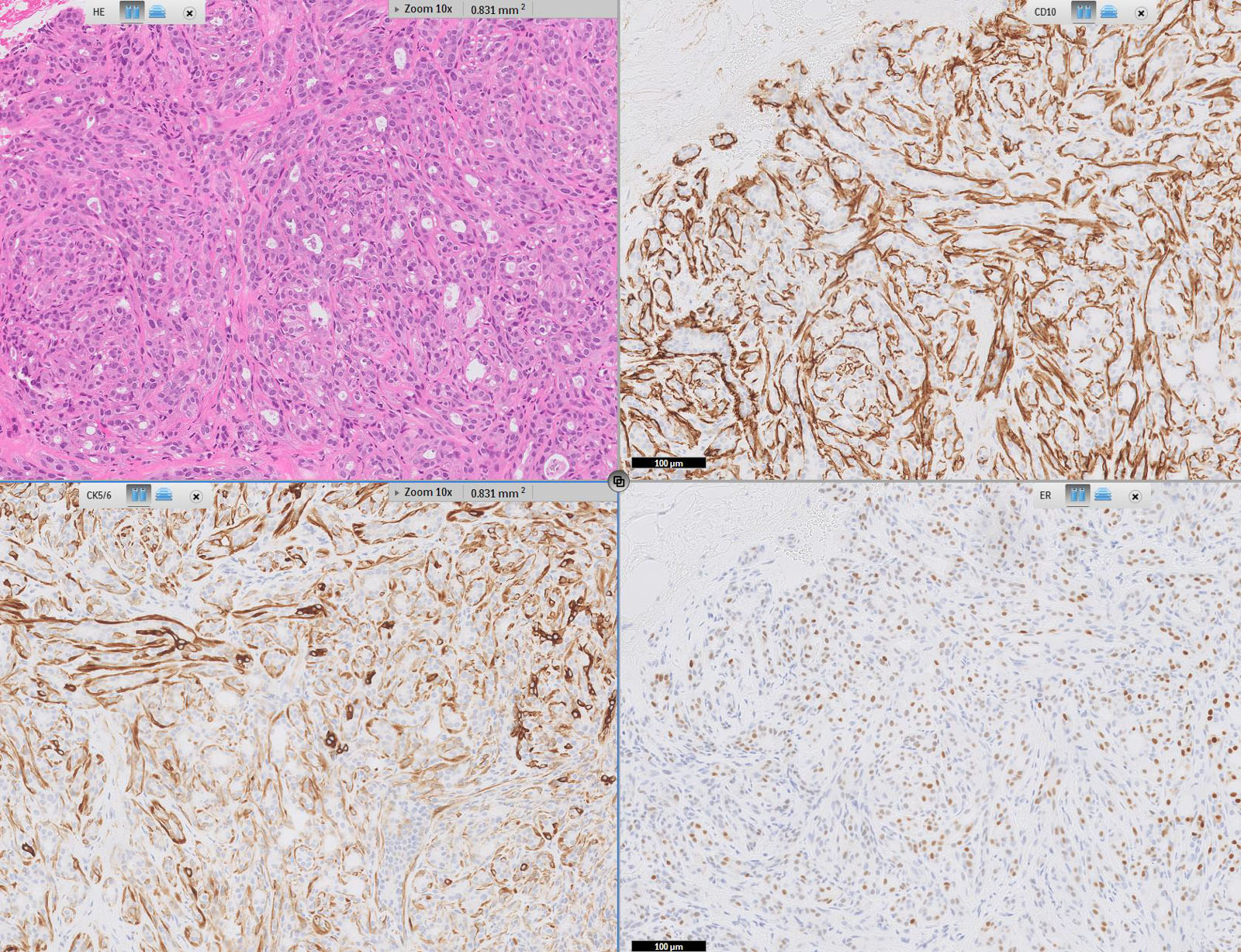
- Expression by myoepithelial cells (Mod Pathol 2002;15:397)
- Positive in mammary myofibroblastoma (Virchows Arch 2007;450:727)
- Rarely positive in:
- Invasive ductal carcinoma (Pathobiology 2015;82:259)
- Papilloma (J Clin Pathol 2007;60:958)
- Benign stroma (Virchows Arch 2006;448:871)
- Mammary sarcoma, NOS (Diagn Pathol 2013;8:14, Am J Surg Pathol 2006;30:450)
- Stromal expression is associated with increased biologic aggressiveness and poor prognosis in invasive ductal carcinoma (Mod Pathol 2007;20:84, Virchows Arch 2002;440:589)
- Stromal expression may change postchemotherapy and correlates with clinical response (Indian J Cancer 2013;50:46)
- Helps differentiate collagenous spherulosis (CD10+, HHF35+) from adenoid cystic carcinoma of the breast (CD10-, HHF35-) (Pathol Res Pract 2012;208:405)
- Ectopic prostatic tissue:
- Used with prostate specific antigen (PSA) and prostate specific alkaline phosphatase (PSAP) to confirm diagnosis in uterus and vagina (CD10+ in cytoplasm of basal cell layer) (Am J Surg Pathol 2006;30:209)
- Endometrial stromal tumors:
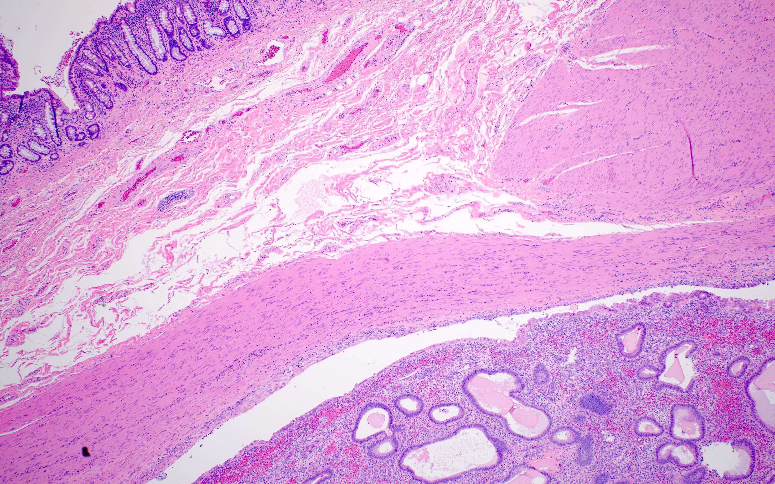
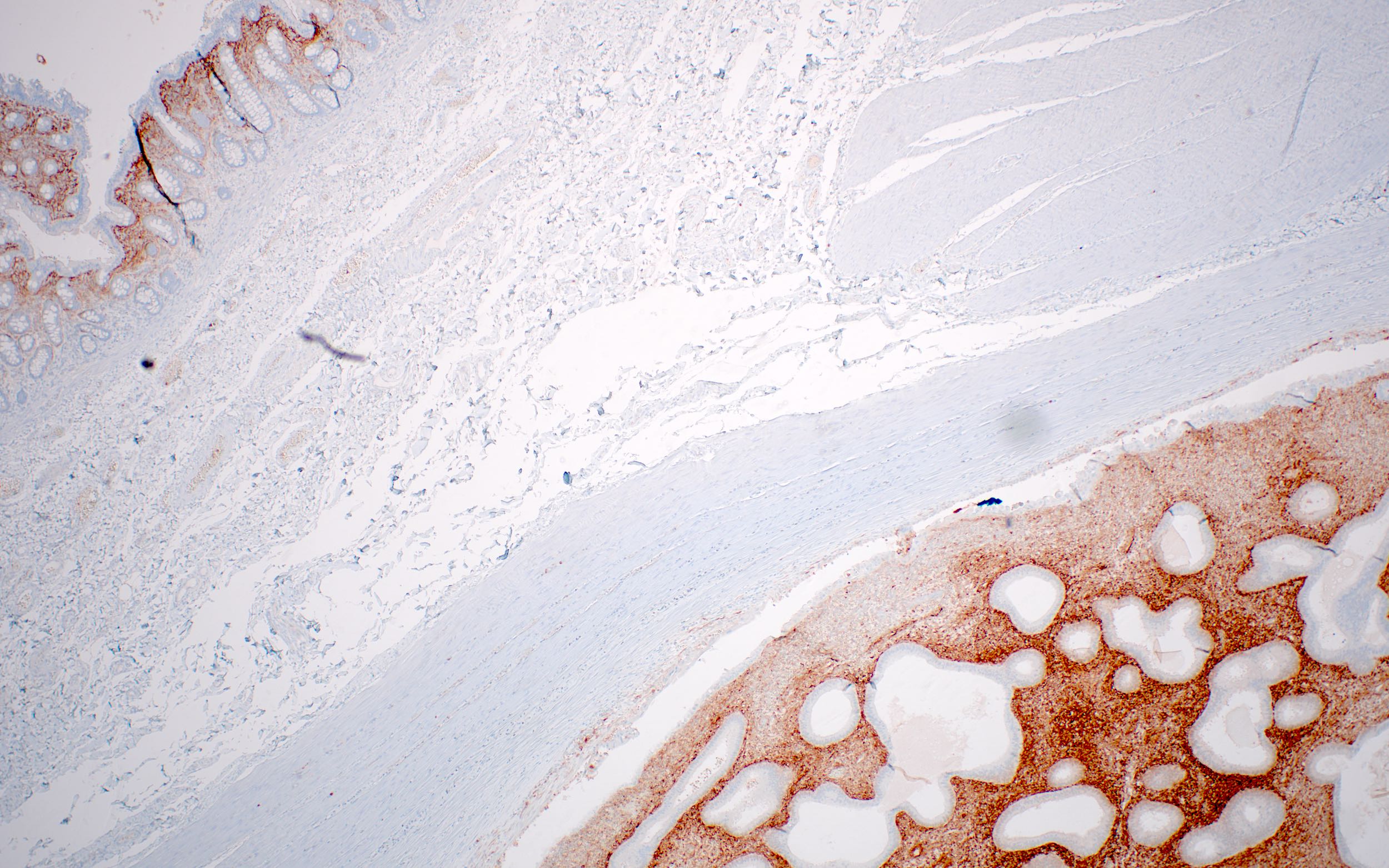
- Used with smooth muscle actin (SMA), muscle specific actin (MSA) and desmin to differentiate endometrial stromal tumors (CD10+) from uterine smooth muscle tumors (CD10-, with rare exceptions) (Mod Pathol 2001;14:465, Mod Pathol 2002;15:923)
- Endometriosis:
- May be useful in diagnosis, except in cervix (Adv Anat Pathol 2004;11:310)
- Gynecologic tumors:
- Mesonephric remnants (e.g., mesonephric remnants of the uterine cervix, epoophoron, rete ovarii) and tumors (e.g., mesonephric adenocarcinoma of the uterine cervix, tumors of wolffian origin of the broad ligament and ovary) are CD10+ (Am J Surg Pathol 2003;27:178)
- Trophoblast tissue of normal gestations, moles, choriocarcinoma and placental site trophoblastic tumors are CD10+ (Am J Surg Pathol 2003;27:178)
- Müllerian epithelium and tumors are CD10- (with the exception of squamous epithelium and tumors with squamous differentiation)
- CD10 differentiates primary clear cell carcinoma (CD10-) from metastatic renal cell carcinoma (CD10+) (Am J Surg Pathol 2003;27:178)
- Müllerian system derived mesenchymal neoplasms, including malignant Müllerian mixed tumor (MMMT) and Müllerian adenosarcoma without sarcomatous overgrowth (MA-NSO) are generally CD10+ (Mod Pathol 2001;14:465, Mod Pathol 2002;15:923, Am J Surg Pathol 2008;32:1013)
- Hepatocellular carcinoma:
- CD10+ is 52 - 82% sensitive and > 95% specific for differentiating hepatocellular carcinoma from metastatic carcinoma when exhibiting a canalicular pattern, though cannot differentiate benign hepatic tissue (Am J Surg Pathol 2001;25:1297, Am J Surg Pathol 2002;26:978, Diagn Cytopathol 2004;30:92)
- Another study recommends use of HepPar1, MOC31 and pCEA but not CD10 (Mod Pathol 2002;15:1279)
- Kidney tumors:
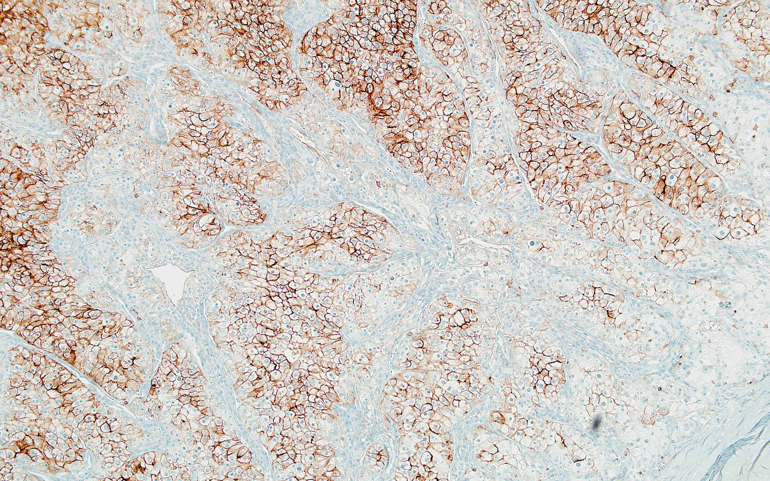
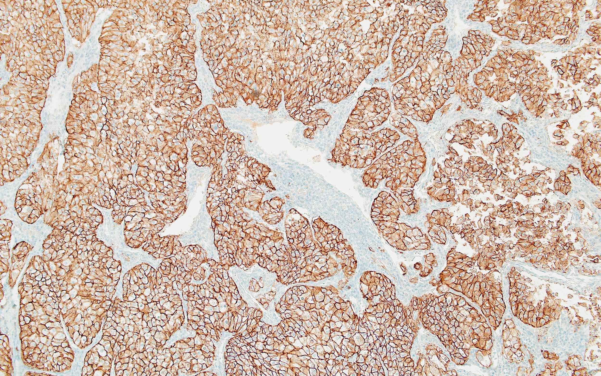
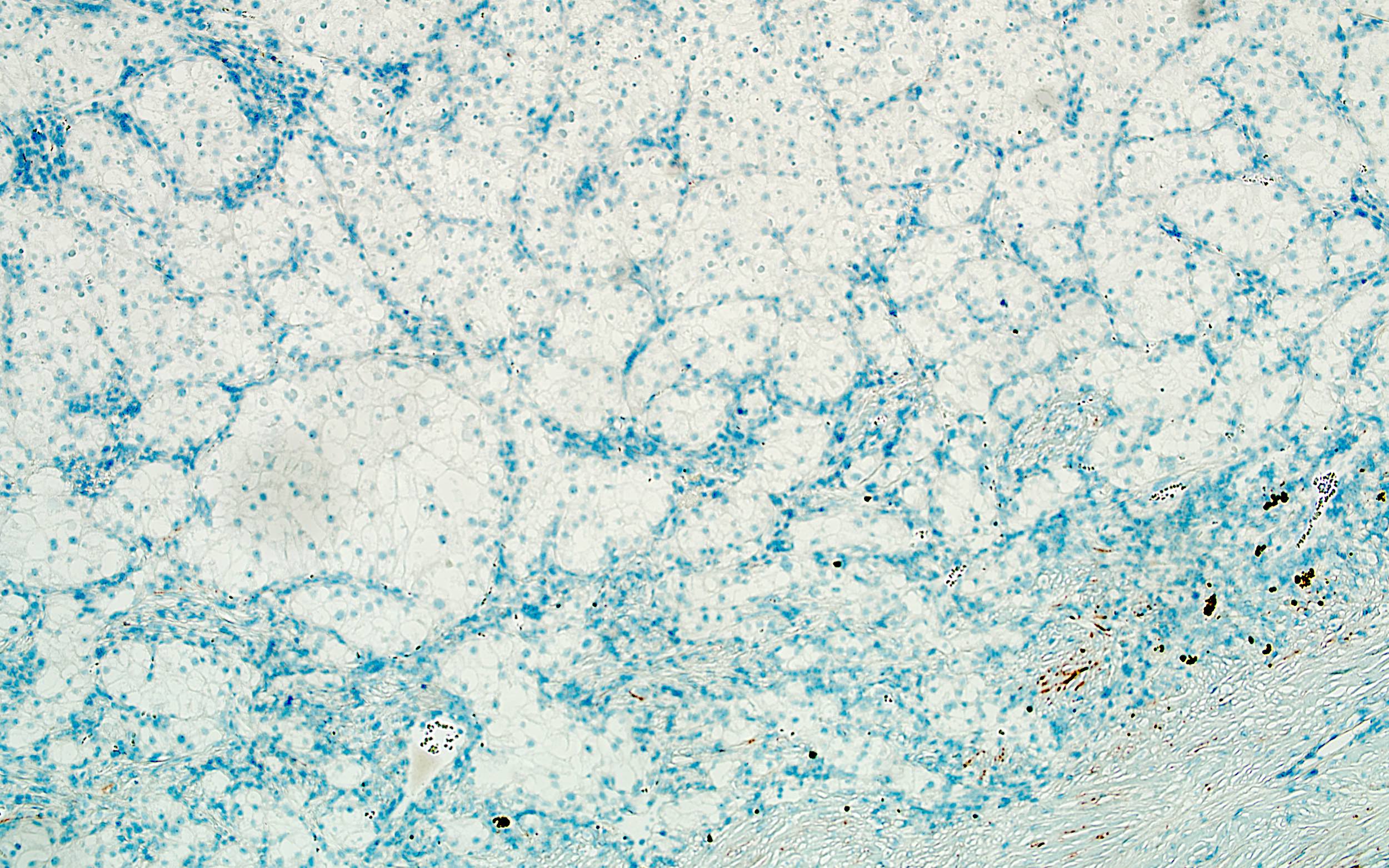
- Distinguish renal cell carcinoma, clear cell type, eosinophilic variant (CD10+) from chromophobe carcinoma, eosinophilic variant or oncocytoma (both CD10-) (Appl Immunohistochem Mol Morphol 2012;20:454)
- Liver:
- Support diagnosis of Alagille syndrome in pediatric patients (CD10+ canalicular surfaces in children > 24 months old; CD10- canalicular surfaces in children aged 2 months to 6 years of age) (Lab Invest 2007;87:1138)
- Lymphoma - angioimmunoblastic T cell (AITL) and other mature T cell lymphomas of T follicular helper (TFH) cell origin:
- Marker for T follicular helper cell phenotype (also BCL6, PD1, CXCL13, CXCR5, ICOS, SAP) in benign and malignant T cells (Mod Pathol 2003;16:879)
- Distinguish AITL and other mature T cell lymphomas of T follicular helper (TFH) cell origin (e.g., follicular T cell lymphoma and nodal peripheral T cell lymphoma with TFH phenotype) (CD10+) at nodal and extranodal sites from other T cell lymphomas (CD10-) (Mod Pathol 2011;24:993, Am J Surg Pathol 2004;28:54, Hum Pathol 2005;36:784)
- Lymphoma - Burkitt:
- CD10+ aids in diagnosis but must exclude CD10+ diffuse large B cell lymphoma, Burkitt-like lymphoma with 11q aberration, high grade B cell lymphoma with MYC and BCL2 / BCL6 rearrangement (double / triple hit lymphoma) (75 - 90% CD10+) and B lymphoblastic leukemia / lymphoma (B ALL / LBL) (Am J Clin Pathol 2012;137:665, Am J Clin Pathol 2010;133:718, Blood 2014;123:1187, Hematology Am Soc Hematol Educ Program 2014;2014:90)
- Lymphoma - diffuse large B cell (Mod Pathol 2005;18:1113, J Hematop 2009;2:187):
- Marker for germinal center phenotype (also HGAL, BCL6, CD38, LMO2, MEF2B), generally considered a favorable prognostic factor, secondary in part to association with cell of origin classification (germinal center B cell versus activated B cell) but see:
- Am J Clin Pathol 2001;116:183 (CD10+, BCL2+ tumors have poorer survival)
- Virchows Arch 2004;445:545 (no difference in survival)
- Sci Rep 2016;6:20465 (CD10+, MUM1+ tumors have similar survival to nongerminal center phenotype)
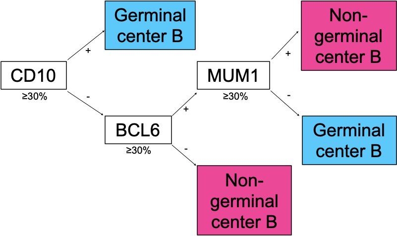
- Hans classification (Blood 2004;103:275, see diagram above)
- Marker for germinal center phenotype (also HGAL, BCL6, CD38, LMO2, MEF2B), generally considered a favorable prognostic factor, secondary in part to association with cell of origin classification (germinal center B cell versus activated B cell) but see:
- Lymphoma - follicular (Am J Clin Pathol 2002;117:291, Hum Pathol 2013;44:1328):
- CD10+ / strong (often stronger than normal germinal center B cells) aids in diagnosis of primary or secondary spread but:
- High grade follicular lymphomas and interfollicular infiltrates may be CD10- (Am J Clin Pathol 2001;115:862)
- Bone marrow involvement by follicular lymphoma may be CD10- (Am J Surg Pathol 2010;34:1266)
- Leukemic presentation by follicular lymphoma often CD10- (Am J Surg Pathol 2010;34:1266)
- Does not distinguish variants (diffuse, testicular, duodenal type follicular lymphoma), primary cutaneous follicle center lymphoma or pediatric type follicular lymphoma (all CD10+)
- Other lymphomas may be CD10+, including:
- Angioimmunoblastic T cell (see above)
- Burkitt (see above)
- Diffuse large B cell lymphoma (see above)
- Mantle cell (Appl Immunohistochem Mol Morphol 2010;18:103)
- Marginal zone (J Clin Pathol 1999;52:849)
- Hairy cell (Am J Clin Pathol 2003;120:228)
- Other rare CD5+, CD10+ lymphomas (Am J Clin Pathol 2003;119:218, Arch Pathol Lab Med 2001;125:951)
- CD10+ / strong (often stronger than normal germinal center B cells) aids in diagnosis of primary or secondary spread but:
- Microvillous inclusion disease:
- Intense CD10+ apical cytoplasmic staining in small bowel enterocytes or focal apical cytoplasmic staining in absorptive colonocytes (versus linear brush border staining of enterocytes and negative colonic staining in normal tissue) (Am J Surg Pathol 2002;26:902, Orphanet J Rare Dis 2006;1:22, Am J Surg Pathol 2010;34:970)
- Mixed phenotype acute leukemia (MPAL), B / myeloid:
- Component of evaluation for assignment of B cell lineage to a single blast population in cases of multiple lineage designation
- Strong CD19 with ≥ 1 of the following strongly expressed: CD10, CD79a, cytoplasmic CD22
- Or
- Weak CD19 with ≥ 2 of the following strongly expressed: CD10, CD79a, cytoplasmic CD22
- Strong CD19 with ≥ 1 of the following strongly expressed: CD10, CD79a, cytoplasmic CD22
- Myeloid lineage assignment requirements include
- MPO (by flow cytometry, immunohistochemistry or cytochemistry)
- Or
- Monocytic differentiation (≥ 2 of the following: nonspecific esterase, CD11c, CD14, CD64, lysozyme)
- MPO (by flow cytometry, immunohistochemistry or cytochemistry)
- Component of evaluation for assignment of B cell lineage to a single blast population in cases of multiple lineage designation
- Müllerian system derived neoplasms - MMMT and Müllerian adenosarcoma:
- CD10+ expression in Müllerian system derived mesenchymal neoplasms, including malignant Müllerian mixed tumor (MMMT) and Müllerian adenosarcoma without sarcomatous overgrowth (MA-NSO) (Mod Pathol 2001;14:465, Mod Pathol 2002;15:923, Am J Surg Pathol 2008;32:1013)
- Pancreas:
- Confirm diagnosis of solid pseudopapillary neoplasm (CD10+) (Am J Surg Pathol 2000;24:1361)
- Differentiates mucinous cystic neoplasms (CD10+ / CK20+) from intraductal papillary mucinous neoplasm of branch duct type (CD10- / CK20-) (Pancreas 2009;38:558)
- Skin tumors:
- CD10 staining patterns may aid in differentiation of basal cell carcinoma (epithelial staining), trichoblastoma (peritumoral stromal staining) and squamous cell carcinoma (strong stromal staining) (Int J Dermatol 2009;48:713, Iran J Med Sci 2013;38:100)
- Differentiates atypical fibroxanthoma (diffuse membranocytoplasmic staining) from spindle cell melanoma and sarcomatoid squamous cell carcinoma (CD10-) (J Cutan Pathol 2010;37:744)
- Membranous expression in majority of other xanthomatous skin lesions (xanthomas, xanthelasmas and xanthogranulomas) versus cytoplasmic expression in metastatic renal cell carcinomas, balloon cell nevi and nodular / clear cell hidradenomas (J Cutan Pathol 2005;32:348)
- Vascular tumors:
- Aids in differentiation of hemangioblastoma (usually CD10-, rarely focal staining) from metastatic renal cell carcinoma (CD10+) (Diagn Pathol 2012;7:39, Mod Pathol 2005;18:788)
- Epithelioid hemangioendothelioma can have strong / diffuse CD10+ (30%), mimicking metastatic renal cell carcinoma (Arch Pathol Lab Med 2009;133:1965)
Prognostic factors
- CD10- B lymphoblastic leukemia / lymphoma is associated with t(v;11q23.3); KMT2A (MLL) rearrangement, infantile (< 1 year of age) presentation and poor prognosis (Leukemia 2002;16:1233)
- CD10 in diffuse large B cell lymphoma is a marker for germinal center phenotype (also HGAL, BCL6, CD38, LMO2), generally considered a favorable prognostic factor, secondary in part to association with cell of origin classification (germinal center B cell versus activated B cell) (Mod Pathol 2005;18:1113, J Hematop 2009;2:187)
- Stromal expression in invasive breast carcinoma, no special type is associated with increased biologic aggressiveness and poor prognosis (Mod Pathol 2007;20:84, Virchows Arch 2002;440:589)
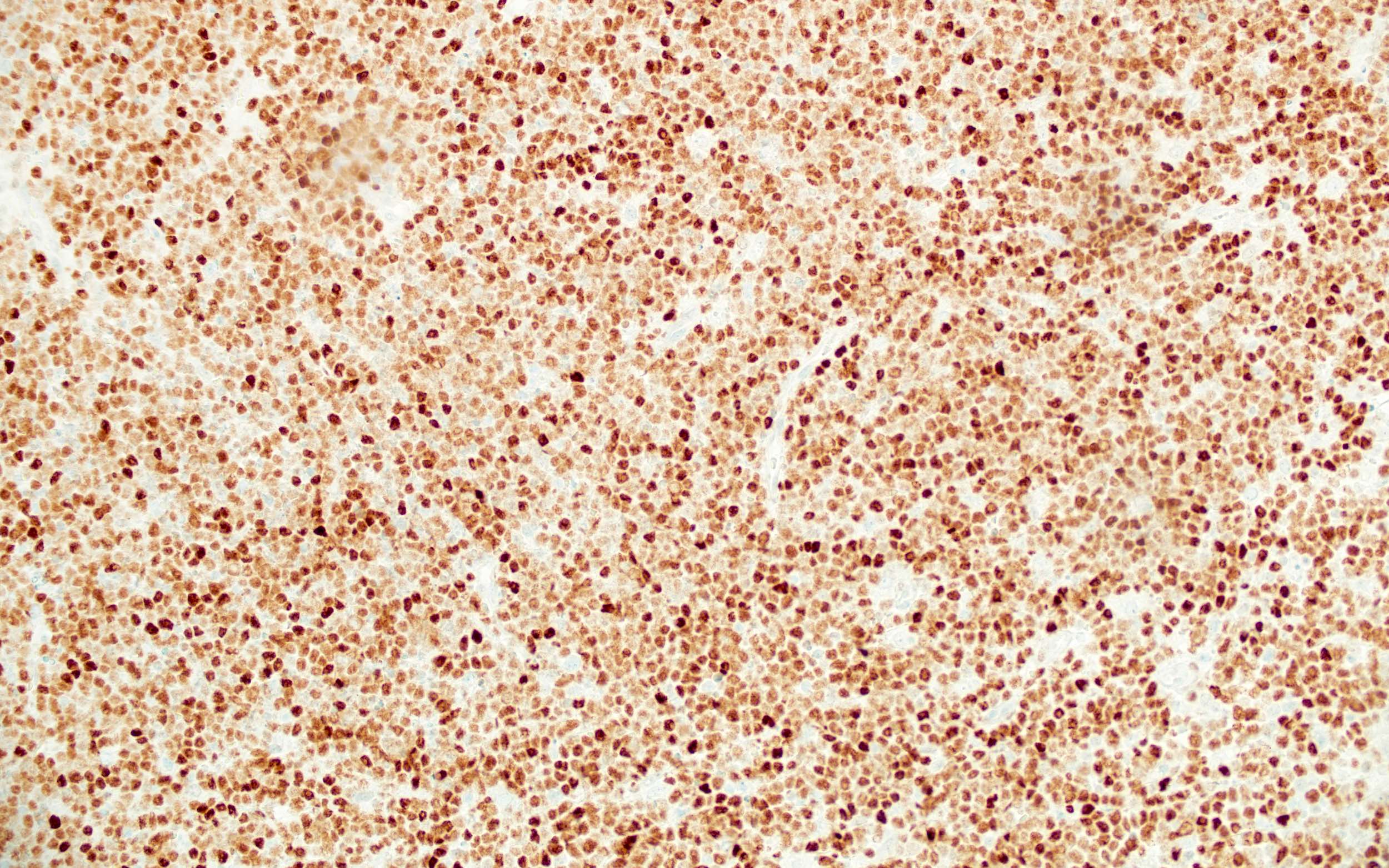
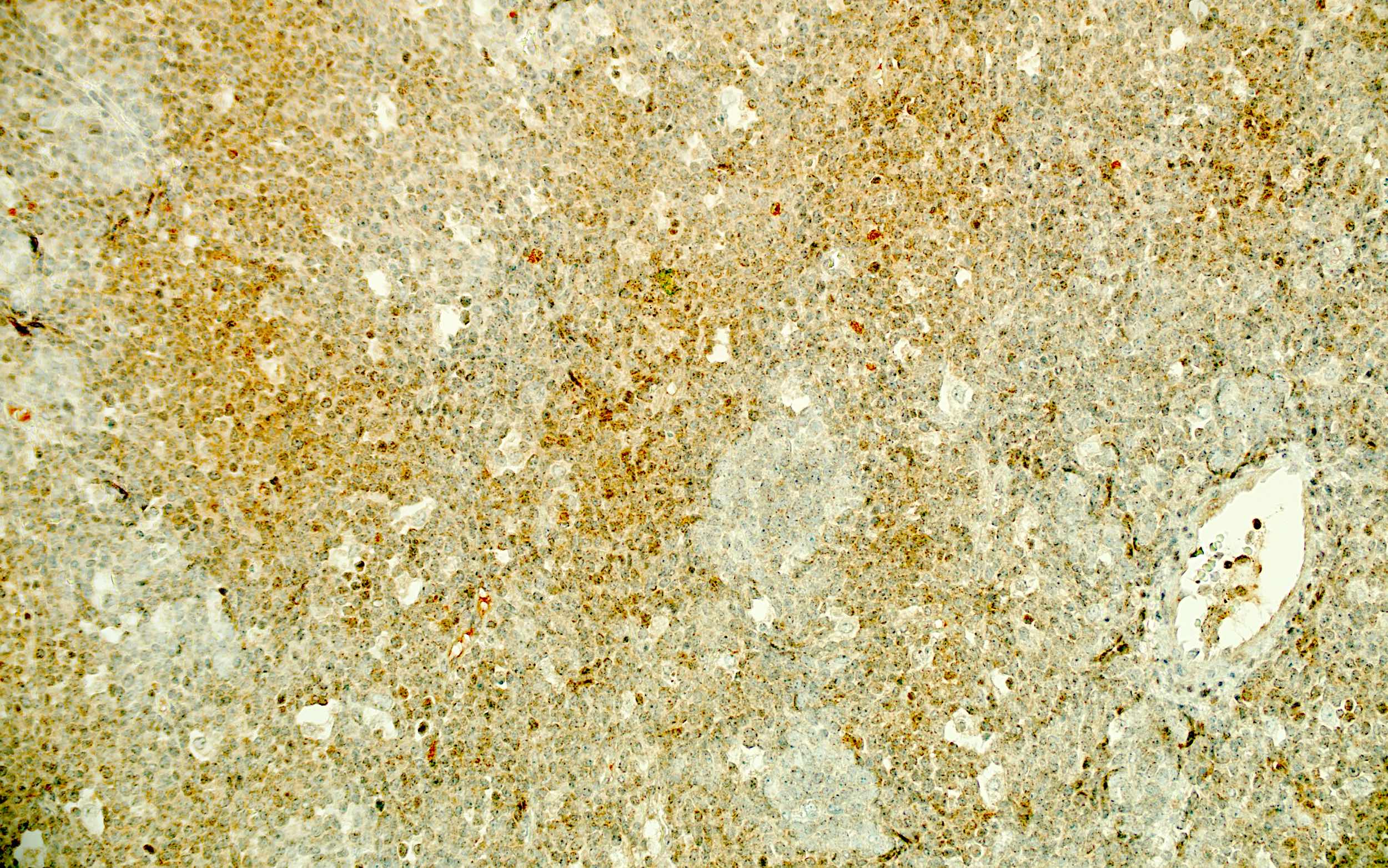
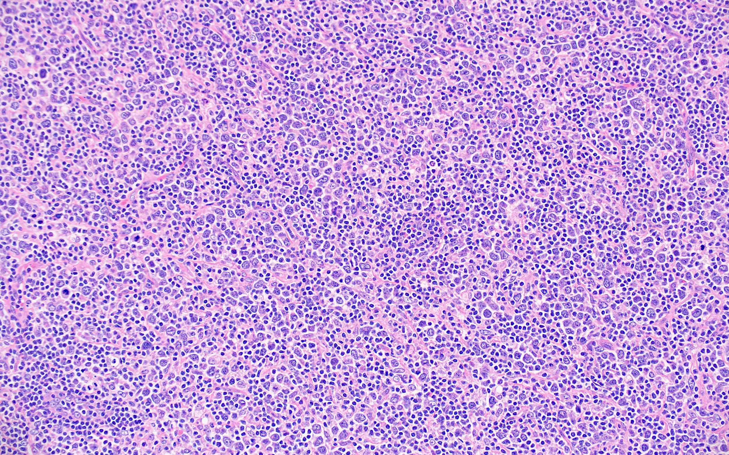
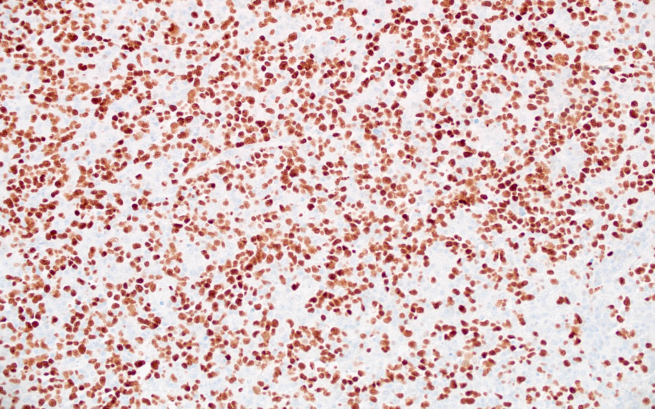
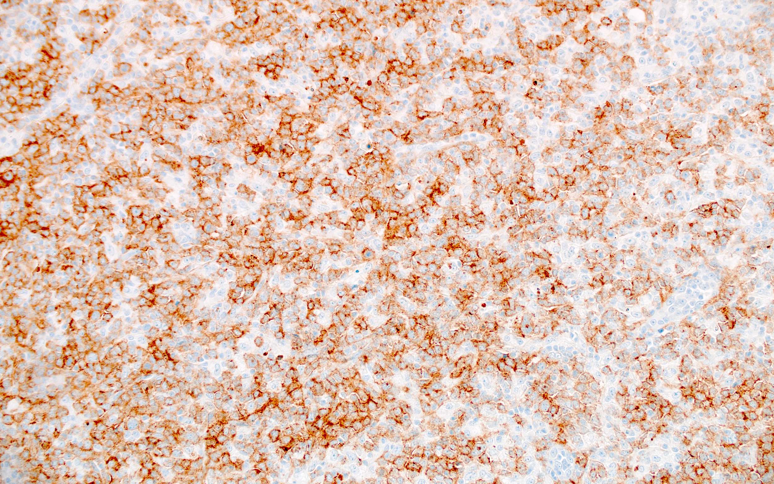
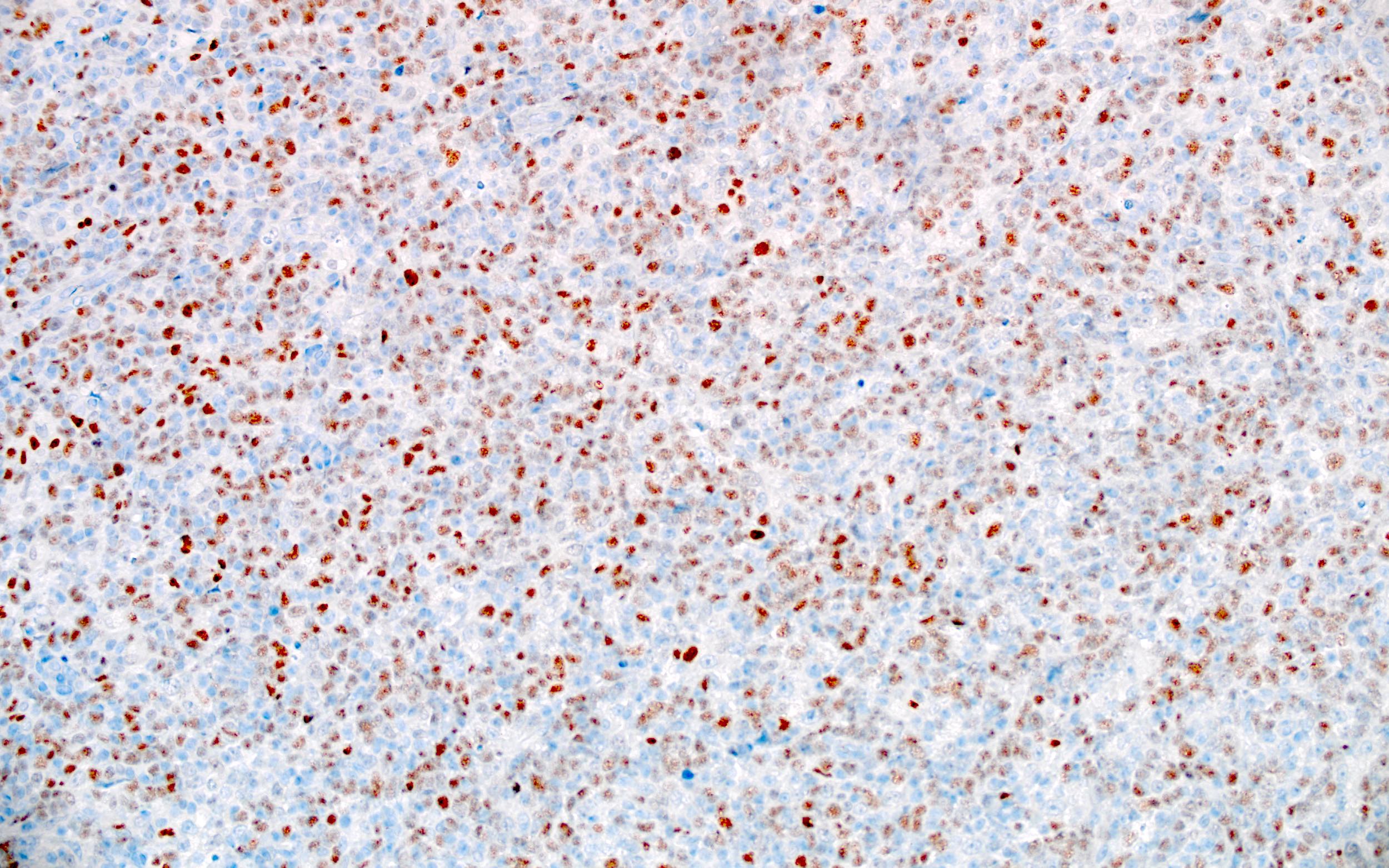
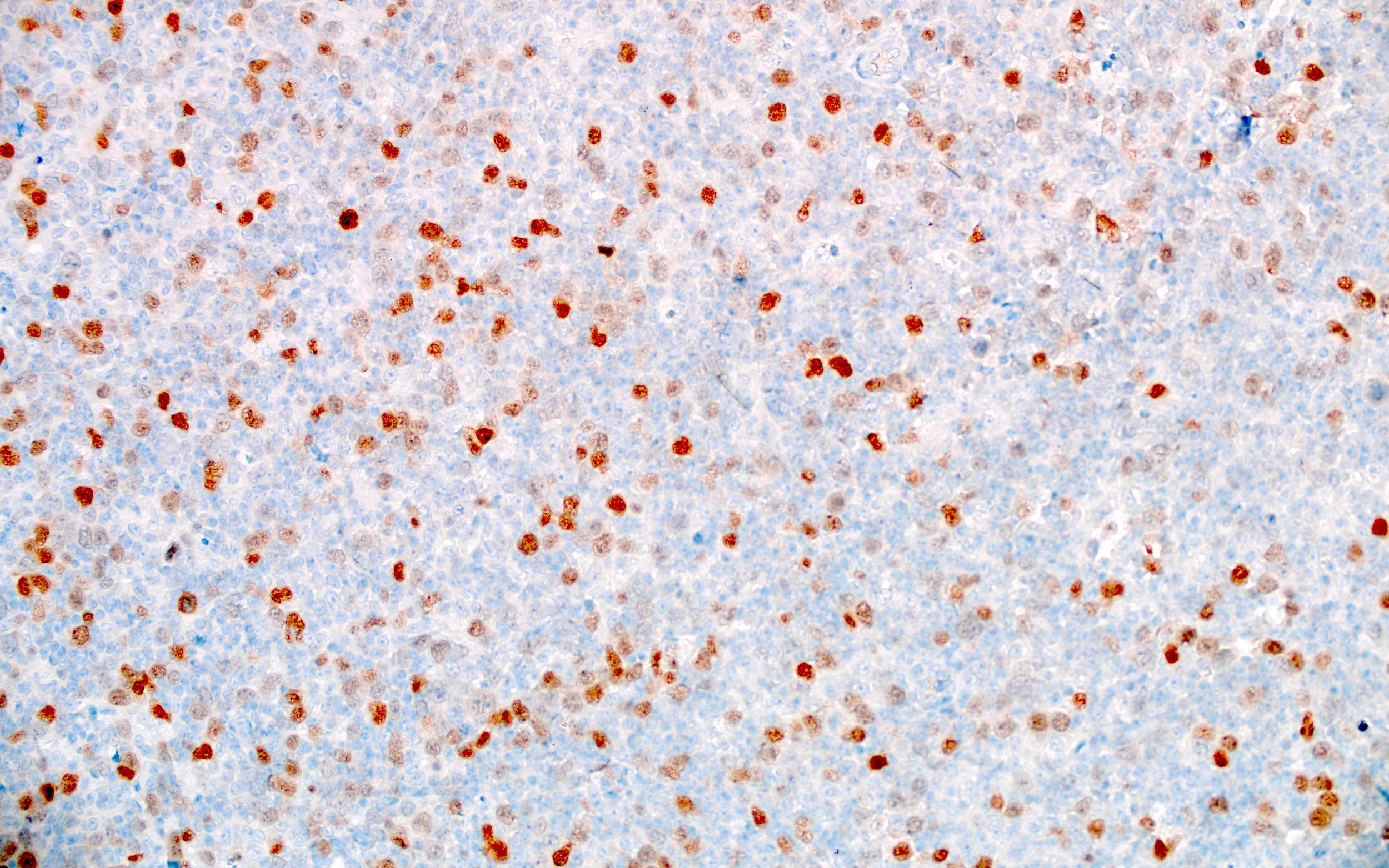
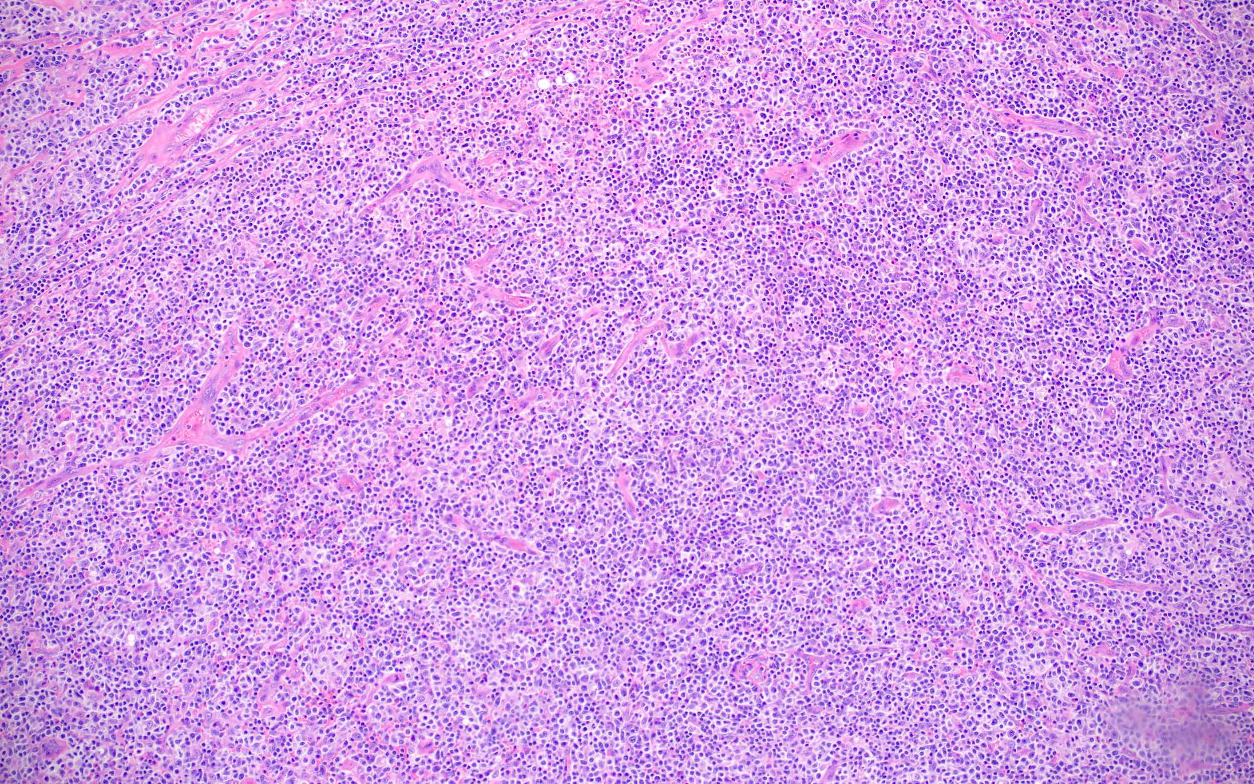
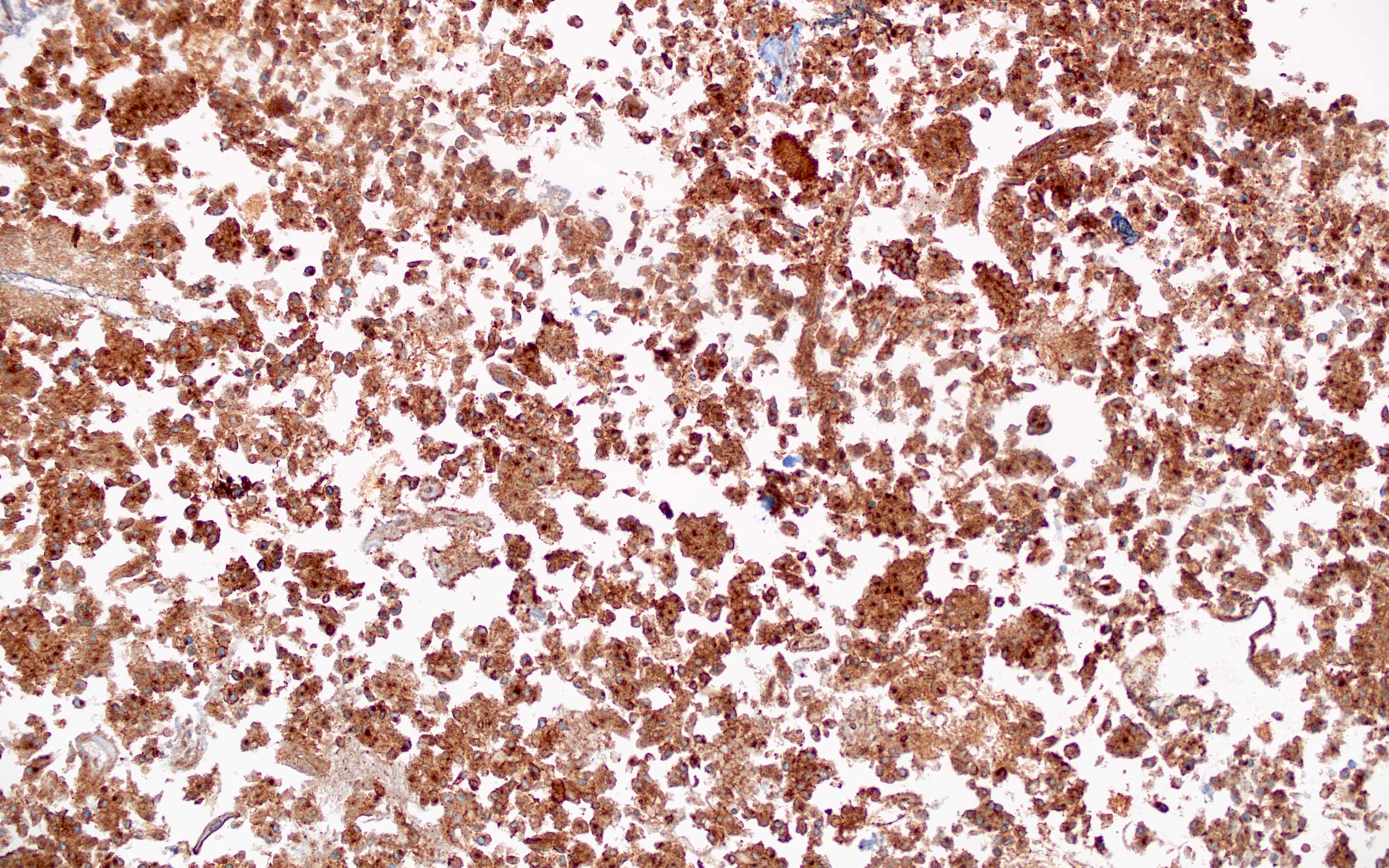
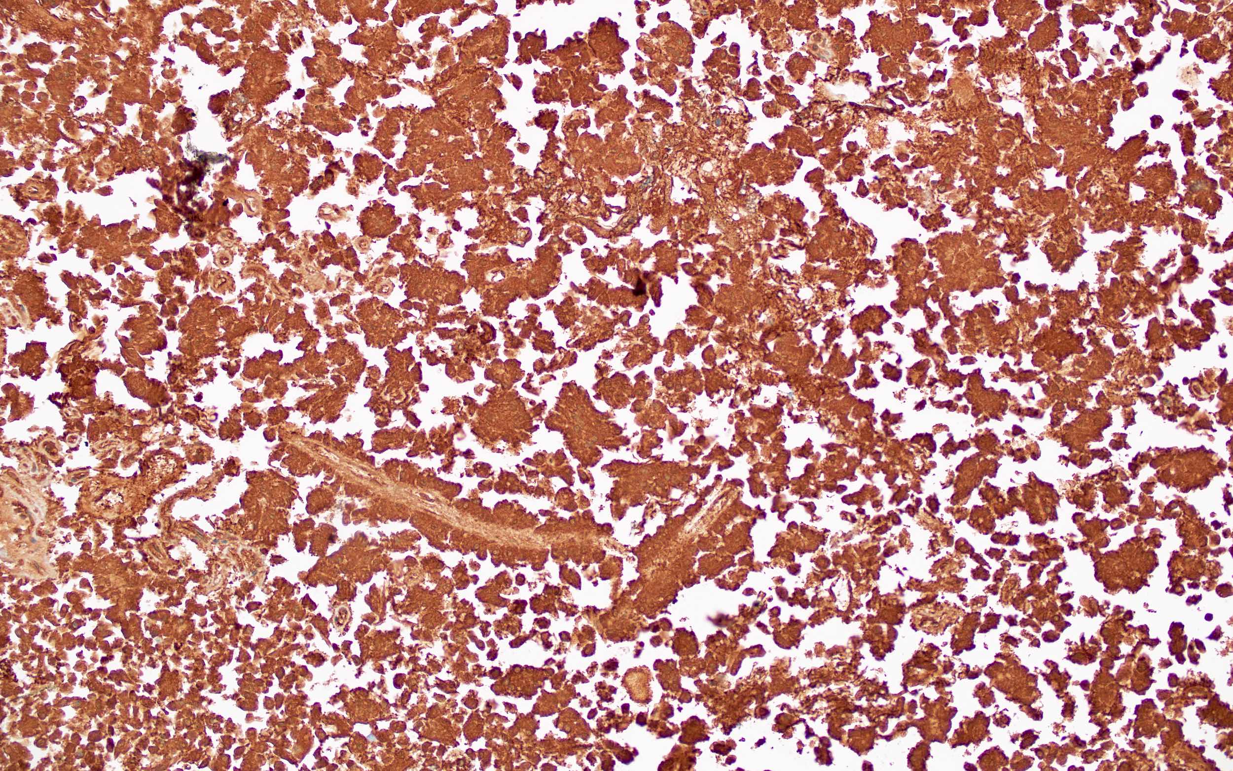
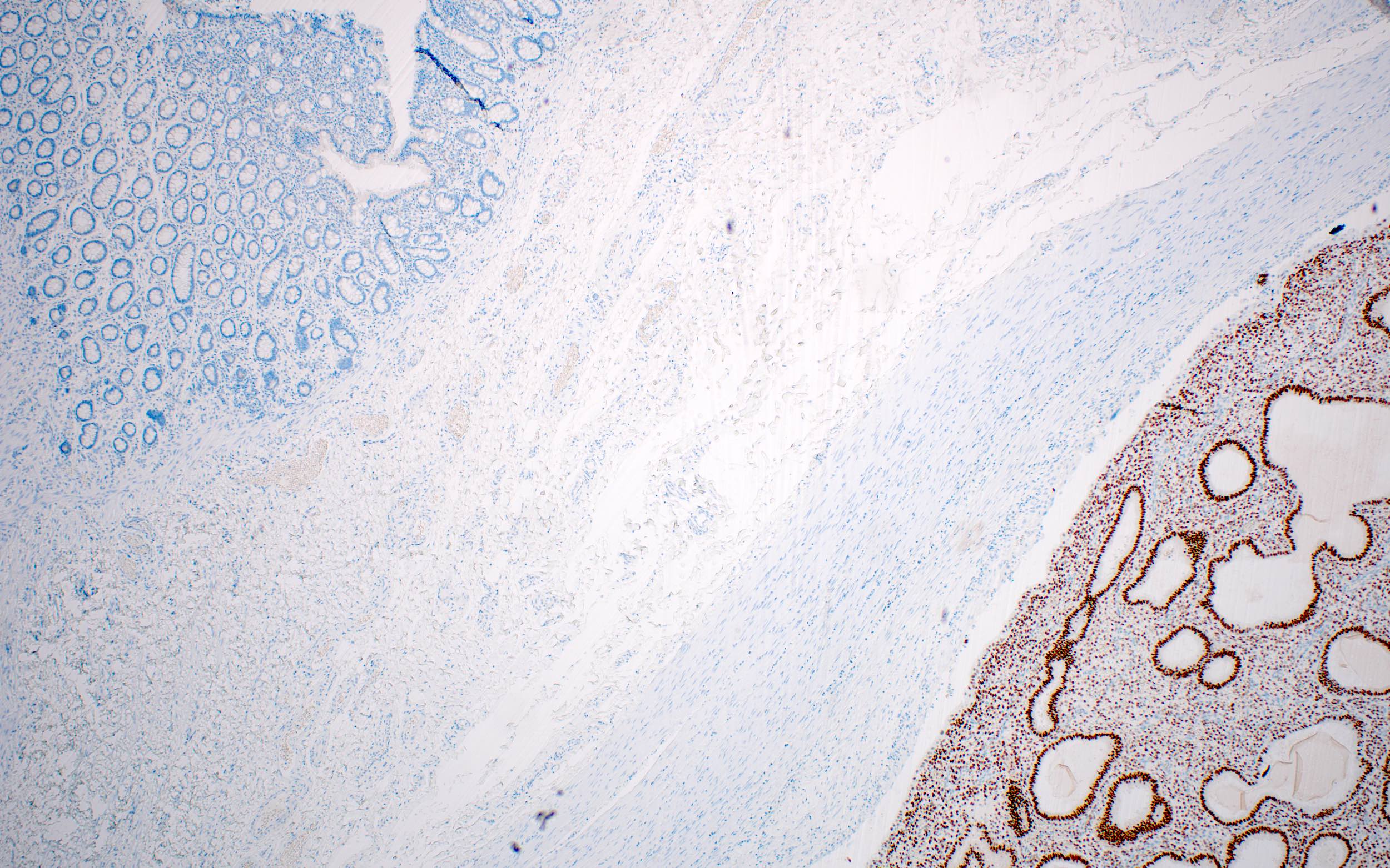
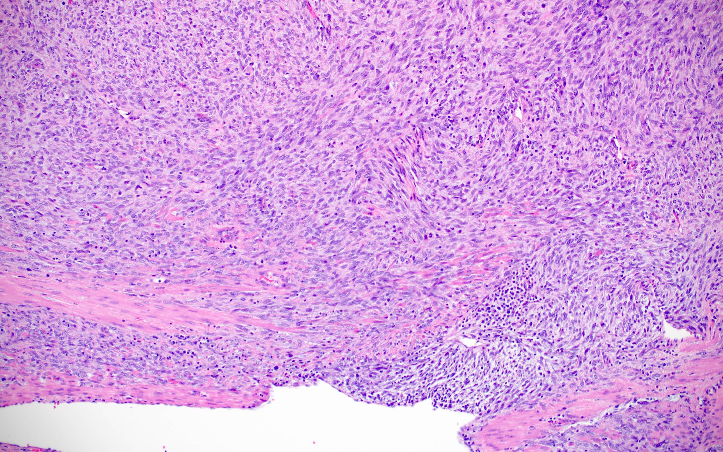
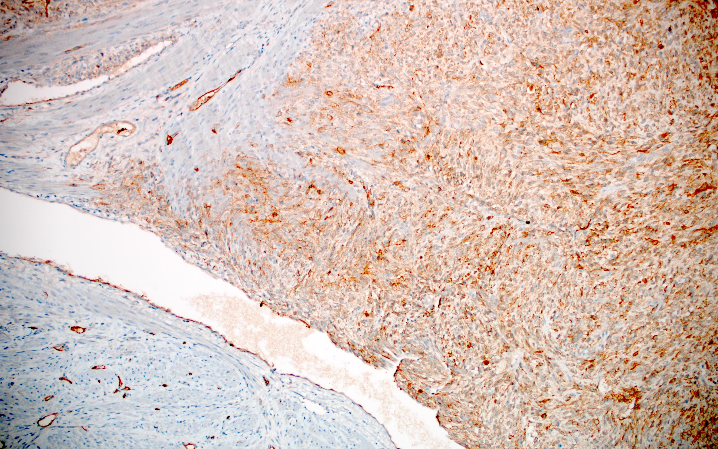
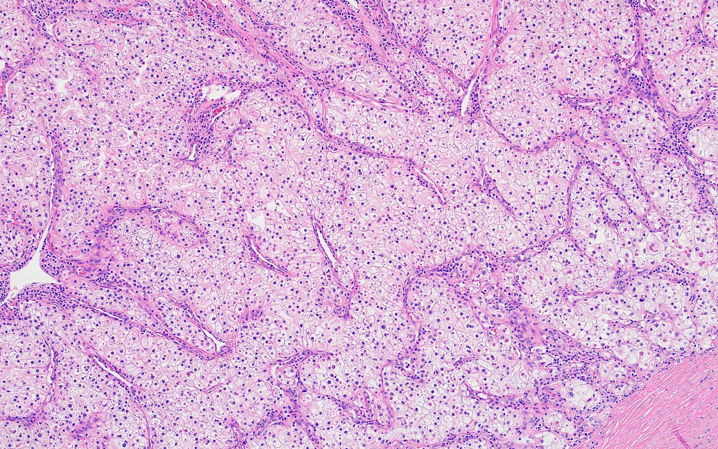
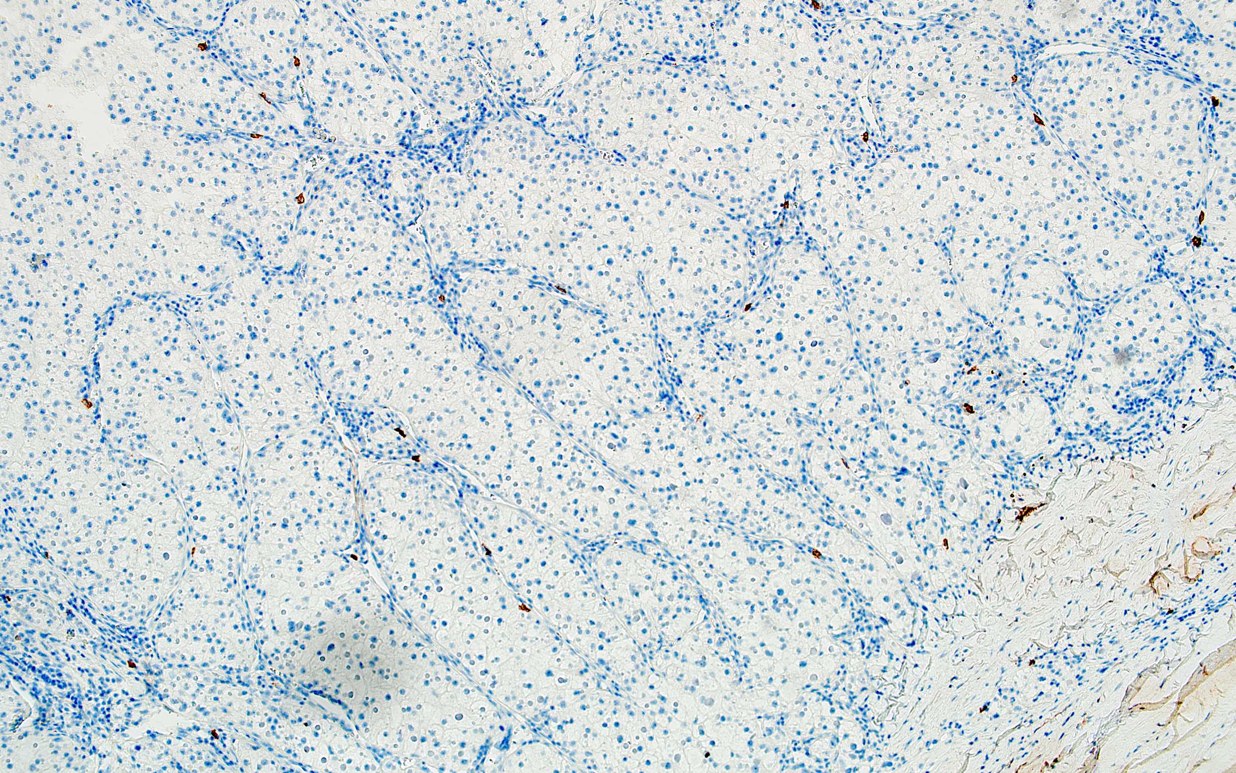
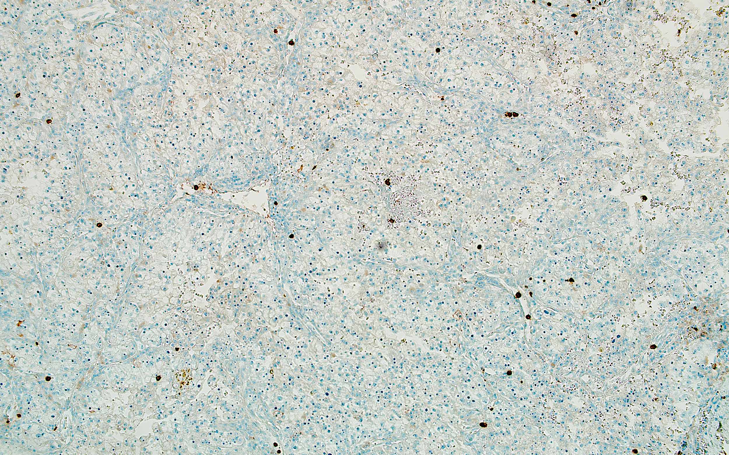
Microscopic (histologic) images
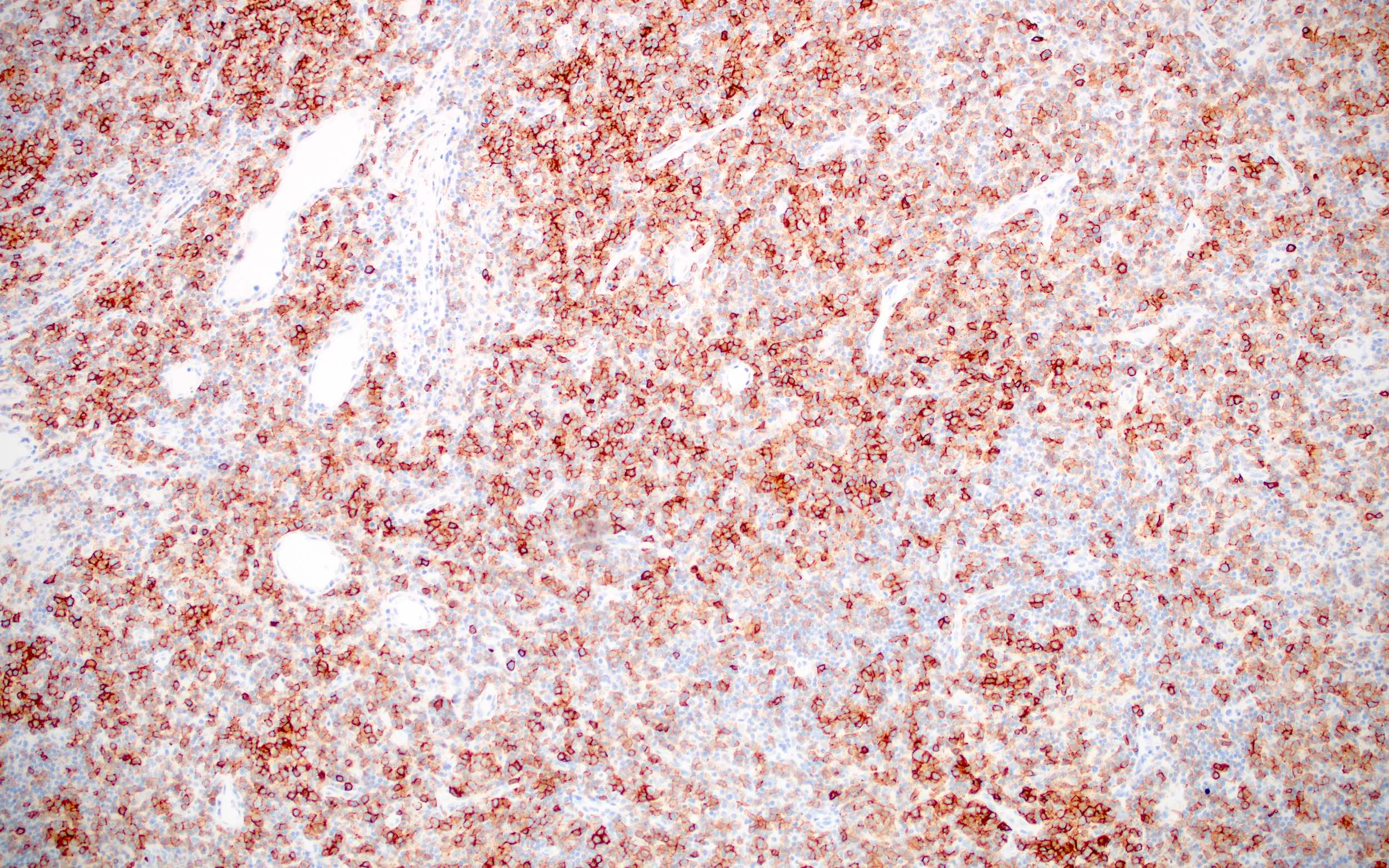
Contributed by Bradford Siegele, M.D., J.D.
Contributed by Jijgee Munkhdelger, M.D., Ph.D. and Andrey Bychkov, M.D., Ph.D.
Virtual slides
Positive staining - normal
- Hematopoietic cells:
- Hematogones, pre-T cells, T follicular helper cells, germinal center B cells, mature granulocytes
- Other cells:
- Adrenal cortex, choroid plexus,
- Endometrial stroma, endothelial cells (some), fibroblasts (Hypertension 1995;26:230)
- GU (male) epithelium, kidney (microvilli)
- Liver (bile canaliculi, biliary epithelium), mesonephric remnants, myoepithelial cells (breast) (Am J Surg Pathol 2003;27:178, Mod Pathol 2002;15:397, J Clin Pathol 2004;57:625)
- Ovary, placenta (cytotrophoblast, intermediate trophoblast, syncytiotrophoblast), prostate basal and secretory cells
- Small intestine (linear brush border staining, variable loss with active enteritis), Wolffian (but not Müllerian) type epithelium (Mod Pathol 2011;24:1627, Am J Surg Pathol 2003;27:178)
Positive staining - disease
- Leukemia / lymphoma:
- Angioimmunoblastic T cell and other nodal lymphomas of T follicular helper cell origin (T follicular helper cell, nodal peripheral T cell lymphoma with TFH phenotype)
- B lymphoblastic leukemia (B ALL / LBL) (75%), Burkitt, Burkitt-like with 11q aberration, chronic myeloid leukemia (CML) in B lymphoid blast crisis (90%) (Arch Pathol Lab Med 2000;124:704, Blood 2014;123:1187)
- Diffuse large B cell (variable; CD10+ is germinal center B cell defining by Hans criteria)
- Follicular lymphoma (70%) (diffuse, testicular and duodenal type variants, primary cutaneous follicle center lymphoma or pediatric type follicular lymphoma generally CD10+; high grade may be CD10-)
- High grade large B cell lymphoma with MYC and BCL2 / BCL6 rearrangements (double hit / triple hit lymphoma) (75 - 90% CD10+) (Hematology Am Soc Hematol Educ Program 2014;2014:90)
- Large B cell lymphoma with IRF4 rearrangement (66%) (Blood 2011;118:139)
- Mixed phenotype acute leukemia (B / T, B / myeloid)
- T lymphoblastic leukemia (T ALL / LBL) (63%) (Arch Pathol Lab Med 2000;124:704)
- Other:
- Breast cancer associated stroma, breast metaplastic carcinoma, breast myofibroblastoma, choriocarcinoma (Virchows Arch 2007;450:727)
- Colonic carcinoma and high grade dysplasia associated stroma, dermatofibroma, dermatofibrosarcoma (Hum Pathol 2002;33:806)
- Epithelioid hemangioendothelioma, Ewing sarcoma, female adnexal tumor of probable Wolffian origin (FATWO) (50 - 100%), gastric carcinoma associated stroma (Arch Pathol Lab Med 2009;133:1965, Jpn J Clin Oncol 2005;35:245)
- Hepatocellular carcinoma (canalicular pattern similar to polyclonal CEA) (Am J Pathol 2001;159:1415)
- Malignant mixed Müllerian tumors (MMMT)
- Malignant peripheral nerve sheath tumor (72%, usually focal) (Pathol Res Pract 2012;208:281)
- Mediastinal germ cell tumors
- Mesonephric tumors (Am J Surg Pathol 2001;25:1540)
- Mesothelioma (malignant) (Arch Pathol Lab Med 2006;130:823)
- Microvillous inclusion disease (strong cytoplasmic staining)
- Müllerian adenosarcoma
- Pancreatic adenocarcinoma (50%)
- Pancreatic solid pseudopapillary neoplasm
- Placental site trophoblastic tumor
- Prostatic adenocarcinoma (some Gleason pattern 4 and 5 cases)
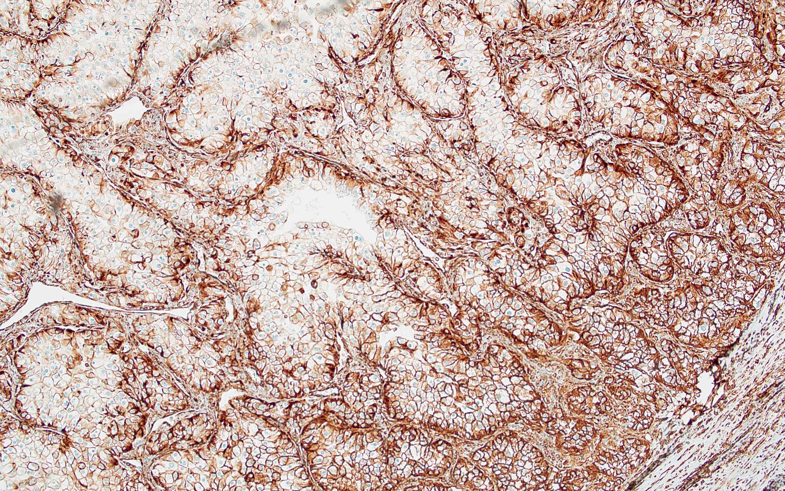
- Renal cell carcinoma (most clear cell [94 - 100%], papillary [variable, 67 - 93%], chromophobe [variable, 0 - 72%], Xp11 translocation [100%]) (Am J Surg Pathol 2000;24:203, Mod Pathol 2005;18:535, Mod Pathol 2004;17:1455, Arch Pathol Lab Med 2011;135:92)
- Rhabdomyosarcoma (60%, particularly pleomorphic) and other sarcomas (variable)
- Seminoma / dysgerminoma (87 - 92%) (Int J Clin Exp Pathol 2013;6:498, Histol Histopathol 2014;29:101)
- Spindle cell lipoma (Virchows Arch 2007;450:727)
- Urothelial carcinoma (54%)
- Uterus: adenomyosis, cellular leiomyoma (50%), endometrial adenocarcinoma (may also be present in desmoplastic stroma), endometrial stromal tumor, leiomyosarcoma, other sarcomas (Am J Surg Pathol 2003;27:786)
- Yolk sac tumor
Negative staining
- Leukemia / lymphomas:
- Acute myeloid leukemia (AML), chronic lymphocytic leukemia / small lymphocytic lymphoma (CLL / SLL), EBV+ lymphoproliferative disorders
- Lymphoplasmacytic (LPL) (usually), mantle cell, marginal zone (MALT, splenic, nodal) (usually), primary cutaneous diffuse large B cell, leg type, pyothorax associated
- Extranodal marginal zone lymphoma of mucosa associated lymphoid tissue (MALT lymphoma) (very rare) (Leuk Lymphoma 2012;53:1032)
- Hairy cell leukemia (10%) (Am J Clin Pathol 2003;120:228)
- Lymphoplasmacytic lymphoma (some), plasma cell myeloma (some), primary cutaneous diffuse large B cell (variable), plasmablastic (variable), primary mediastinal B cell (32%) (Am J Surg Pathol 2001;25:1277)
- Other:
- Atypical polypoid adenomyoma (stromal cells), clear cell carcinoma of female genital tract
- Colon (normal, diseased), erythroid and myeloid precursors, hemangioblastoma, high grade prostatic intraepithelial neoplasia (PIN) and basal cell hyperplasia, prostatic adenocarcinoma (all Gleason patterns 2 and 3, some 4 and 5) (Hum Pathol 2003;34:450)
- Melanoma (40%) (Mod Pathol 2004;17:1251)
- Schwannoma (45%)
Flow cytometry images
Sample pathology report
- Lymph node, right neck level 5, excision:
- Large B cell lymphoma (see comment)
- Morphology: diffuse large B cell type
- Hans criteria: germinal center B cell phenotype (CD10+, BCL6+, MUM1-)
- Double expressor status: double expressor phenotype (BCL2+, MYC+)
- Ki67 proliferation index: high (60 - 70%)
- EBER in situ hybridization studies: negative
- Comment: The findings are consistent with a diagnosis of a CD10+ large B cell lymphoma, with diffuse large B cell morphology. Ancillary studies, including fluorescence in situ hybridization studies to evaluate for MYC, BCL2 and BCL6 rearrangement status, are pending and will be reported in an addendum.
Board review style question #1
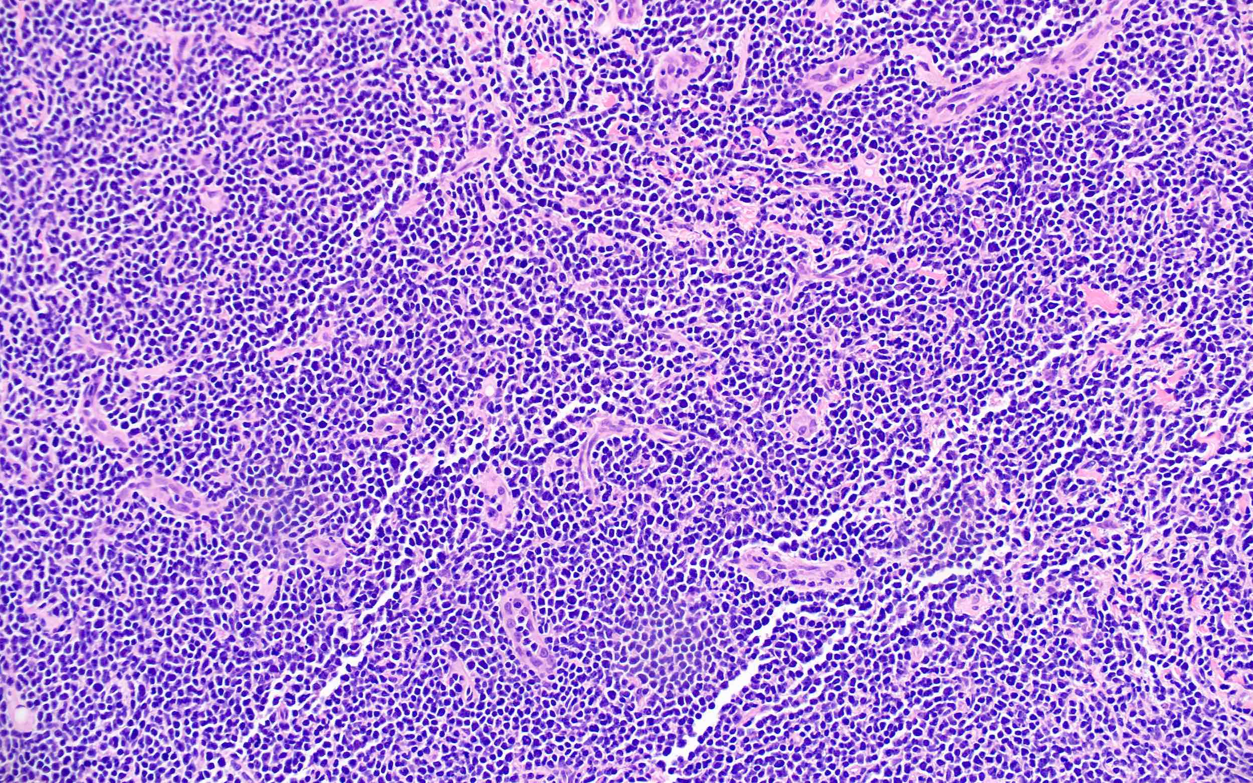
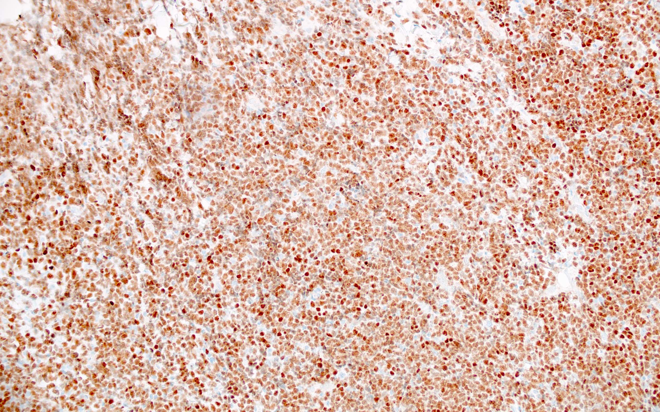
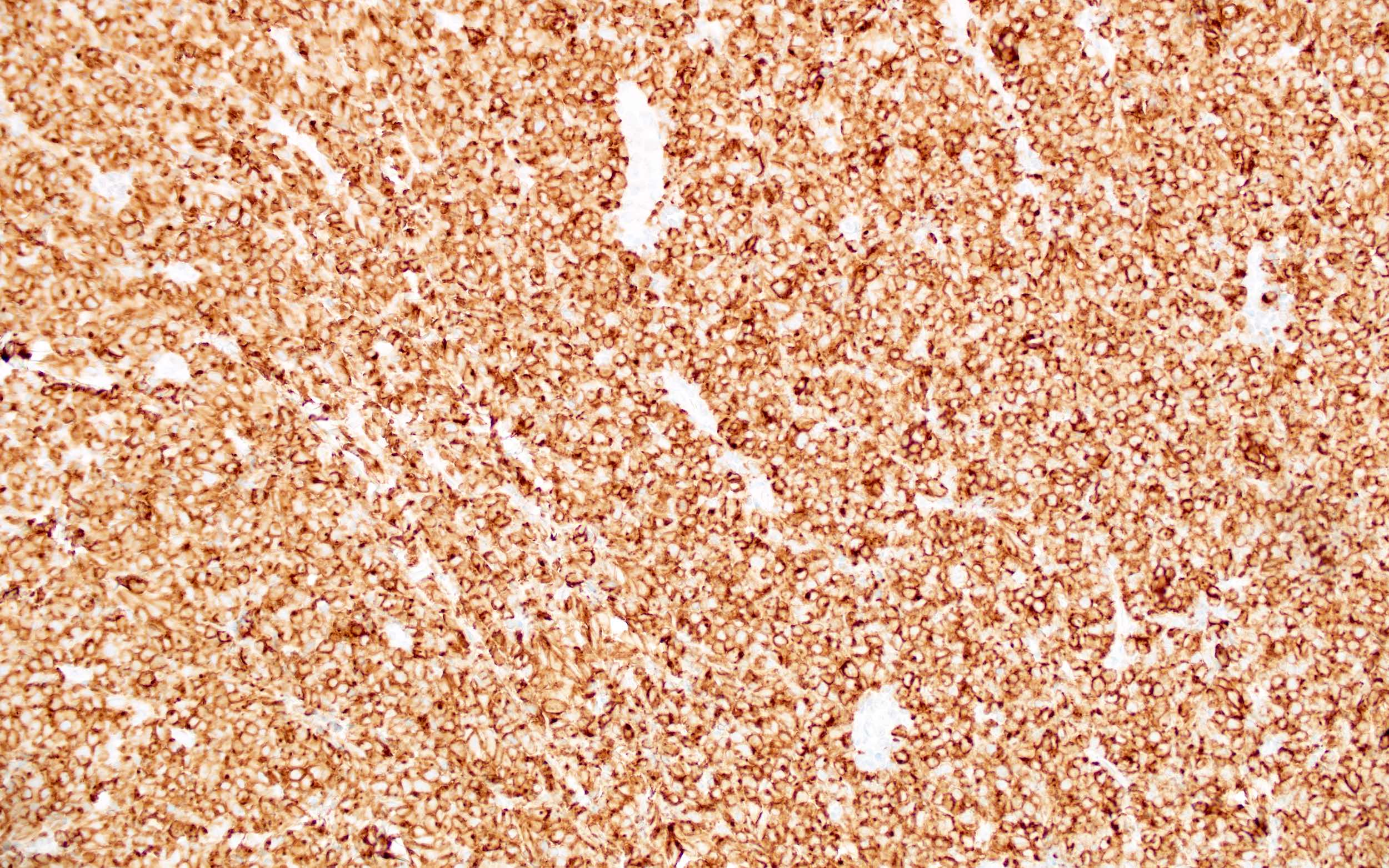
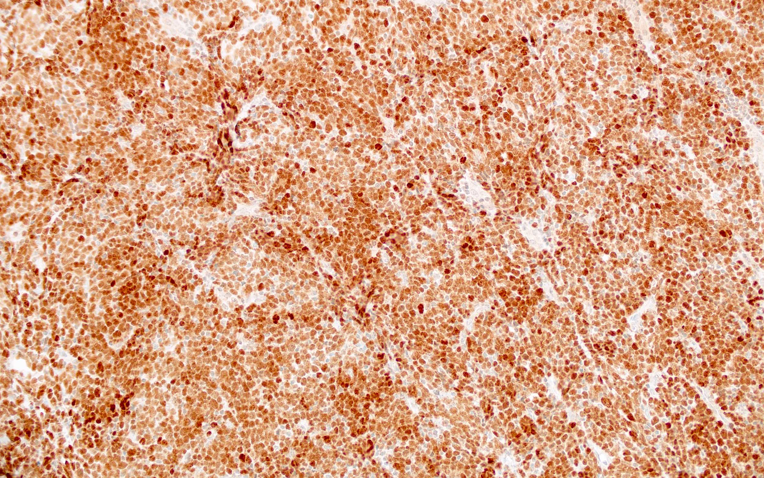
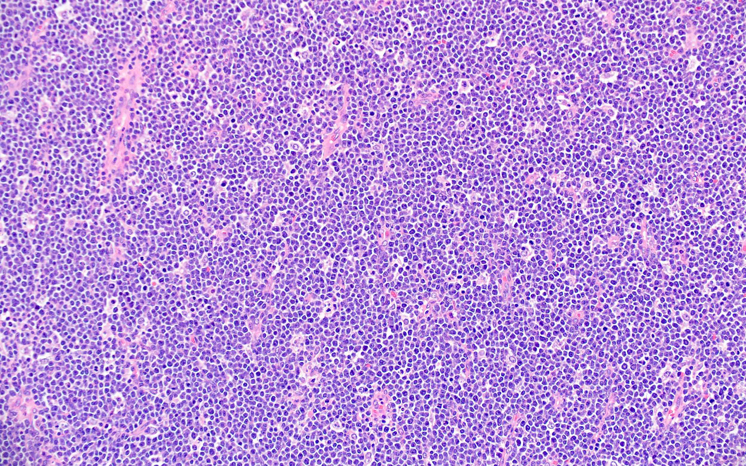
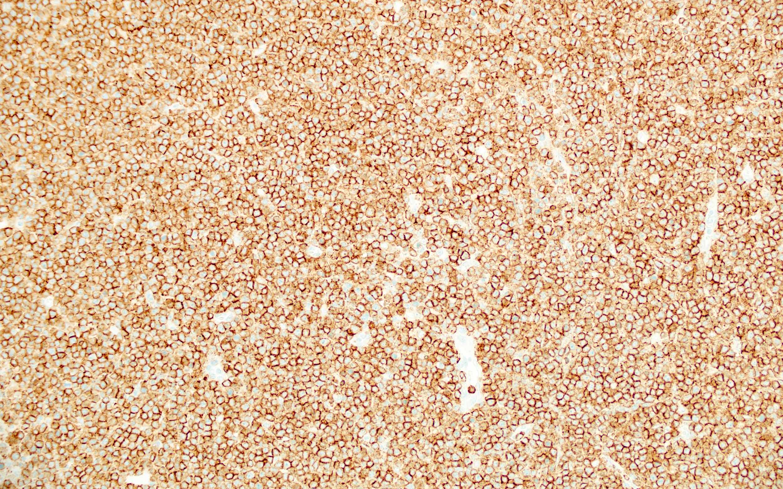
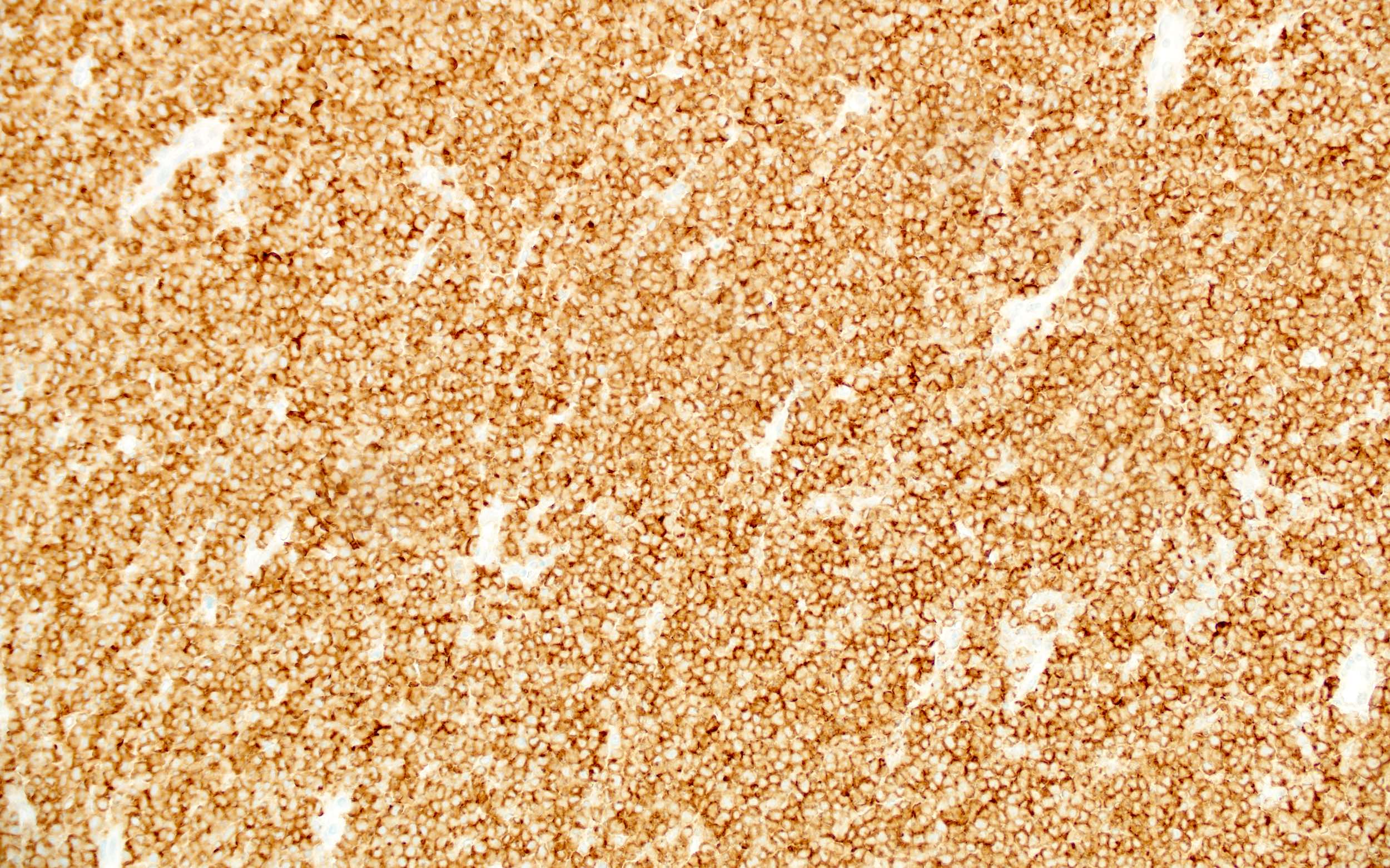
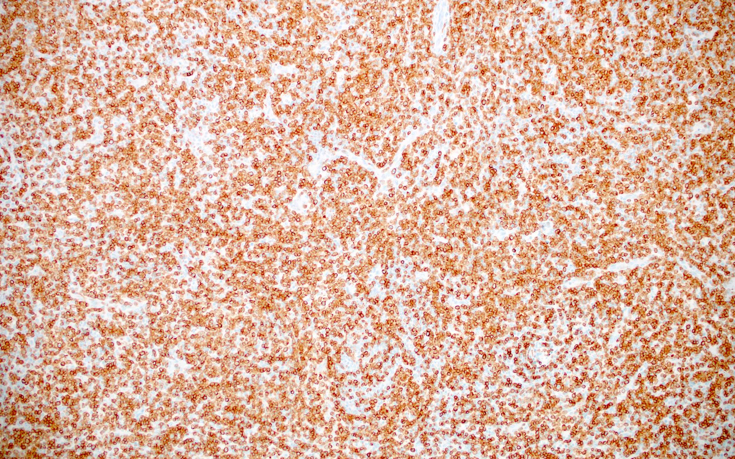
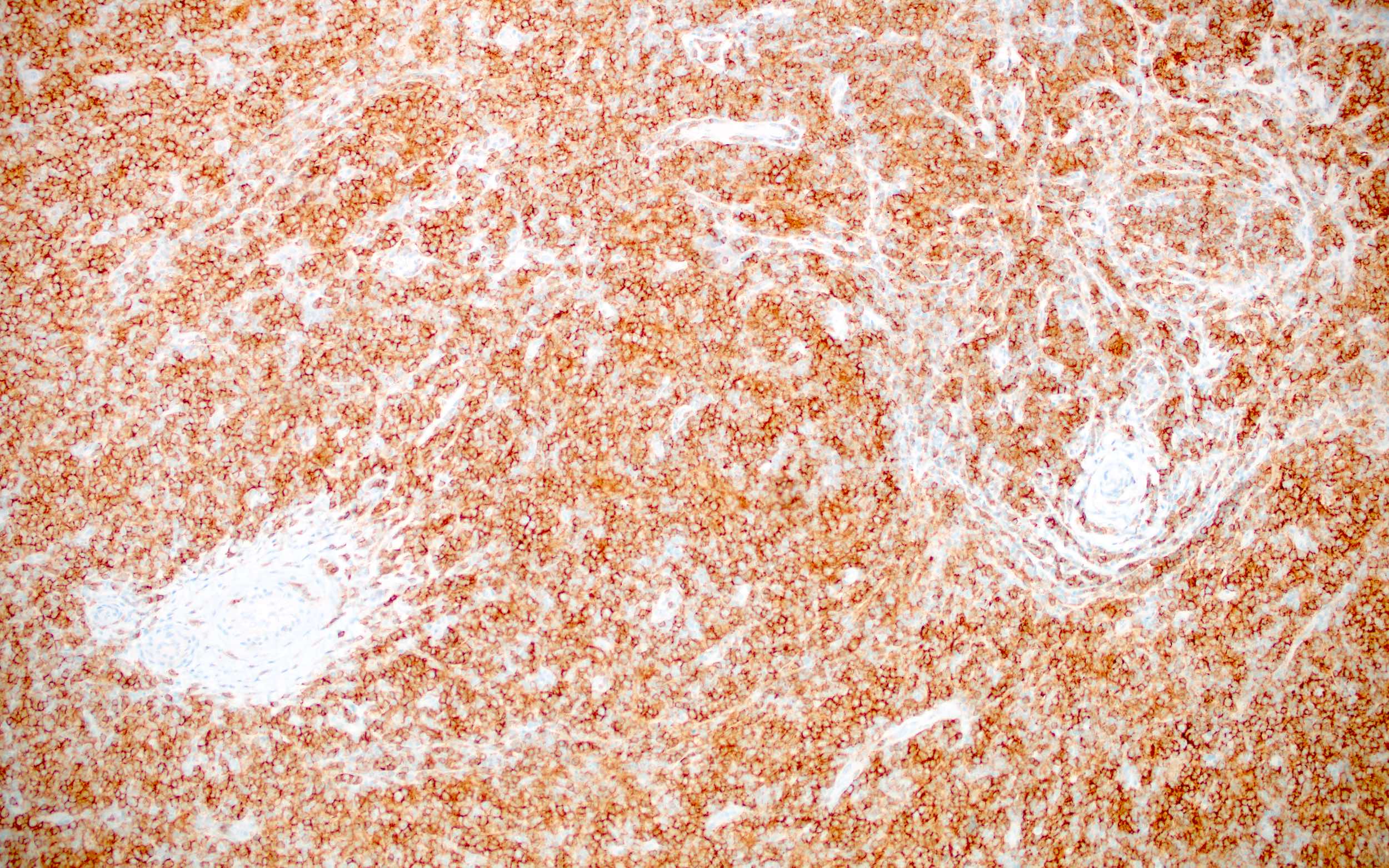
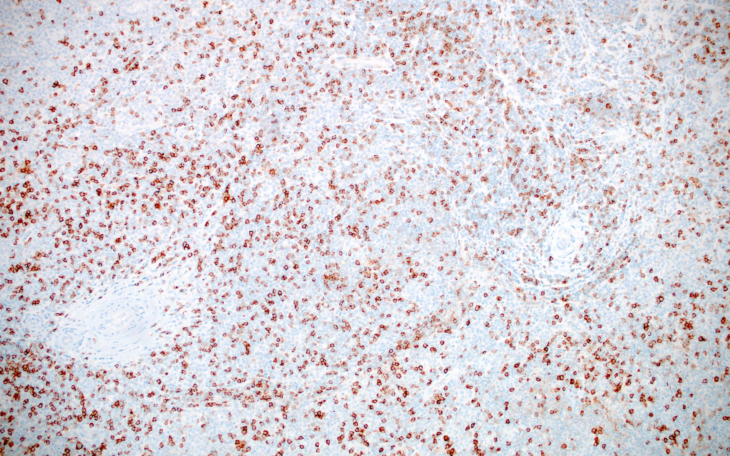
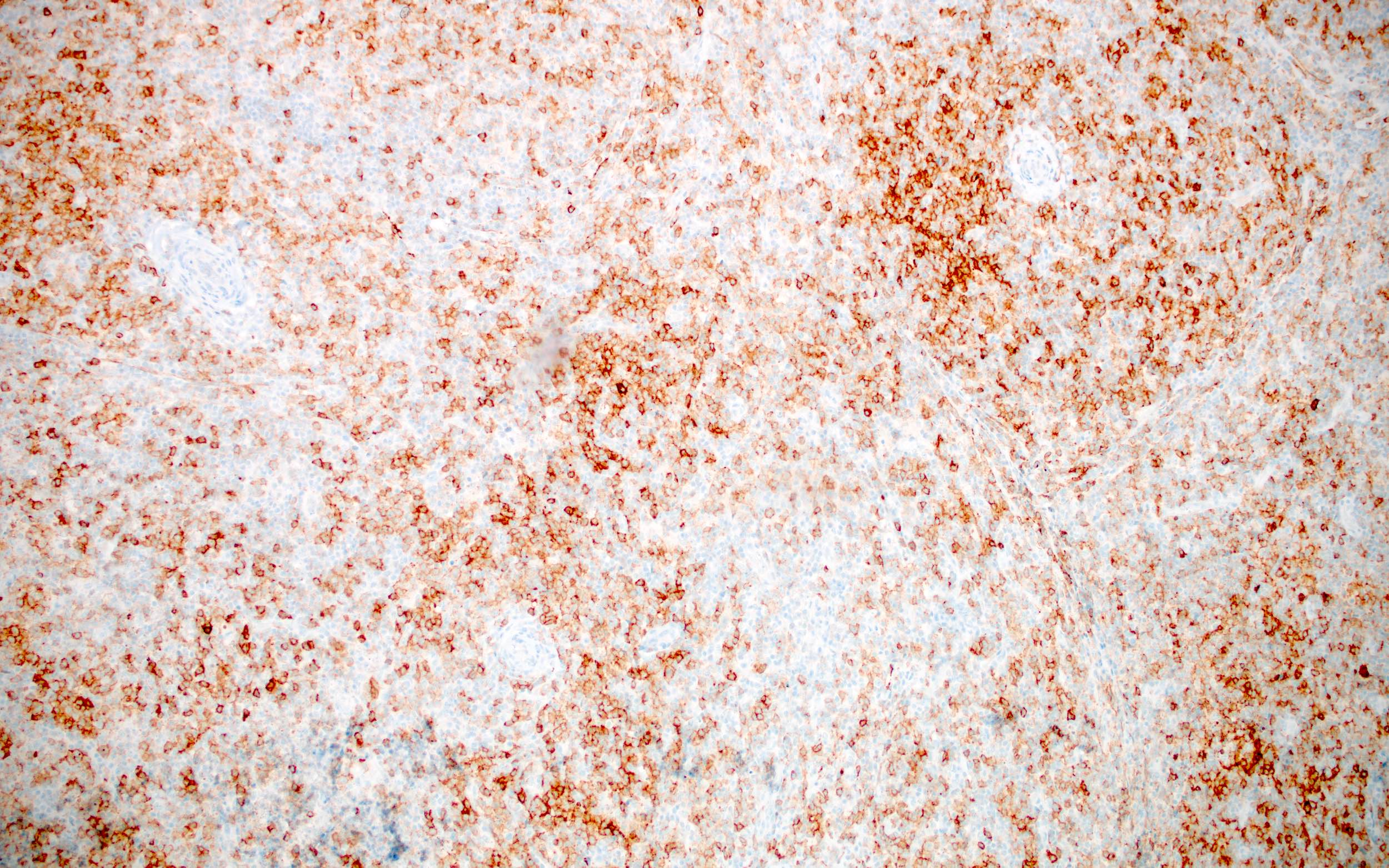
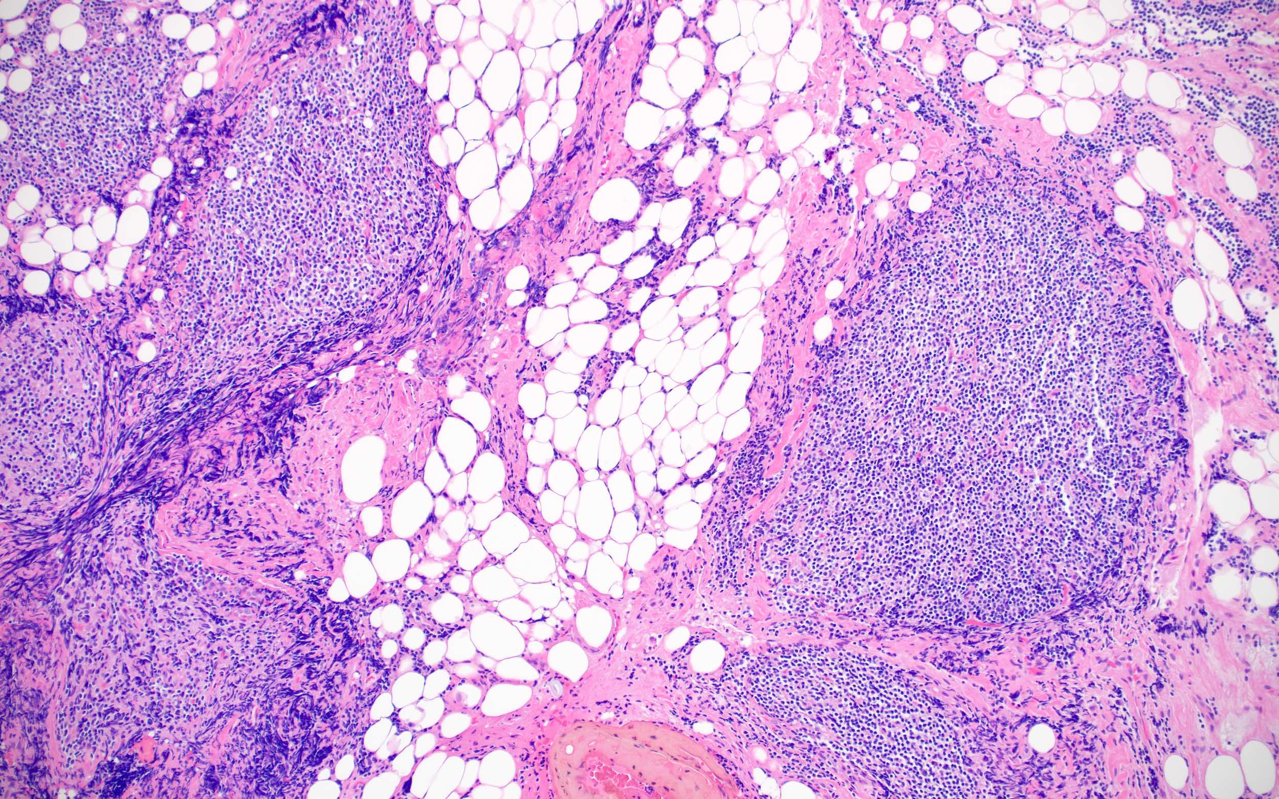
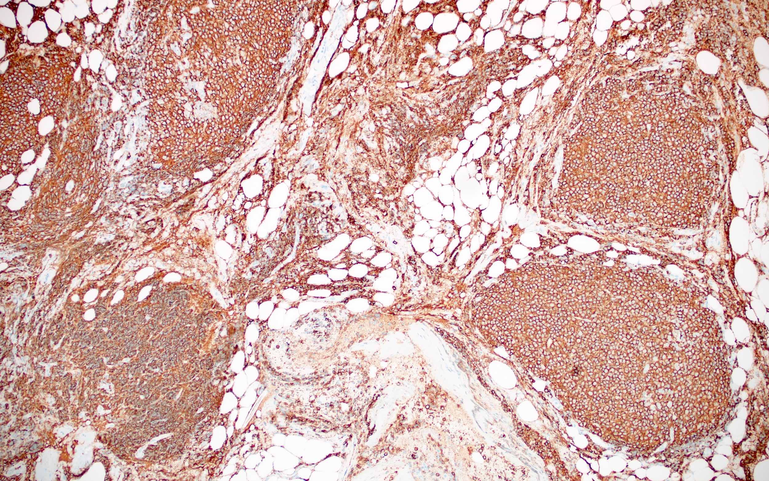
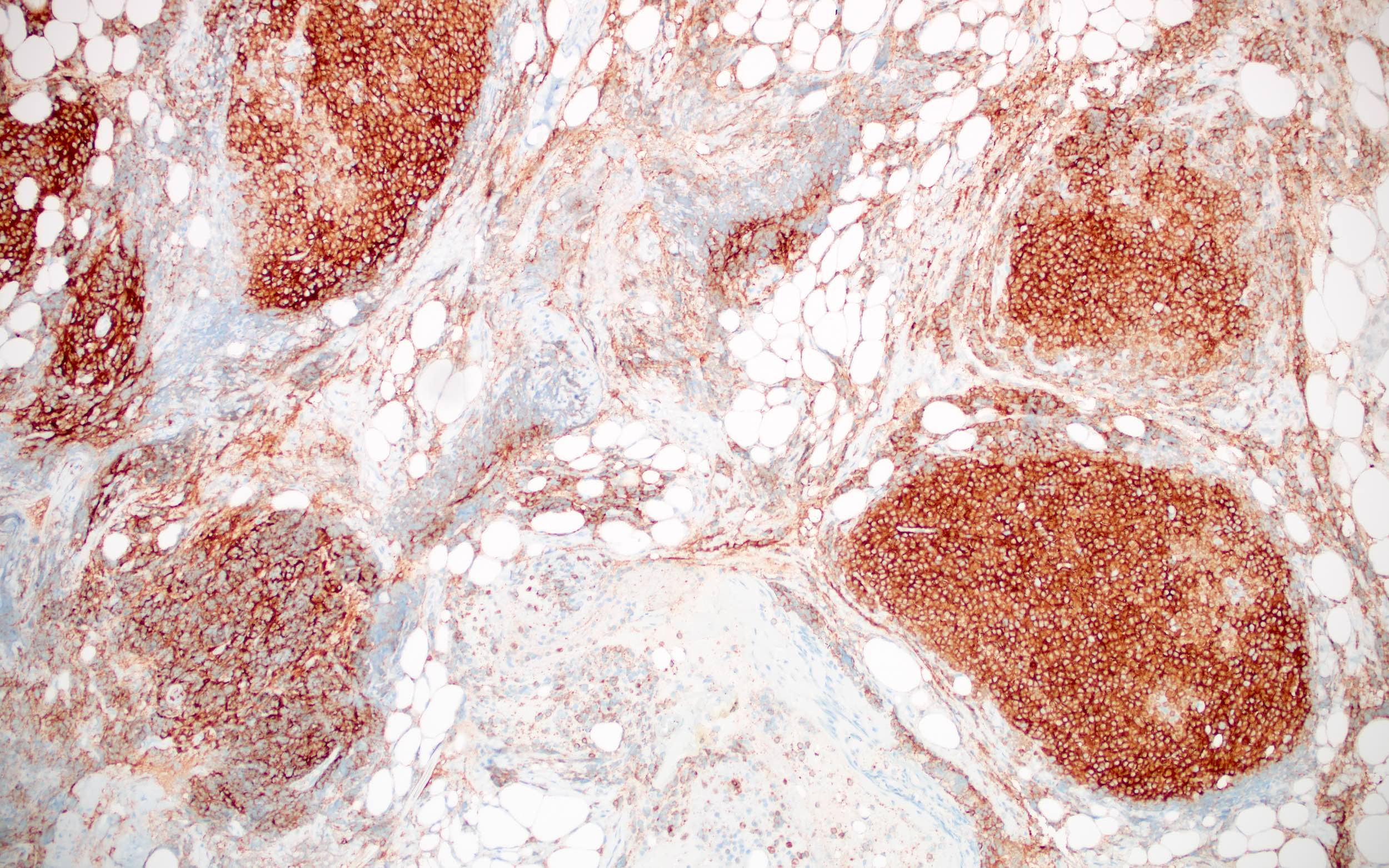
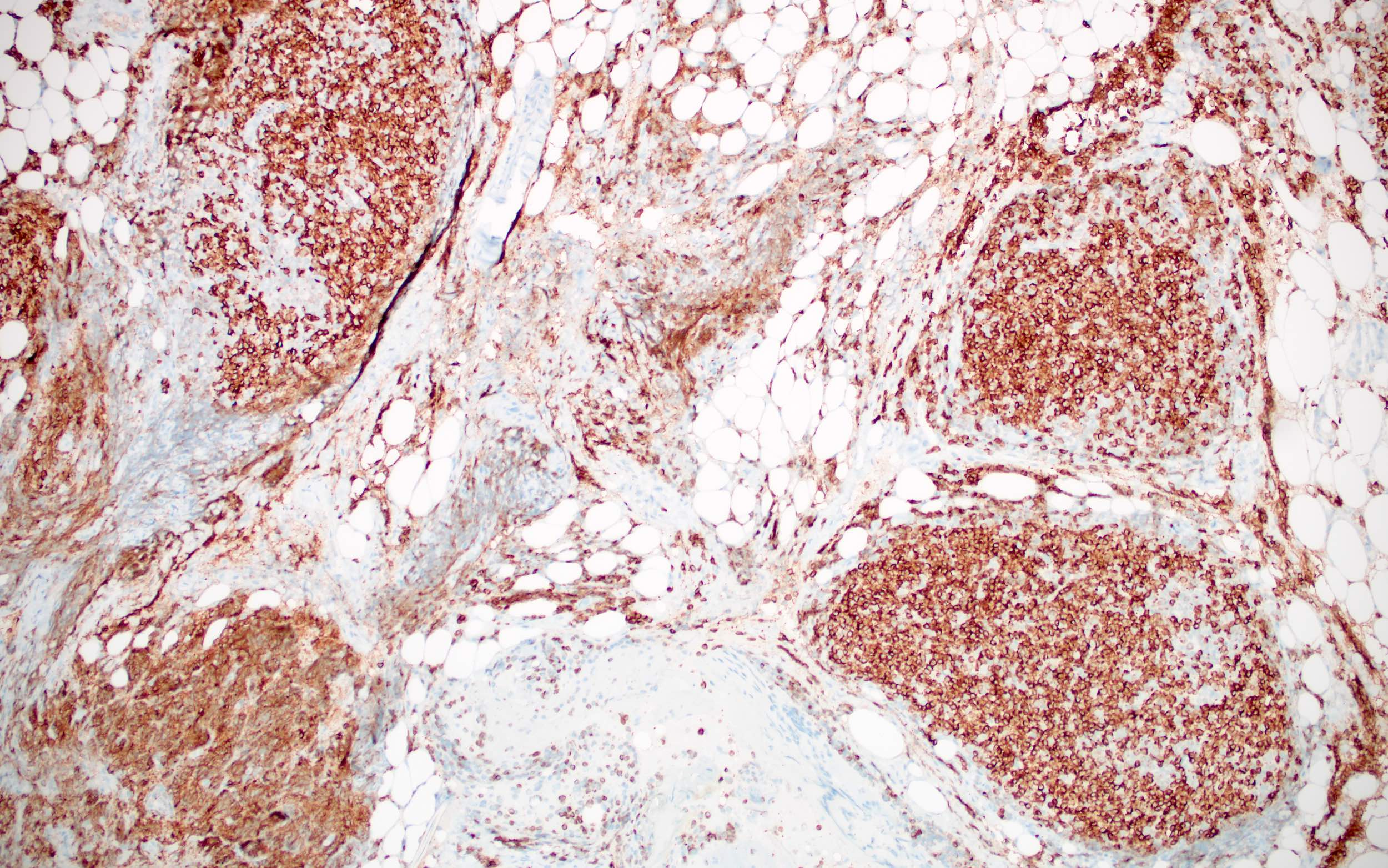
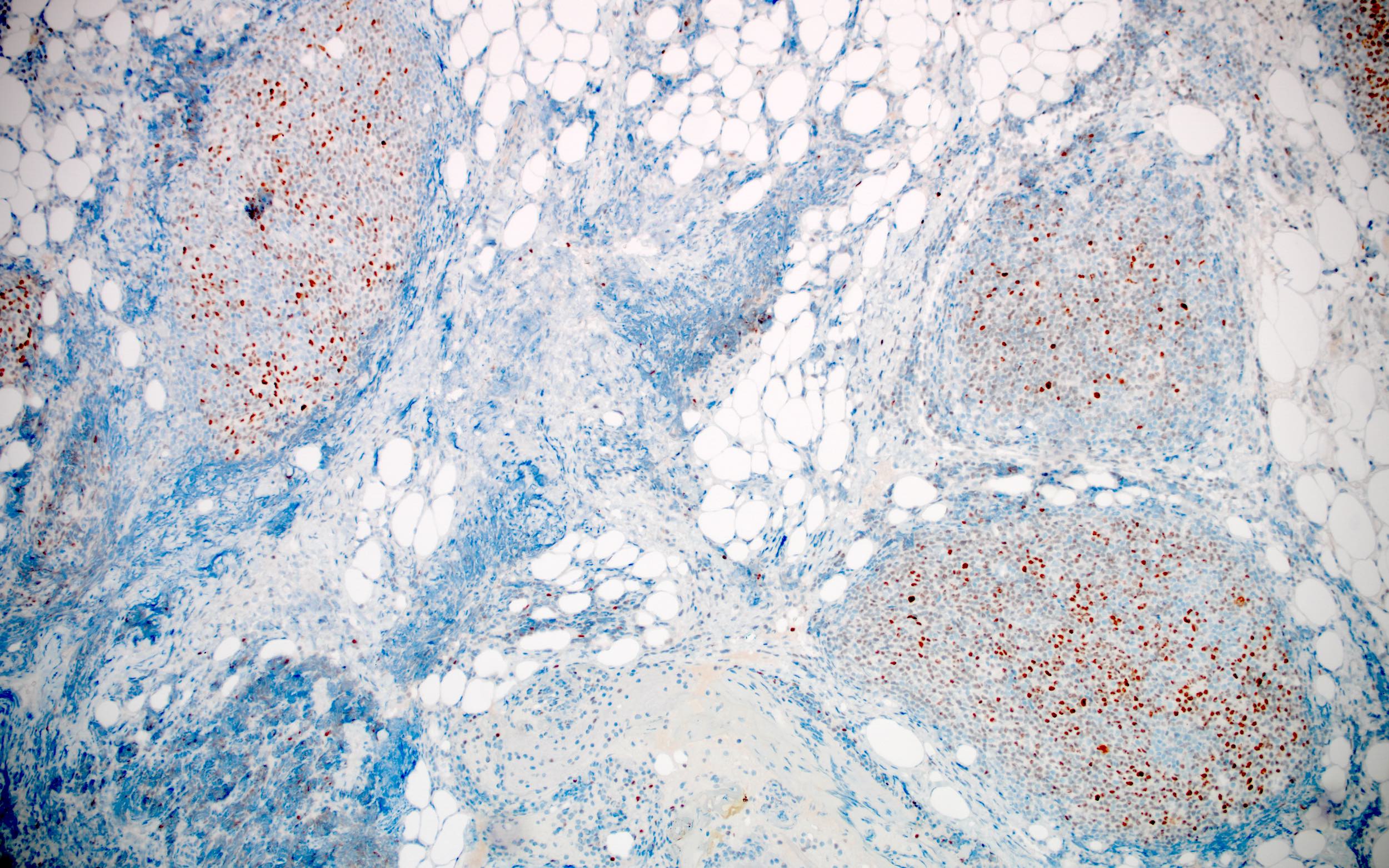
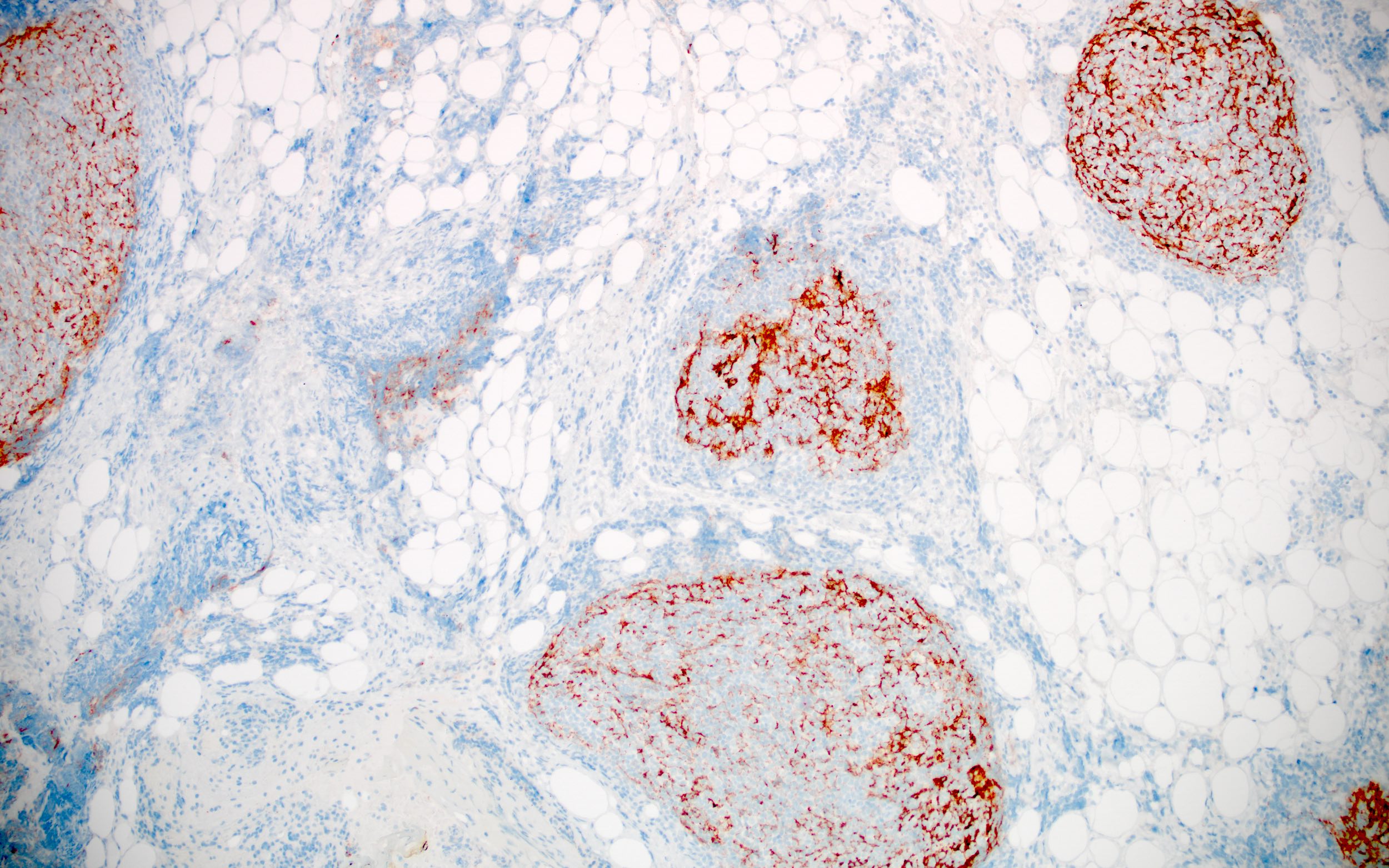
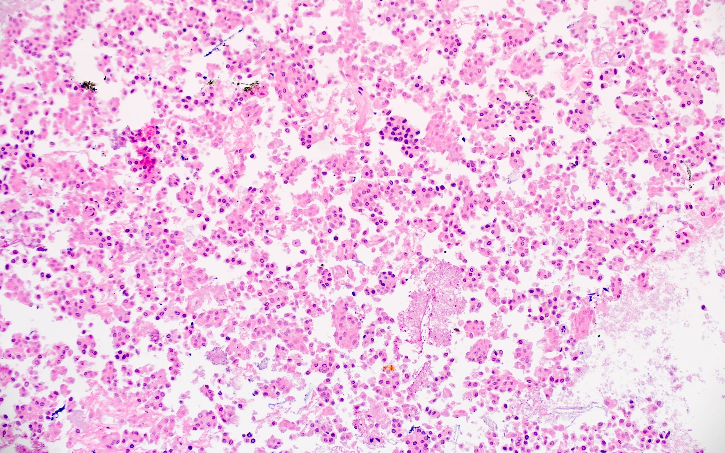
A 72 year old man presented for evaluation of a distal gastrointestinal tract adenocarcinoma, with staging computed tomography (CT) radiography that incidentally revealed bulky confluent mesenteric lymphadenopathy. Excision of mesenteric lymph nodes show effacement of nodal architecture as well as infiltration of mesenteric fat by a nodular infiltrate of small lymphoid cells with cleaved nuclei and condensed chromatin. The tumor cells are positive for CD45, CD20, CD10, BCL6 and BCL2. The images above show H&E, CD20, CD10 and BCL2 staining. The Ki67 proliferation index is ~5%. The tumor cells are negative for CD5 and CD23. What molecular / cytogenetic abnormality may be associated with this lymphoma?
- Deletion of 13q14.3
- MYD88 L265P mutation
- t(11;14)(q13;q32)
- t(11;18)(q21;q21)
- t(14;18)(q32;q21)
Board review style answer #1
E. t(14;18)(q32;q21). The morphologic appearance, including nodular infiltrates of small lymphoid cells with cleaved nuclei and condensed chromatin, in conjunction with the immunophenotype showing lymphoid cell positivity for CD20, CD10, BCL6 and BCL2, with a low Ki67 proliferation index, is compatible with a diagnosis of a low grade (grade 1 - 2) follicular lymphoma. The t(11;14)(q13;q32) translocation between the IGH and BCL2 genes is present in approximately 90% of grade 1 - 2 follicular lymphomas. Other small B cell lymphomas demonstrate different immunophenotypes and characteristic cytogenetic or molecular abnormalities, including chronic lymphocytic leukemia / small lymphocytic lymphoma (CD20+, CD10-, CD5+, CD23+; deletion of 13q14.3), lymphoplasmacytic lymphoma (CD20+, CD10-, CD5-; MYD88 L265P mutation) and extranodal marginal zone lymphoma of mucosa associated lymphoid tissue (MALT lymphoma) (CD20+, CD10-, CD5-, t(11;18)(q21;q21) [pulmonary and gastric] and t(14;18)(q32;q21) [thyroid, ocular adnexa, orbit and skin]), aiding in diagnostic differentiation.
Comment Here
Reference: CD10
Comment Here
Reference: CD10


