Table of Contents
Definition / general | Essential features | Terminology | ICD coding | Epidemiology | Sites | Etiology | Clinical features | Diagnosis | Radiology description | Radiology images | Prognostic factors | Case reports | Treatment | Clinical images | Gross description | Gross images | Frozen section description | Microscopic (histologic) description | Microscopic (histologic) images | Virtual slides | Cytology description | Positive stains | Negative stains | Molecular / cytogenetics description | Videos | Sample pathology report | Differential diagnosis | Board review style question #1 | Board review style answer #1 | Board review style question #2 | Board review style answer #2 | Board review style question #3 | Board review style answer #3Cite this page: Sabljic T, Mrkonjic M. Nipple adenoma. PathologyOutlines.com website. https://www.pathologyoutlines.com/topic/breastnipplead.html. Accessed March 30th, 2025.
Definition / general
- Nipple adenoma is a benign epithelial proliferation with a variety of morphological patterns that arise near the collecting ducts of the nipple and may be continuous with the overlying epidermis
Essential features
- Benign, often florid, epithelial proliferation with retained myoepithelial cell layer involving superficial ducts of the nipple; the proliferation may extend and replace the overlying squamous epithelium resulting in erosion, hemorrhage and inflammation
- Histologic patterns include a mix of sclerosing adenosis, papillary hyperplasia and usual ductal hyperplasia
Terminology
- Synonym: nipple duct adenoma
- Not recommended: florid papillomatosis of the nipple, erosive adenomatosis of the nipple, papillomatosis of the nipple, superficial papillary adenomatosis, papillary adenoma of the nipple
ICD coding
Epidemiology
- Rare; occurs in males and females
- Most common in women in the fifth decade of life but has a wide age range (5 months to 89 years)
Sites
- Breast; superficial duct orifices of the nipple, the associated stroma and often the adjacent overlying epidermis
Etiology
- Unknown
Clinical features
- Detected when small due to skin changes
- Clinically, epidermal involvement mimics the appearance of Paget disease
- On physical examination, small, palpable dermal nodule
- Nipple may appear eroded and erythematous as glandular epithelium replaces squamous epithelium
- Bleeding and exudative crust may be interpreted as nipple discharge
- If left untreated for years, may develop into large pedunculated mass (World J Surg Oncol 2014;12:91)
Diagnosis
- Diagnosis can be made on biopsy (often of the skin due to clinical presentation) or cytology specimen and confirmed on excision
- Imaging findings are generally nonspecific (see Radiology description)
Radiology description
- Mammography: undetected in 33% of cases due to superficial location and small size (World J Surg Oncol 2014;12:91)
- Ultrasound: nonspecific; may detect a circumscribed mass for larger lesions, with or without cystic components, hypervascular on Doppler (Chin Med J (Engl) 2016;129:2386)
- MRI: nipple may show enhancement (J Comput Assist Tomogr 2006;30:148)
Radiology images
Prognostic factors
- Can recur if not completely excised
- Risk of developing a subsequent breast cancer is comparable to other proliferative lesions of the breast (Am J Surg Pathol 1986;10:87)
Case reports
- 22 year old woman with a papillomatous nodule on the left nipple from childhood (Clin Case Rep 2020;8:3254)
- 46 year old woman with pedunculated papillomatous mass on top of the nipple of the right breast (BMJ Case Rep 2019;12:e231516)
- 49 year old woman with a reddish nodule with surface crust on the nipple of the right breast (Dermatol Online J 2021;27)
- 82 year old woman with small painful mass in the right axilla (Diagn Pathol 2012;7:162)
Treatment
- Surgical excision depending on the size and extent of lesion (ANZ J Surg 2015;85:444)
- Alternative treatments:
- Mohs micrographic surgery (Dermatol Surg 2016;42:684)
- Cryotherapy (Breast J 2016;22:584, Ann Dermatol 2021;33:182)
Clinical images
Gross description
- Rubbery; white to gray; 0.5 cm to > 4 cm
Frozen section description
- Glandular nests of various sizes with diffuse growth within ducts lined by myoepithelial cells; complex and irregular structure may falsely mimic invasion (Chin Med J (Engl) 2007;120:630)
Microscopic (histologic) description
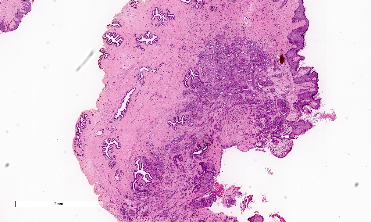
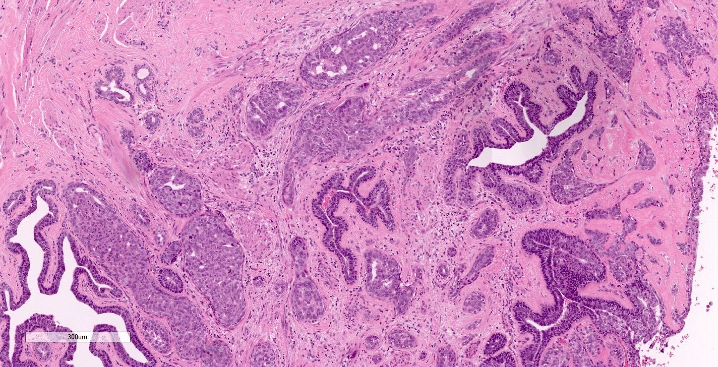
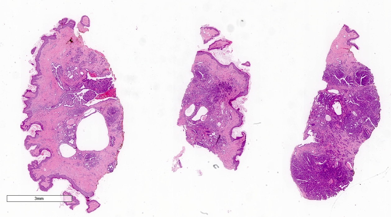
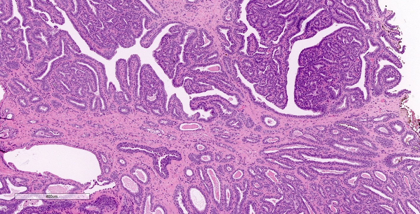
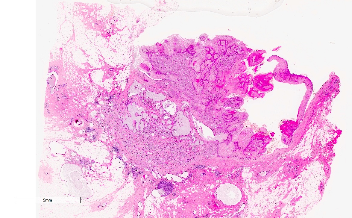
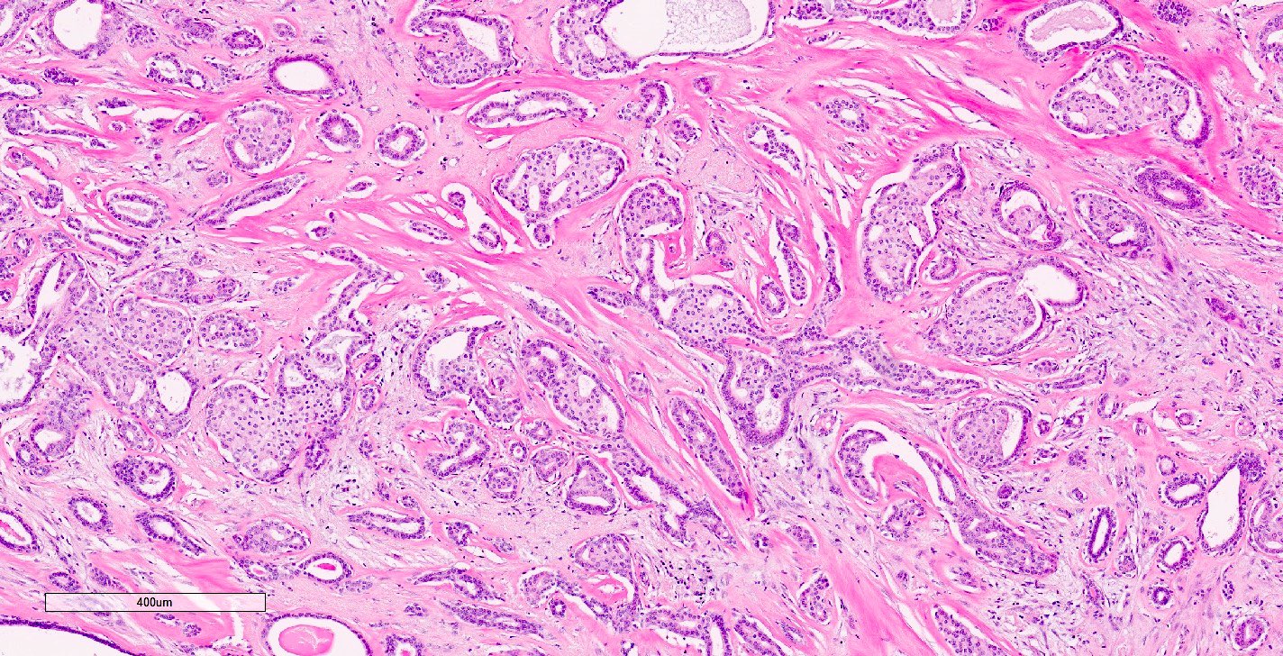
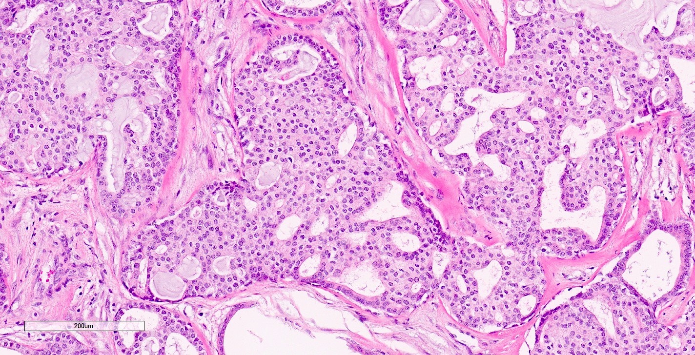
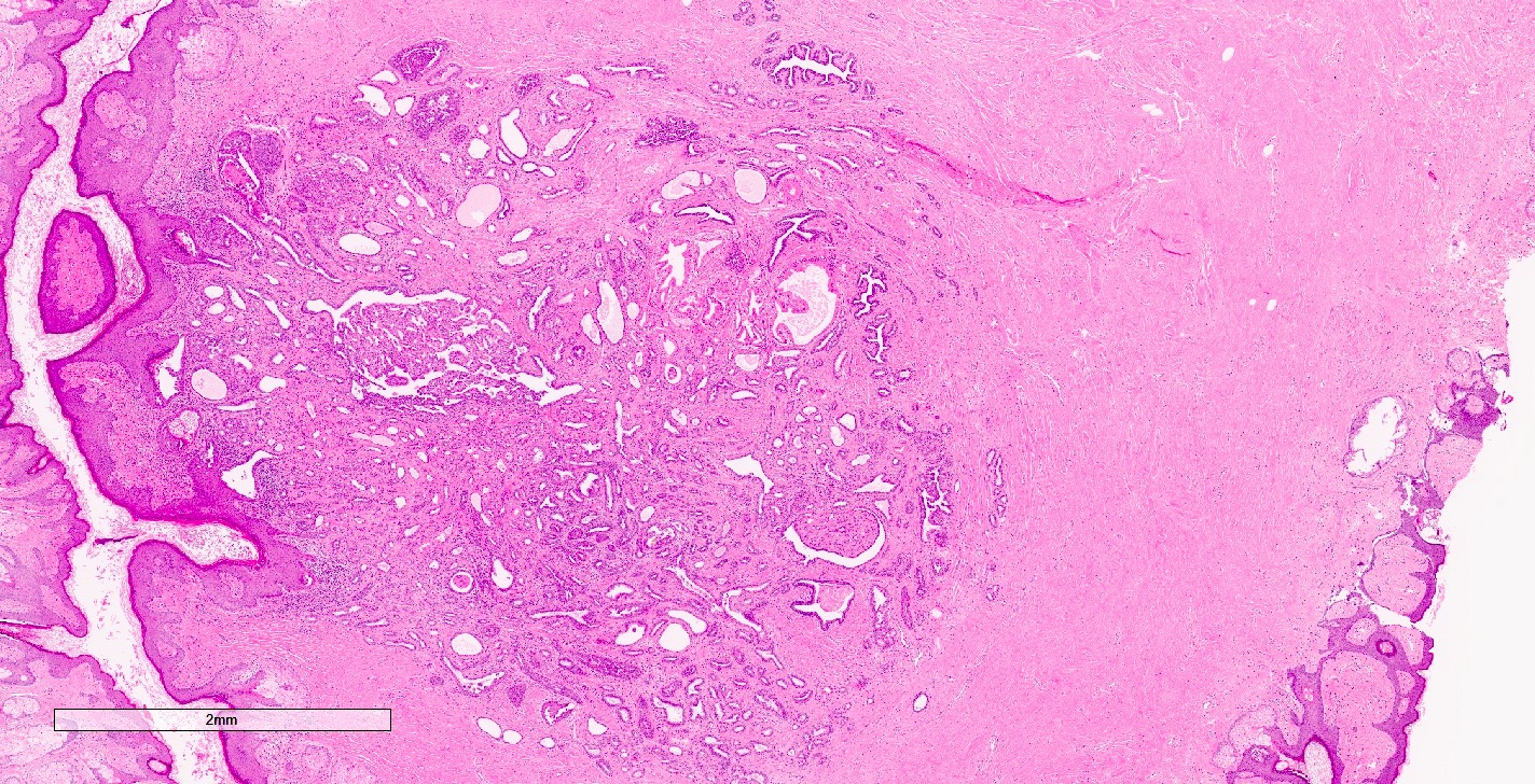
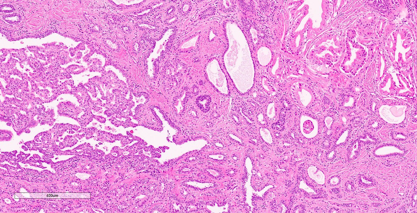
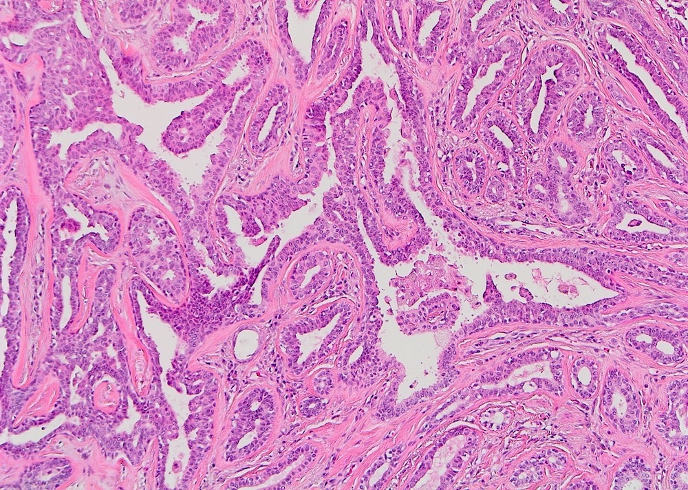
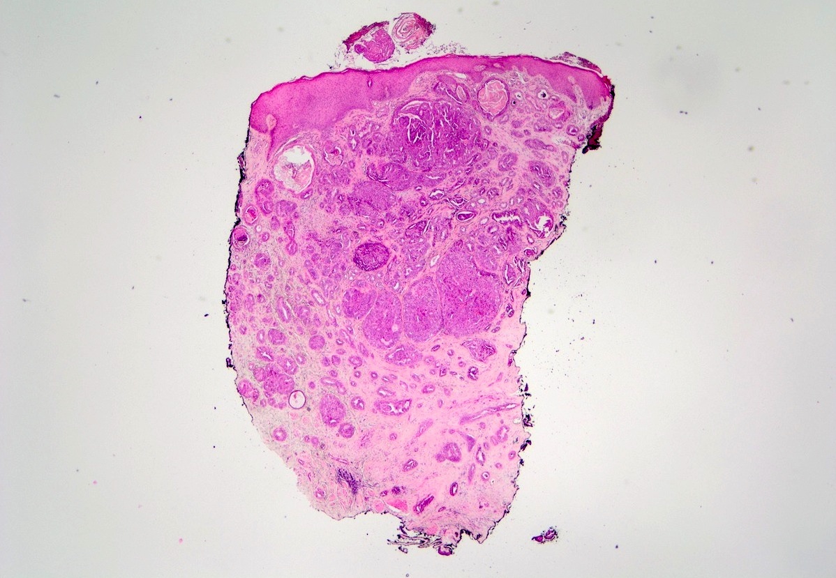
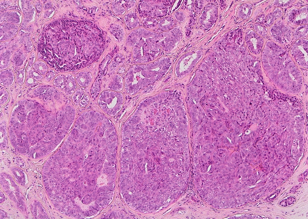
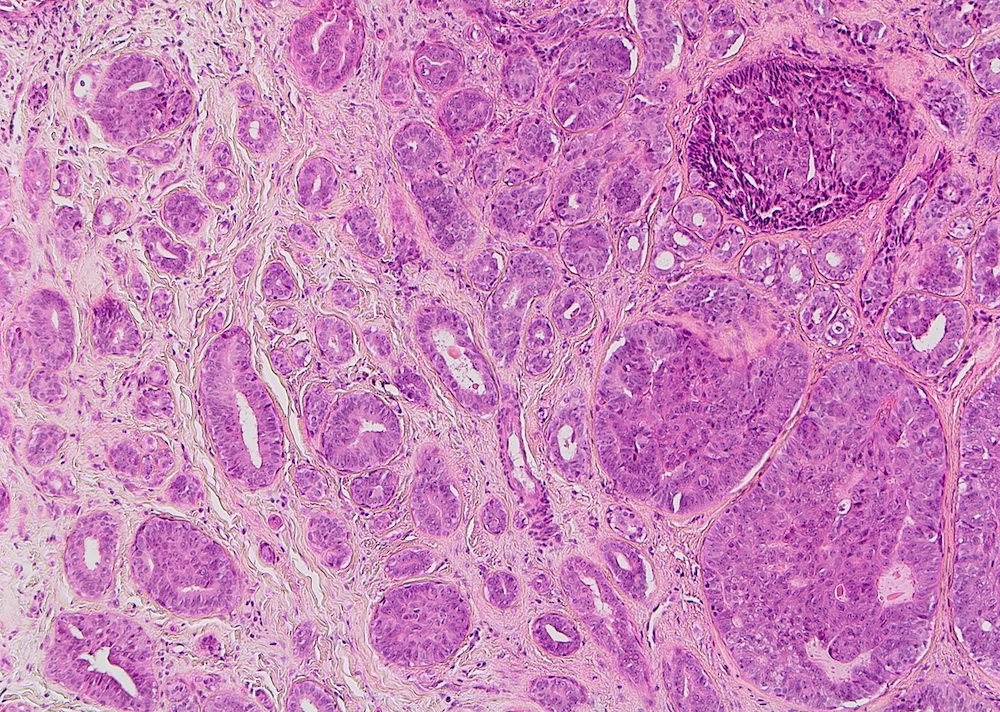
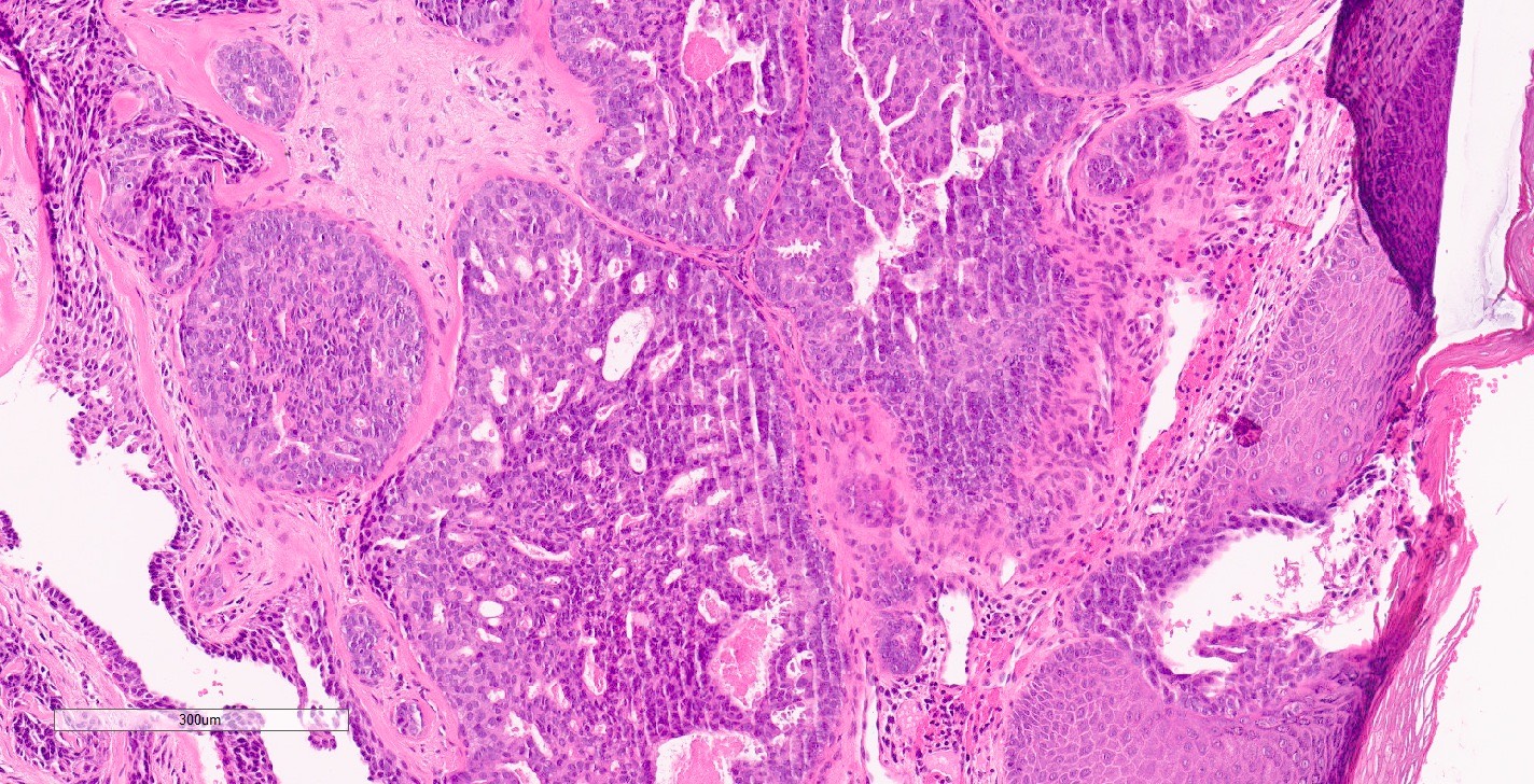
- Nodular florid epithelial proliferation of the lactiferous ducts of the nipple with papillary hyperplasia / adenosis / sclerosis / mixed pattern
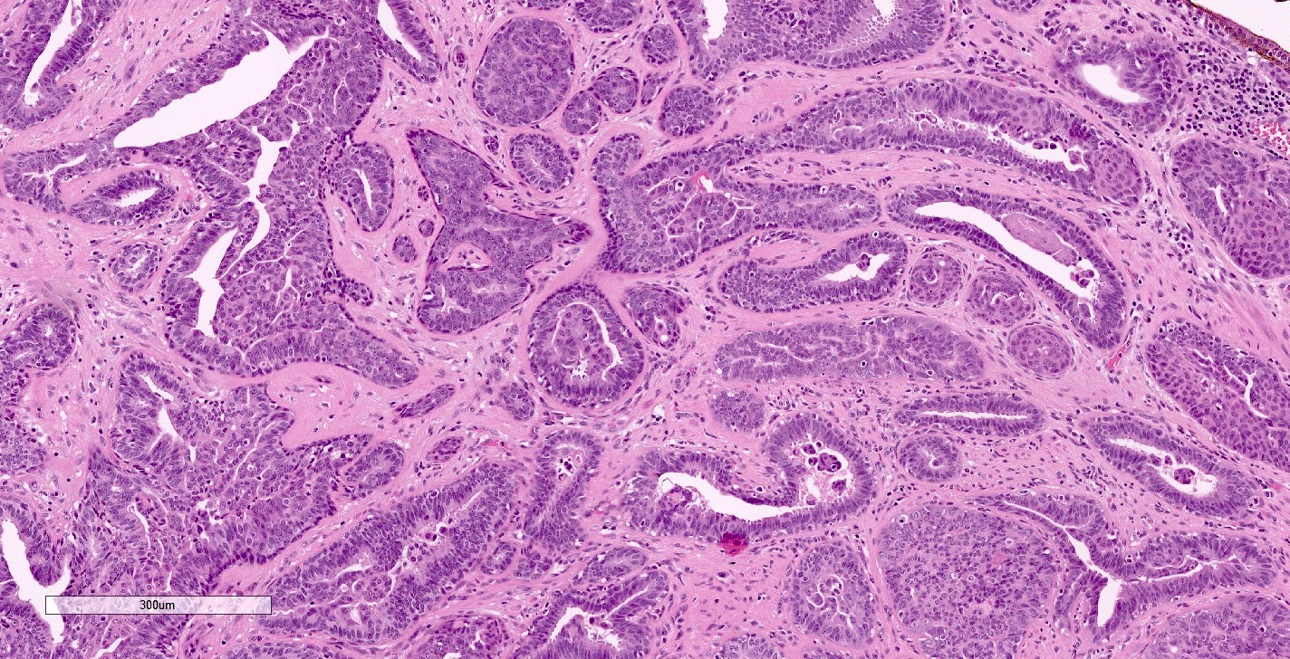
- Epithelium is cytologically bland
- Myoepithelial cell layer is preserved uniformly (Mod Pathol 1990;3:288)
- 4 major morphologic patterns:
- Sclerosing papillary hyperplasia pattern:
- Intraductal papillary growth with prominent stromal proliferation (collagenous bands, myxoid change or elastosis)
- Focal central necrosis may be present
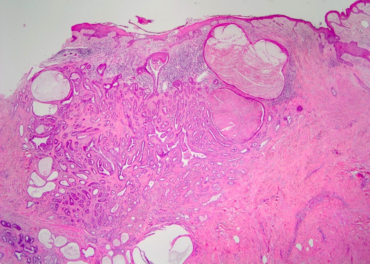
- Squamous cysts can be present in duct orifices
- Papillary hyperplasia pattern:
- Papillary growth in large ducts with less prominent stromal proliferation and focal necrosis
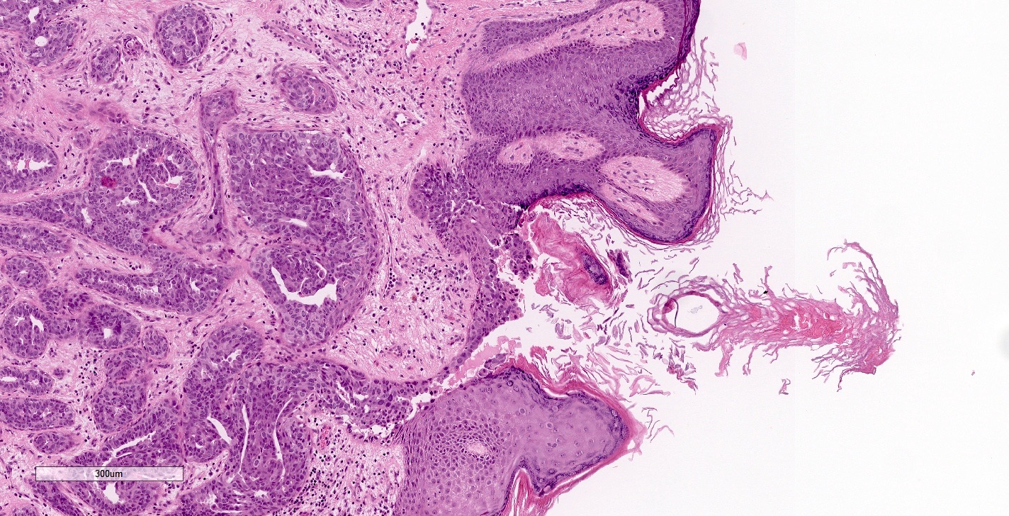
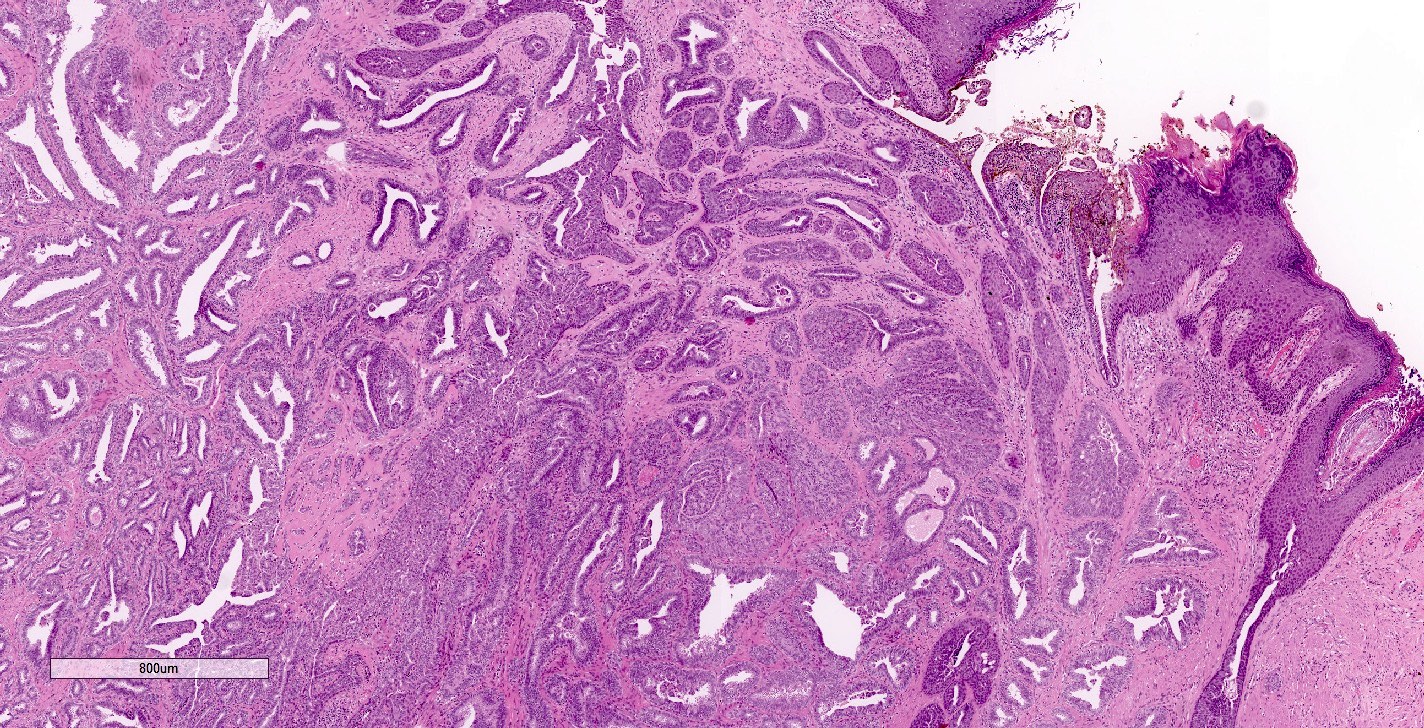
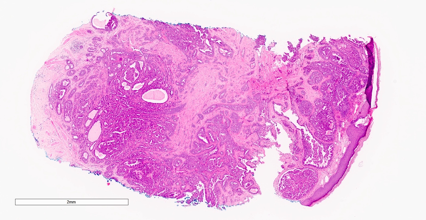
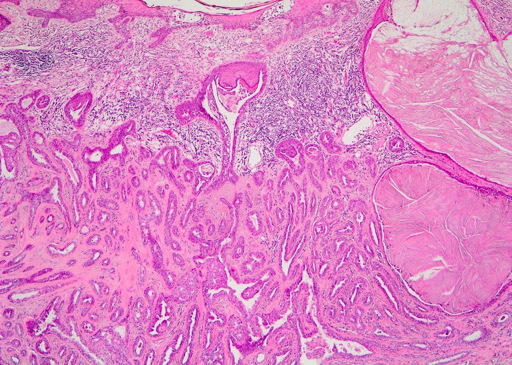
- Replacement of epidermis with glandular epithelium
- Adenosis pattern:
- Small duct proliferation with a pattern resembling sclerosing adenosis and may be pseudoinfiltrative
- Necrosis is uncommon
- Mixed pattern:
- Any combination of the above described patterns (Am J Surg Pathol 1986;10:87)
- Sclerosing papillary hyperplasia pattern:
- May be in continuity with the skin surface, which may appear eroded or ulcerated
- Fairly well circumscribed but unencapsulated (Br Med J 1963;1:563)
- Other features often seen:
- Squamous metaplasia and superficial keratin cysts
- Acanthosis
- Toker cell hyperplasia in the epidermis
- Multinucleated giant cells
- Apocrine metaplasia (Oncol Lett 2014;7:1839)
- In rare cases, ductal carcinoma in situ involving an adenoma or a contiguous invasive carcinoma have been reported (Mod Pathol 1995;8:633)
Microscopic (histologic) images
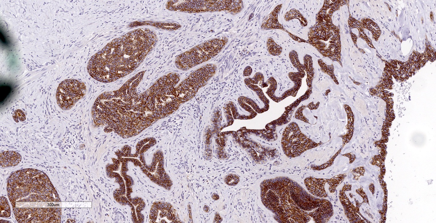
Contributed by Hal Berman, M.D., Ph.D., Miralem Mrkonjic, M.D., Ph.D. and Mary Ann Gimenez Sanders, M.D., Ph.D.
Virtual slides
Cytology description
- Obtained by fine needle aspiration (FNA), brush cytology or tumor imprint cytology
- Reported features vary; benign when myoepithelial cells are detected
- FNA:
- Large cell clusters with a papillary or sheet structure, as well as small clusters and solitary epithelial cells in a necrotic background
- Duct epithelial and myoepithelial 2 cell pattern in the clusters
- Round to oval or spindle nuclei, fine and bland chromatin
- Brush cytology (Diagn Cytopathol 2015;43:664):
- Small papillary clusters with attached myoepithelial cells and solitary epithelial cells in a hemorrhagic or neutrophilic background
- Round to oval nuclei, with smooth contour, bland and granular chromatin
- Squamous metaplasia-like cells present
- Cytology often classified as atypical or suspicious, especially in the presence of coagulative necrosis (ANZ J Surg 2015;85:444)
Positive stains
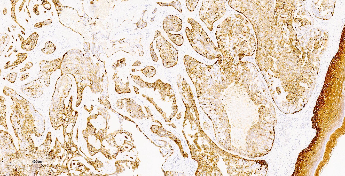
- AE1 / AE3, CK7: positive in epithelium (Mod Pathol 2003;16:893, Oncol Lett 2014;7:1839)
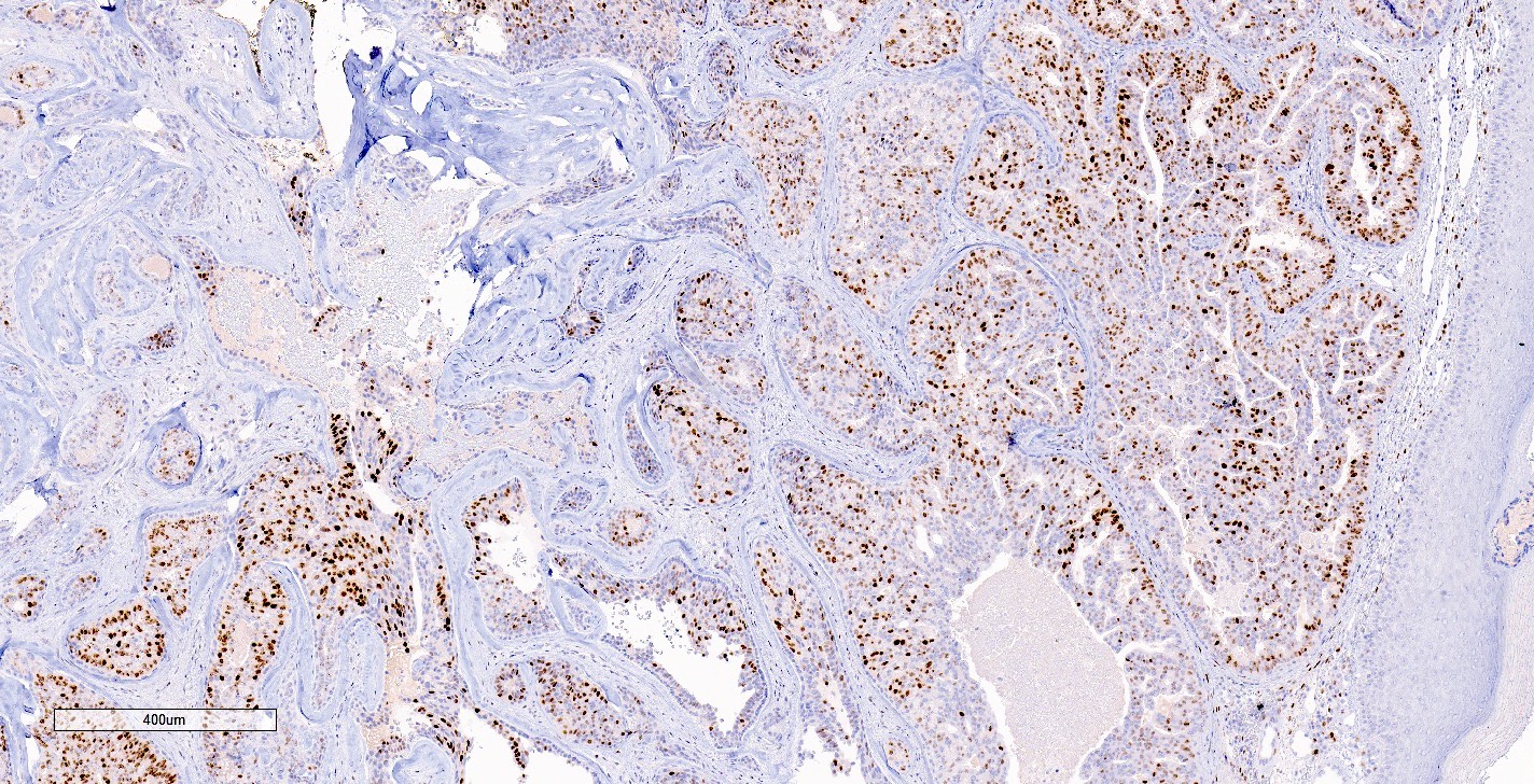
- p63, smooth muscle myosin, calponin: positive in myoepithelium (Oncol Lett 2014;7:1839, Am J Surg 2010;200:e39)
- ER and CK5/6: heterogeneous, mosaic type staining of the epithelium with florid usual ductal hyperplasia (Am J Surg Pathol 2010;34:896, Oncol Lett 2014;7:1839)
Negative stains
Molecular / cytogenetics description
- Activating PIK3CA mutations reported in 12/24 cases
- KRAS (1/24 cases) and BRAF mutations (2/24 cases) (Histopathology 2017;70:195)
Videos
Dr. Jerad Gardner presents normal nipple histology and 2 cases of nipple adenoma
3 minute overview of the presentation and histology of nipple adenomas
Sample pathology report
- Right breast (nipple), punch biopsy:
- Nipple adenoma (see comment)
- Comment: Sections show ductal proliferation involving a large nipple duct with areas of papillary growth and usual ductal hyperplasia in continuity with the epidermis. Ductal epithelial cells are cuboidal to columnar with eosinophilic cytoplasm and bland nuclei. Focal areas of stromal sclerosis are identified. Immunohistochemistry for smooth muscle myosin and p63 highlights the myoepithelial cell layer while ER and CK5/6 show heterogeneous staining patterns of epithelial cells. Taken together, the histomorphology and immunohistochemistry are consistent with a nipple adenoma.
Differential diagnosis
- Syringomatous tumor:
- Ducts with teardrop, comma and branching shapes
- Infiltrative growth with desmoplastic stroma, may see perineural invasion
- Does not involve the epidermis
- Invasive ductal carcinoma, no special type (low grade):
- Negative for myoepithelial cell markers (p63, smooth muscle myosin, calponin)
- Infiltrative growth pattern
- DCIS:
- Intraductal papilloma:
- Similar to nipple adenoma
- Papillary projections with fibrovascular cores
- Arises below the skin surface, involves lactiferous ducts within deeper breast parenchyma and is unlikely to be a palpable subdermal mass
- Tubular adenoma:
- Well circumscribed
- Uniform glands with scant stroma
- Encapsulated papillary carcinoma:
- Circumscribed mass found deeper in the breast compared with nipple adenomas
- Cytologic atypia
- Myoepithelial cells absent in papillae and along the periphery
Board review style question #1
Board review style answer #1
C. Nipple adenoma. Nipple adenoma is a benign epithelial proliferation in the breast, often in continuity with the epidermis of the nipple. It forms a nodular mass that can have a number of different morphologic patterns including sclerosing adenosis, papillary hyperplasia and epithelial hyperplasia. Myoepithelial cells are retained in nipple adenoma. Focal necrosis can be seen and squamous cysts can be seen superficially.
Comment Here
Reference: Nipple adenoma
Comment Here
Reference: Nipple adenoma
Board review style question #2
A fairly well circumscribed lesion associated with the nipple of a 55 year old woman and consisting of papillary growth within ducts with areas of focal central necrosis in areas of florid epithelial hyperplasia is suspected to be a nipple adenoma on needle core biopsy. What stain can be used to confirm this diagnosis?
- CK7
- ER
- HER2
- p53
- p63
Board review style answer #2
E. p63. Nipple adenoma is a benign epithelial proliferation of the breast that retains a myoepithelial cell layer. p63, a marker of myoepithelial cells, can be used to rule out invasive carcinoma and support the diagnosis of nipple adenoma.
Comment Here
Reference: Nipple adenoma
Comment Here
Reference: Nipple adenoma
Board review style question #3
A 54 year old woman presents with erythema, erosion and crusting of the nipple. A punch biopsy is performed showing nipple adenoma. Which of the following statements is true regarding nipple adenomas?
- HER2 positivity is an essential characteristic of nipple adenomas
- Myoepithelial cells are absent in nipple adenoma
- Sclerosing adenosis, papillary hyperplasia and epithelial hyperplasia can be seen in mixed patterns in a nipple adenoma
- The presence of necrosis excludes nipple adenoma in the differential diagnosis
- The skin changes that can be seen in nipple adenoma are clinically distinct from Paget disease
Board review style answer #3
C. Sclerosing adenosis, papillary hyperplasia and epithelial hyperplasia can be seen in mixed patterns in a nipple adenoma. Nipple adenoma is a benign breast lesion and can consist of sclerosing adenosis, papillary hyperplasia and epithelial hyperplasia, which can be seen in mixed patterns. Necrosis can sometimes be seen in areas of florid epithelial hyperplasia. The myoepithelial cell layer is retained and HER2 IHC is negative. Nipple adenoma can present clinically with skin changes that are very similar to Paget disease.
Comment Here
Reference: Nipple adenoma
Comment Here
Reference: Nipple adenoma


