
Bone marrow nonneoplastic
Infectious / inflammatory
Parvovirus (erythrovirus) B19
Author: Nat Pernick, M.D.

Last author update: 1 December 2006
Last staff update: 2 October 2023
Copyright: 2002-2019, PathologyOutlines.com, Inc.
PubMed Search: Bone marrow [title] parvovirus B19

Table of Contents
Definition / general | Case reports | Microscopic (histologic) description | Microscopic (histologic) images | Positive stains | Electron microscopy images | Differential diagnosis | Additional referencesCite this page: Pernick N. Parvovirus (erythrovirus) B19. PathologyOutlines.com website. https://www.pathologyoutlines.com/topic/bonemarrowparvovirusB19.html. Accessed April 2nd, 2025.
Definition / general
- Associated with HIV but serology may be negative because patients cannot produce IgG antibodies (Hum Pathol 2000;31:161)
- Affects bone marrow erythroid precursors, causing transient aplastic crisis in patients with hemolytic anemia or chronic anemia
- Affects placenta and other tissues in fetuses, causing fetal hydrops or death
- Also causes erythema infectiosum (Fifth disease), arthropathy (note: B19 DNA is common in rheumatic patients but clinical significance is unclear, J Clin Virol 2005;33:71)
- B19 DNA found in bone marrow of 2% of healthy individuals (J Clin Microbiol 2002;40:933)
- Erythrovirus V9 causes similar pathology (J Clin Microbiol 1999;37:2483)
- Chronic parvovirus infection: associated with immunodeficiency, erythroid hyperplasia, numerous inclusions, particularly in basophilic and polychromatic erythroblasts
Case reports
- Newborn with leukoerythroblastosis (Haematologica 2005;90:ECR38)
- 7 year old boy with coinfection with falciparum malaria (Haematologica 2005;90:ECR41)
- 22 year old woman with sickle cell-like crisis and bone marrow necrosis associated with parvovirus B19 infection and heterozygosity for haemoglobins S and E (J Intern Med 1999;245:103)
- 29 year old woman with fatal fungal superinfection, sickle cell disease and massive bone marrow necrosis (Haematologica 2006;91:ECR18)
- 41 year old HIV+ man with fulminant parvovirus infection following erythropoietin treatment (Arch Pathol Lab Med 2000;124:441)
- 42 year old woman with systemic lupus erythematosus (Intern Med 2003;42:538)
- 50 year old man with chronic anemia (University of Pittsburgh: Chronic Anemia)
- 63 year old man with hereditary spherocytosis (Postgrad Med J 2003;79:244)
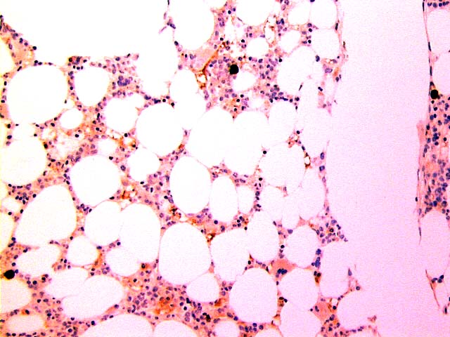
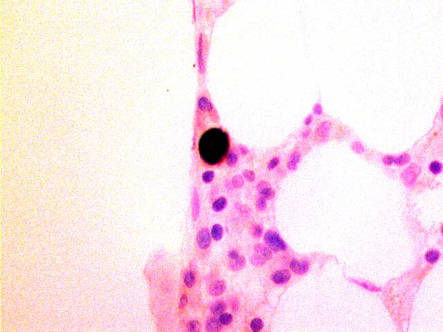
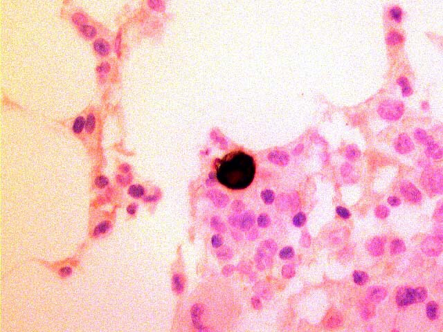
Microscopic (histologic) description
- Marked erythroblast hypoplasia, immature giant erythroblasts
- Occasional erythroblasts may have intranuclear inclusions with surrounding rim of residual chromatin
- Rarely hemophagocytosis (Jpn J Infect Dis 2005;58:149)
Microscopic (histologic) images
Images hosted on other servers:


50 year old man: rare pronormoblast forms


Large and megaloblastic pronormoblast forms

Giant cell with eosinophilic nuclear inclusion

B19+
B19+ cells in bone marrow biopsy
Positive stains
- Hemoglobin A, B19
Electron microscopy images
Images hosted on other servers:

Native parvovirus B19
Differential diagnosis
- Congenital dyserythropoietic anemia (J Pediatr Hematol Oncol 2004;26:133)
Additional references
