
Bone marrow nonneoplastic
Infectious / inflammatory
CMV
Author: Dragos C. Luca, M.D.

Last author update: 1 December 2014
Last staff update: 6 November 2020
Copyright: 2002-2025, PathologyOutlines.com, Inc.
PubMed Search: CMV [title] bone marrow [title]

Table of Contents
Definition / general | Clinical presentation and diagnosis | Case reports | Microscopic (histologic) description | Microscopic (histologic) images | Electron microscopy description | Differential diagnosis | Additional referencesCite this page: Luca DC. CMV. PathologyOutlines.com website. https://www.pathologyoutlines.com/topic/bonemarrowcmv.html. Accessed April 1st, 2025.
Definition / general
- Suppresses hematopoiesis (Blood 1990;75:1965)
- Monocyte and granulocyte precursors are major site for latent CMV (Intervirology 1999;42:308)
- Rarely CMV can induce clonal T cell proliferations
Clinical presentation and diagnosis
- Clinical and hematologic features show considerable overlap with EBV infection
- Intrauterine infection is associated with thrombocytopenia and hemolytic anemia in neonates
- CMV pp65 antigenemia assay is more specific in immunocompromised patients than antibody titers
Case reports
- 57 year old woman with T gamma gene rearrangement and CMV mononucleosis (Am J Hematol 2001;66:64)
Microscopic (histologic) description
- Generally nonspecific findings in bone marrow including myeloid and megakaryocytic suppression
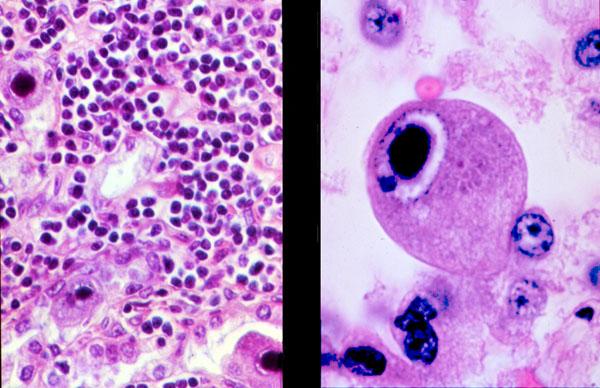
- Infected cells with intranuclear inclusions (not identifiable in many cases); may be highlighted via IHC or ISH
- Nuclear inclusions: single, round or oval, deep blue or amphophilic, surrounding halo, margination of nuclear chromatin
- Cytoplasmic inclusions: multiple, irregular in shape, basophilic
- Occasional hemophagocytosis (Hum Pathol 1998;29:1074), granulomas (Postgrad Med J 1987;63:277), lymphoid aggregates or hematogones (Leuk Res 2003;27:193)
- Atypical lymphocytosis in peripheral blood (mostly increased transformed large granular lymphocytes and NK cells, infrequent infectious mononucleosis-like classic Downey type 2 cells)
- May have circulating CMV infected endothelial cells at feathered edge of peripheral blood smear in immunosuppressed individuals
Microscopic (histologic) images
Images hosted on other servers:
Site unknown (not bone marrow)
Electron microscopy description
- Virus particles with electron dense icosahedral core containing capsomeres with a large pleomorphic lipid envelope
- Particles composed of filamentous or amorphous material detached from nuclear membrane
Differential diagnosis
- Pre B ALL
- T cell lymphoma
