
Bone & joints
Other tumors
Infantile myofibromatosis
Author: Nat Pernick, M.D.

Last author update: 1 June 2005
Last staff update: 17 January 2022
Copyright: 2003-2025, PathologyOutlines.com, Inc.
PubMed search: infantile myofibromatosis [title] bone

Table of Contents
Definition / general | Radiology description | Radiology images | Case reports | Microscopic (histologic) description | Microscopic (histologic) images | Positive stains | Differential diagnosis | Additional referencesCite this page: Pernick N Infantile myofibromatosis. PathologyOutlines.com website. https://www.pathologyoutlines.com/topic/boneinfantilemyofibromatosis.html. Accessed April 1st, 2025.
Definition / general
- Also called congenital fibromatosis, solitary infantile myofibromatosis
- Rare; median age 16 months, range 6 months to 16 years
- Solitary lesion in craniofacial bone, single nodule in soft tissue, multiple nodules in bone and soft tissue or diffuse involvement of viscera
Radiology description
- Well circumscribed bone lucency
- Spontaneous resolution with good prognosis unless visceral involvement
Radiology images
Contributed by Mark R. Wick, M.D.
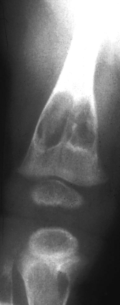
Multifocal congenital Xray
Case reports
- 11 month old boy with solitary lesion of parietal bone (Am J Surg Pathol 1993;17:308)
Microscopic (histologic) description
- Same as soft tissue myofibroma - proliferation of spindle cells with pink cytoplasm, myxoid background
- Cells arranged in nodules with slit-like vascular spaces or hemangiopericytomatous pattern
Microscopic (histologic) images
Contributed by Mark R. Wick, M.D.
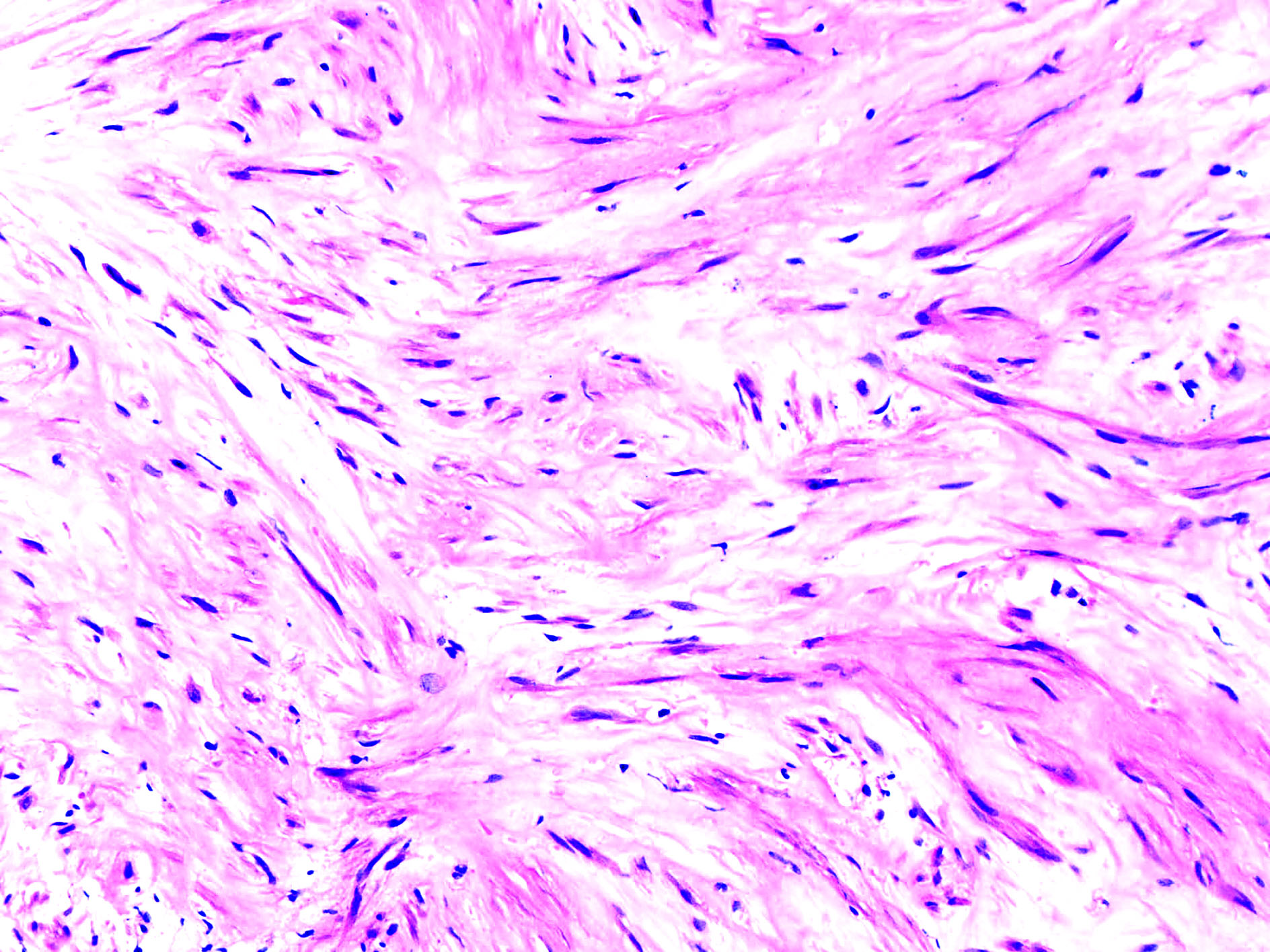
Multifocal congenital
Positive stains
Differential diagnosis
- Sarcoma
Additional references
