
Bone & joints
Vascular tumors
Hemangioma
Editorial Board Member: Farres Obeidin, M.D.
Deputy Editor-in-Chief: Borislav A. Alexiev, M.D.

Last author update: 21 January 2025
Last staff update: 21 January 2025
Copyright: 2003-2025, PathologyOutlines.com, Inc.
PubMed Search: Hemangioma

Table of Contents
Definition / general | Essential features | Terminology | ICD coding | Epidemiology | Sites | Pathophysiology | Etiology | Clinical features | Diagnosis | Radiology description | Radiology images | Prognostic factors | Case reports | Treatment | Gross description | Gross images | Microscopic (histologic) description | Microscopic (histologic) images | Positive stains | Negative stains | Molecular / cytogenetics description | Sample pathology report | Differential diagnosis | Additional references | Board review style question #1 | Board review style answer #1 | Board review style question #2 | Board review style answer #2Cite this page: Abrahao Machado LF. Hemangioma. PathologyOutlines.com website. https://www.pathologyoutlines.com/topic/bonehemangioma.html. Accessed April 1st, 2025.
Definition / general
- Hemangioma is a benign neoplasm of bone showing endothelial differentiation (Skeletal Radiol 2000;29:63)
- Among the most common primary benign tumors of bone (Crit Rev Oncol Hematol 2024;195:104268, Semin Diagn Pathol 2014;31:30)
Essential features
- Benign tumor of bone composed of small or large caliber vascular channels (Skeletal Radiol 2000;29:63)
- Vessels are thin walled and lined by a single layer of bland endothelial cells
- Majority arise in vertebral bodies and are incidental findings (Crit Rev Oncol Hematol 2024;195:104268)
- Multifocal occurrence is frequent (Surg Pathol Clin 2021;14:645)
- Known as angiomatosis when involving a large region or if widespread throughout the skeleton (Skeletal Radiol 2000;29:63)
Terminology
- Hemangioma; conventional hemangioma; intraosseous hemangioma
- Gorham-Stout syndrome (also known as vanishing bone disease): diffuse angiomatosis with massive osteolysis (Skeletal Radiol 2000;29:63)
ICD coding
- ICD-O: 9120/0 - hemangioma, NOS
- ICD-11
- EE6Y - other specified fibromatous disorders of skin and soft tissue
- 2E81.0Y & XH5AW4 - neoplastic hemangioma of other specified site & hemangioma, NOS
Epidemiology
- Most often diagnosed in middle aged adults (peak incidence in fifth decade) (Surg Pathol Clin 2021;14:645)
- Rare in children (Crit Rev Oncol Hematol 2024;195:104268)
- M:F = 0.7:1 (Crit Rev Oncol Hematol 2024;195:104268)
Sites
- Vertebral bodies are the most commonly affected sites (Semin Diagn Pathol 2014;31:30)
- Other sites: craniofacial skeleton and the long bones of extremities (Semin Diagn Pathol 2014;31:30)
Pathophysiology
- Benign neoplastic proliferation of endothelial cells (Histopathology 2022;80:19, Crit Rev Oncol Hematol 2024;195:104268)
Etiology
- Exact etiology and inciting factor of osseous hemangioma genesis are not well known
- Although there is still controversy, there is growing evidence that it is a true neoplasm rather than a malformation or hamartoma (Histopathology 2022;80:19, Am J Surg Pathol 2013;37:613, Am J Surg Pathol 2024;48:487)
Clinical features
- Most cases detected as an incidental radiographic finding (Crit Rev Oncol Hematol 2024;195:104268)
- Clinically significant hemangiomas are rare (< 1% of primary bone tumors) (Skeletal Radiol 2000;29:63, Crit Rev Oncol Hematol 2024;195:104268)
- Large vertebral tumors may cause cord compression, pain and neurological symptoms
- Uncommonly appears with mass-like deformity and pathological fracture
- Massive progressive osteolysis involving contiguous bones is observed in cases of Gorham-Stout syndrome (a rare nonhereditary disorder also known as vanishing bone disease) (Skeletal Radiol 2000;29:63)
Diagnosis
- Tissue sampling is the gold standard for a definitive diagnosis (J Orthop Surg Res 2024;19:310)
- Essential diagnostic criteria according to the WHO classification of tumors: bone tumor with compatible imaging; thin walled blood vessels lined by a single layer of nonatypical endothelial cells
Radiology description
- Plain radiograph: vertically oriented, thickened trabeculae in vertebral lesions (corduroy sign) and irregular and lytic lesions with a honeycomb appearance in long bones (J Neuroradiol 2010;37:37, BMC Musculoskelet Disord 2021;22:27)
- Computed tomography (CT): lucent lesion containing fat and coarse, vertically oriented trabeculae (polka dot sign on axial images and corduroy sign on coronal and sagittal images) (Skeletal Radiol 2022;51:1743)
- Magnetic resonance imaging (MRI): typical high signal intensity on both T1 and T2 because of abundant adipocytes, blood vessels and interstitial edema, although intermediate to low signal intensity is seen in lipid poor hemangiomas (BMC Musculoskelet Disord 2021;22:27, Skeletal Radiol 2022;51:1743)
Radiology images
Images hosted on other servers:

Medullary hemangioma in the tibia (Xray)

Vertebral hemangioma (CT and MRI)

Aggressive vertebral hemangioma (MRI)

Spinal MRI shows nodular lesion in the vertebral body

Lumbar CT showing a vertebral hemangioma
Prognostic factors
- Prognosis is excellent
- Low rate of local recurrence (Crit Rev Oncol Hematol 2024;195:104268)
Case reports
- 10 year old girl with hemangioma in the maxilla (Asian J Surg 2023;46:2822)
- 13 year old boy with multifocal Gorham-Stout disease (Radiol Case Rep 2024;19:5066)
- 23 year old man with hemangioma of the hyoid bone (Medicine (Baltimore) 2024;103:e37137)
- 27 year old woman with cavernous hemangioma of rib (Front Oncol 2023;13:1164331)
- 45 year old woman with a locally aggressive vertebral hemangioma (Medicine (Baltimore) 2024;103:e37885)
- 57 year old woman with intraosseous hemangioma of the clivus (Surg Neurol Int 2024;15:177)
Treatment
- Asymptomatic, incidentally detected lesions require no treatment (Skeletal Radiol 2000;29:63)
- In symptomatic tumors, different interventions might be used (Crit Rev Oncol Hematol 2024;195:104268)
- Curettage and bone grafting
- Endovascular embolization
- Percutaneous vertebroplasty
- Transpedicular ethanol injection
- Radiation therapy
Gross description
- Dark red or brown, sponge-like appearance
- Tumor is usually centered in medullary cavity (Semin Diagn Pathol 2014;31:30)
- Bone sclerosis and periosteal reaction may be present
Gross images
Contributed by Lucas F. Abrahao Machado, M.D., Ph.D.

Hemangioma of the proximal femur

Cavernous hemangioma of the femur
Microscopic (histologic) description
- Proliferation of thin walled, blood filled vessels lined by a single layer of flat, cytologically bland endothelial cells
- Usually of capillary or cavernous type
- Vessels surround pre-existing bone trabeculae and permeate the medullary canal
- Lumina may be filled with red blood cells, although they are frequently empty or contain only proteinaceous material
- Variable amounts of surrounding loose connective tissue and fat
- Secondary changes: thrombosis, calcification, papillary endothelial hyperplasia, reactive sclerosis of involved bone trabeculae
- References: Skeletal Radiol 2000;29:63, Semin Diagn Pathol 2014;31:30, Surg Pathol Clin 2021;14:645
Microscopic (histologic) images
Contributed by Lucas F. Abrahao Machado, M.D., Ph.D.

Vascular channels around bone trabeculae

Capillary type

Vessels filled with red blood cells

Vessels with empty lumina

Cavernous type

Surrounding stroma

Thrombosis

CD31

ERG
Positive stains
Negative stains
Molecular / cytogenetics description
- Specific genetic alterations of hemangioma of bone are still under investigation
- NFATC1 and NFATC2 fusions (mostly with EWSR1) have been described in few hemangiomas of bone, although they will most likely soon be considered a distinct group of vascular neoplasms (Am J Surg Pathol 2013;37:613, Genes Chromosomes Cancer 2021;60:762, Am J Surg Pathol 2024;48:487)
- Hemangiomas with NFATC related fusions show epithelioid phenotype and cytologic atypia, with potential for local aggressive behavior and recurrence
Sample pathology report
- Lesion in thoracic vertebra, biopsy:
- Benign vascular lesion, consistent with hemangioma (see comment)
- Comment: The tumor shows a proliferation of thin walled capillary vessels lined by a single layer of nonatypical endothelial cells, coursing through the marrow spaces and encasing pre-existing bony trabeculae. Hemangioma of bone is the most common vascular tumor of bone and asymptomatic or incidentally found cases generally do not require treatment. Radiological and clinical correlation is recommended to confirm the diagnosis.
Differential diagnosis
- Reactive process:
- Fewer blood vessels
- Vascular channels not as haphazardly arranged as in hemangioma
- Often shows other reactive changes such as fibrosis, inflammation and woven bone
- Epithelioid hemangioma:
- Endothelial cells are large, epithelioid and have abundant eosinophilic cytoplasm
- Intracytoplasmic vacuoles are commonly observed
- Tumor cells can grow in solid cords and sheets
- Stroma may contain eosinophils and plasma cells
- FOSB positivity in the majority of cases (Int J Surg Pathol 2023;31:280)
- Epithelioid hemangioendothelioma:
- Epithelioid endothelial cells arranged in cords or clusters in myxohyaline stroma
- Intracytoplasmic vacuoles are prominent
- Vascular lumina are usually not observed
- CAMTA1 positivity in the majority of cases (Am J Surg Pathol 2016;40:94)
- Angiosarcoma:
- Tumor cells show a greater degree of atypia
- Blood vessels demonstrate irregular and complex architecture, sometimes with anastomosing channels
- Cells often grow in solid clusters
- Tumor can cause bone destruction and necrosis of surrounding trabeculae
Additional references
Board review style question #1
Which of the following is one of the most common tumors of the vertebral column and is rarely clinically significant?
- Conventional hemangioma
- Enchondroma
- Epithelioid hemangioma
- Plasmacytoma
Board review style answer #1
A. Conventional hemangioma. Hemangioma is the most common type of vascular tumor of the skeleton and one of the most common tumors of vertebrae in general. However, the vast majority are asymptomatic; clinically significant tumors account for < 1% of primary bone tumors. Answer D is incorrect because, in plasmacytoma, the vertebrae are more commonly involved as part of a diffuse process (multiple myeloma) by which the bone marrow is affected; therefore, it is usually a symptomatic and clinically significant tumor. Answer C is incorrect because epithelioid hemangioma is an uncommon variant of hemangioma and the majority arises in long tubular bones. Answer B is incorrect because enchondroma of the spine is very rare. Chondroid lesions occurring in the vertebrae should be carefully investigated as they are generally aggressive, if they are not reactive or metaplastic in nature.
Comment Here
Reference: Hemangioma
Comment Here
Reference: Hemangioma
Board review style question #2
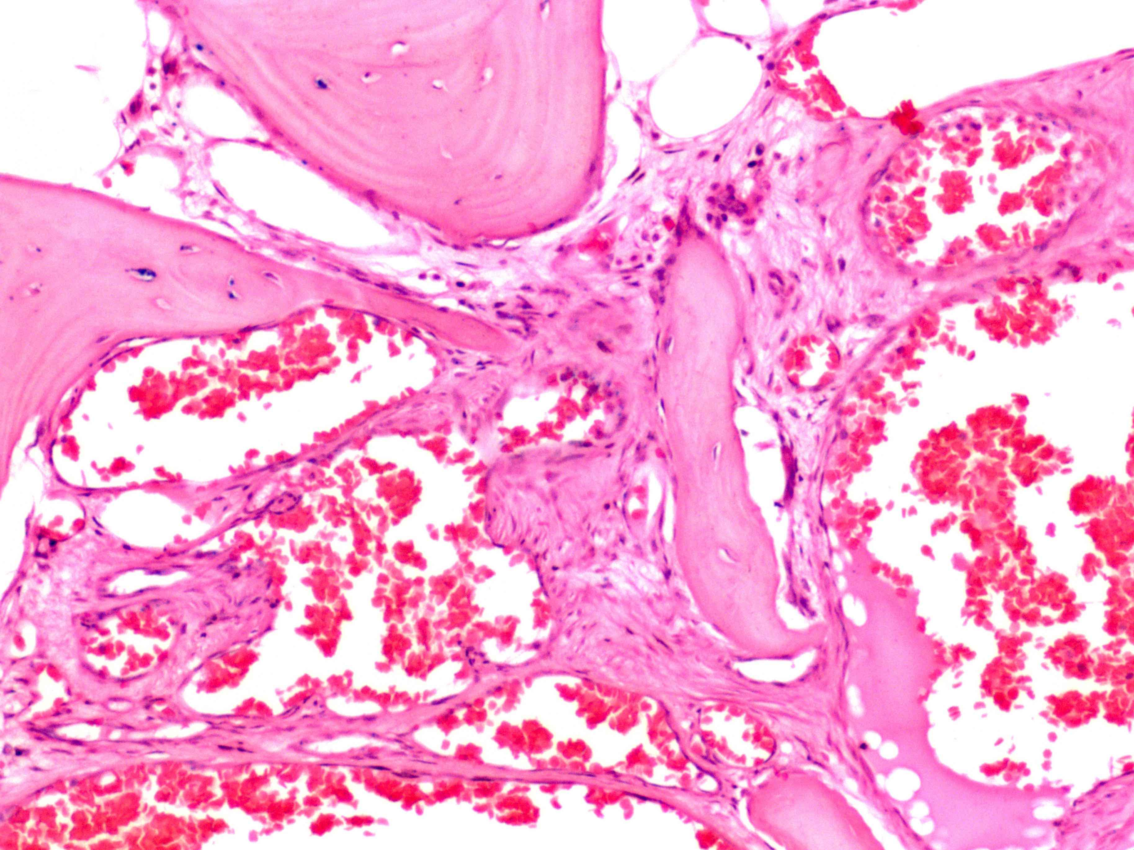
A 34 year old man underwent biopsy of a lesion in the third lumbar vertebra found incidentally after an abdominal CT scan. Microscopically, as shown in the image above, the lesion revealed numerous vascular channels lined by a single layer of endothelial cells without nuclear atypia, interspersing the pre-existing trabeculae. What is the diagnosis?
- Angiosarcoma
- Epithelioid hemangioendothelioma
- Epithelioid hemangioma
- Hemangioma of bone
Board review style answer #2
D. Hemangioma of bone. The image and microscopic description depict a hemangioma of bone, a benign and generally asymptomatic vascular tumor. Answer B is incorrect because epithelioid hemangioendothelioma usually presents with symptoms of pain and swelling and it is histologically characterized by epithelioid cells arranged in cords or clusters within a distinctive myxohyaline stroma, without the formation of vascular lumina. Answer A is incorrect because angiosarcoma usually arises as an ill defined and destructive mass, showing atypical endothelial cells lining irregular vascular channels.
Answer C is incorrect because epithelioid hemangioma has a different morphology from conventional hemangioma, revealing larger, epithelioid cells with abundant eosinophilic cytoplasm.
Comment Here
Reference: Hemangioma
Comment Here
Reference: Hemangioma
