Table of Contents
Definition / general | Essential features | Terminology | ICD coding | Epidemiology | Sites | Pathophysiology | Etiology | Clinical features | Diagnosis | Laboratory | Radiology description | Radiology images | Prognostic factors | Case reports | Treatment | Gross description | Gross images | Frozen section description | Microscopic (histologic) description | Microscopic (histologic) images | Cytology description | Positive stains | Negative stains | Molecular / cytogenetics description | Sample pathology report | Differential diagnosis | Board review style question #1 | Board review style answer #1 | Board review style question #2 | Board review style answer #2Cite this page: Arora K, Velez Torres JM. Epithelioid hemangioma of bone. PathologyOutlines.com website. https://www.pathologyoutlines.com/topic/boneepithelioidhemangioma.html. Accessed April 2nd, 2025.
Definition / general
- Variant of hemangioma that is a benign neoplasm composed of plump, epithelioid cells of endothelial cell differentiation
- Frequently arises in long tubular bones (40%) (Am J Surg Pathol 2009;33:270)
- Can arise in skin, lymph nodes and soft tissue
Essential features
- Most commonly arises in long tubular bones
- Rearrangement of FOS is common
- Symptomatic tumors treated with curettage
- On imaging, lesions are well defined and lucent but can show aggressive features like soft tissue extension
- Microscopically, lobulated growth pattern, composed of epithelioid endothelial cells, numerous inflammatory cells, including eosinophils and plasma cells
- No hyalinized or myxoid matrix
Terminology
- Angiolymphoid hyperplasia with eosinophilia
- Not recommended to use: histiocytoid hemangioma, hemorrhagic epithelioid and spindle cell hemangioma
ICD coding
- ICD-10: D18.09 - hemangioma of other sites
Epidemiology
- Occurs in all age groups, most commonly in the fourth decade (Am J Surg Pathol 2009;33:270)
- More frequent in males (M:F = 1.4:1)
Sites
- Frequently arises in long tubular bones (40%), followed by flat bones (18%), vertebrae (16%) and small bones of hand and feet (8%) (Am J Surg Pathol 2009;33:270)
- ~20% of cases involve multiple bones
- Can involve bones of craniofacial skeleton
Pathophysiology
- 70% of osseous hemangiomas harbor either FOS or FOSB gene fusion (Genes Chromosomes Cancer 2015;54:565)
- Leads to truncation of FOS or FOSB protein, loss of transactivation domain and tumorigenesis
Etiology
- Unclear
Clinical features
- Patients usually present with pain localized to the involved bone
Diagnosis
- Requires interpretation of clinical, radiological and histopathological findings
Laboratory
- No specific abnormality
Radiology description
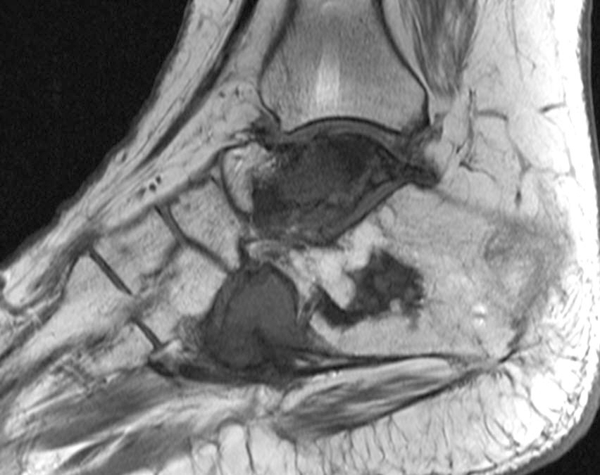
- On plain radiograph, the lesions are lucent with well defined expansile margins but may show a mixed lytic and sclerotic appearance with cortical destruction and periosteal reactive bone formation (Skeletal Radiol 2000;29:63)
- CT scan displays an expansile, lytic lesion with cortical destruction and bone expansion (Skeletal Radiol 2000;29:63)
- On MRI, lesions are hypointense or isointense relative to muscle on T1 weighted images and hyperintense on T2 weighted images; markedly enhanced by gadolinium contrast (Skeletal Radiol 2000;29:63)
Prognostic factors
- Recurrences are uncommon
- Spontaneous resolution has been reported
- Multicentric disease may present as regional lymph node or other organ involvement
- Reference: Am J Surg Pathol 2009;33:270
Case reports
- 14 year old girl with left superior pubic ramus expansile lytic lesion (Int J Surg Pathol 2023;31:667)
- 35 year old man with multiple expansile lesion involving left tibia and left fibula (Indian J Pathol Microbiol 2022;65:401)
- 43 year old man with a lucent mass in T3 vertebral body (J Spinal Cord Med 2019;42:800)
Treatment
- Asymptomatic patients can be followed
- Curettage or en bloc excision for symptomatic tumors
Gross description
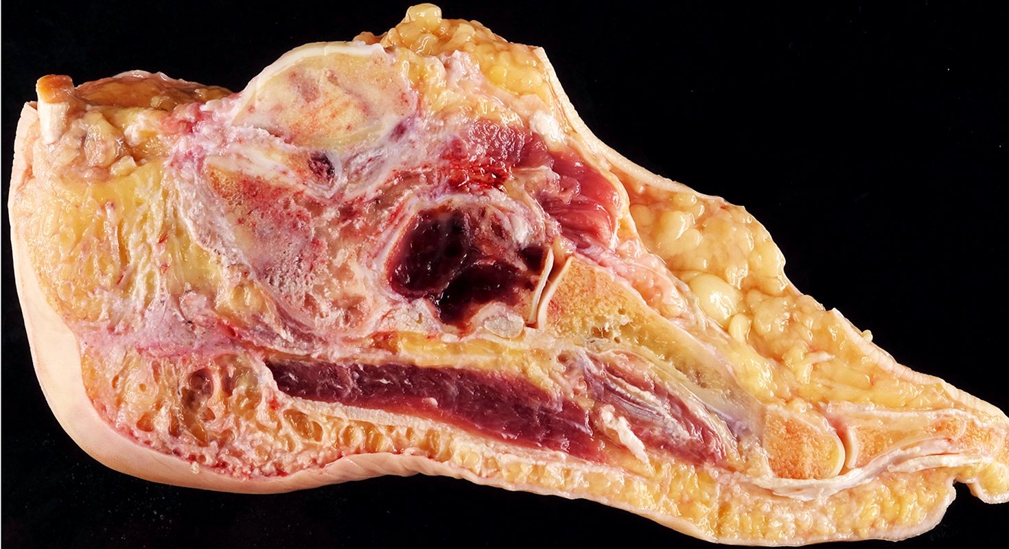
- Soft, solid, red and hemorrhagic
- Tumor is excised intact and is relatively well circumscribed, solid, red and frequently expands the cortex
- Uncommonly, the tumor erodes the cortex and extends into the soft tissue with pushing margins (Am J Surg Pathol 2009;33:270)
Frozen section description
- Irregular infiltrative neoplasm with nodular / lobular growth pattern
- Cytologically, tumor cells are large polyhedral, have eosinophilic cytoplasm and frequently contain intracytoplasmic vacuoles
- These tumors may appear very cellular and also can have a background of lymphocytes, eosinophils, fibrosis and hemorrhage, making it difficult to appreciate the lumen formation
- Frozen section diagnosis can be challenging and can be misdiagnosed as metastatic carcinoma (Cureus 2021;13:e15371)
Microscopic (histologic) description
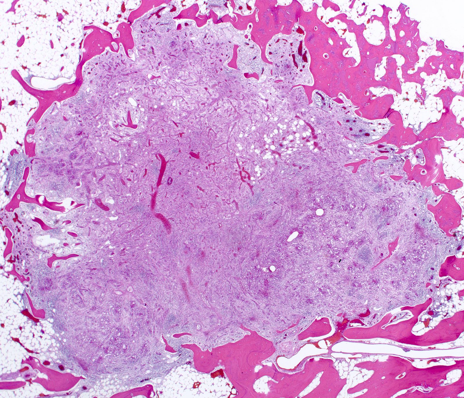
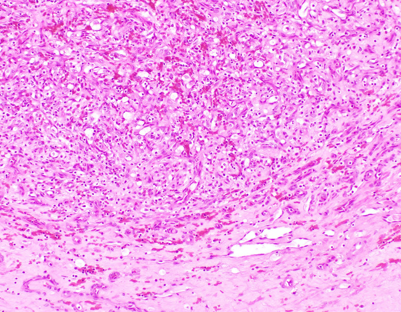
- Lobulated growth pattern
- Organoid architecture
- Well demarcated or infiltrative
- Nodules of the tumor cells are surrounded by loose connective tissue
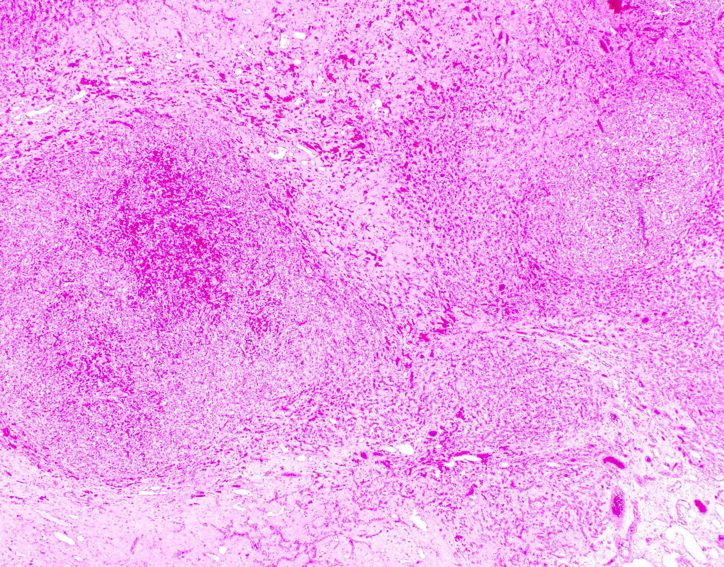
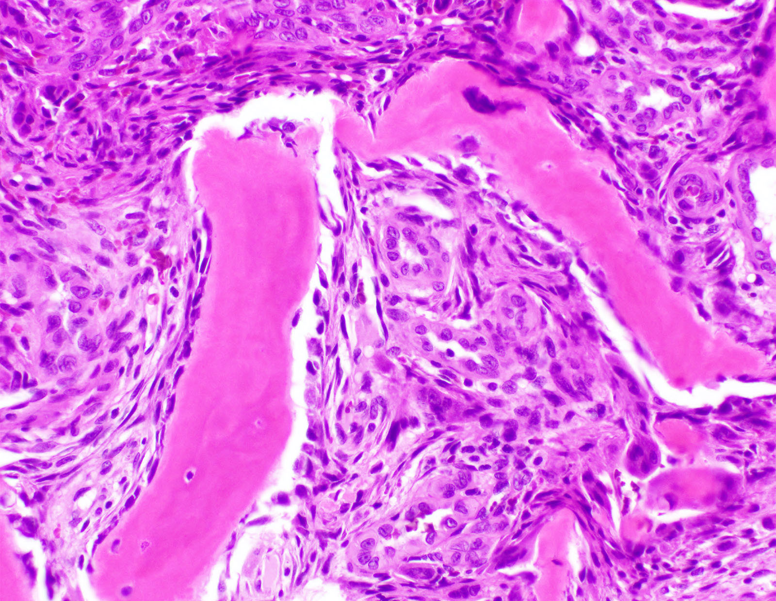
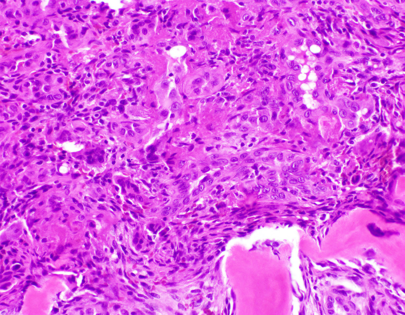
- Tumor cells are large polyhedral, have eosinophilic cytoplasm and frequently contain intracytoplasmic vacuoles
- Mitoses may be present
- Well formed vessels are present and tumor cell cytoplasm may bulge into lumen in a tombstone or hobnail-like pattern
- Cells can grow in solid cords and sheets
- Stroma may contain eosinophils and plasma cells
- Background of hemorrhage, fibrosis and patchy woven bone formation may be present
- No myxohyalinized stroma is present (Am J Surg Pathol 2009;33:270)
Microscopic (histologic) images
Cytology description
- Cytology is not routinely used for diagnosis of epithelioid hemangioma of bone
Positive stains
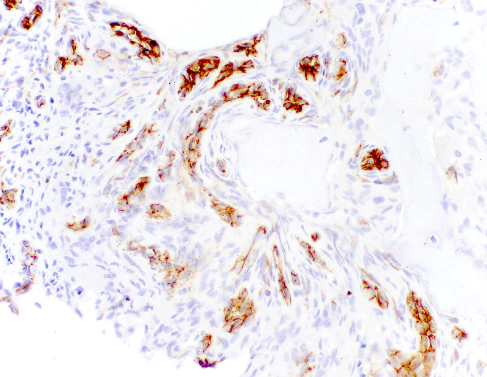
- CD31
- CD34
- ERG
- FLI1
- EMA and keratin AE1 / AE3 (50%)
- FOS and FOSB related to genetics (J Cutan Pathol 2018;45:395)
- SMA in pericytes
Molecular / cytogenetics description
- 70% of osseous hemangiomas harbor either FOS or FOSB gene fusion (Genes Chromosomes Cancer 2021;60:17)
- Fusion partner genes reported are ZFP36 and WWTR1 (Int J Surg Pathol 2023;31:667)
Sample pathology report
- Left foot lesion, curettage:
- Epithelioid hemangioma (see comment)
- Comment: H&E stain slides show lobules and sheets of minimally atypical polyhedral cells with eosinophilic cytoplasm and intracytoplasmic vacuoles. The background stroma shows lymphoplasmacytic and eosinophilic infiltrates and peripheral reactive woven bone formation. Immunohistochemistry shows that tumor cells are positive for CD31, ERG and FOS, while negative for keratin AE1 / AE3. SATB2 stains background osteoblasts and is negative in the tumor cells.
Differential diagnosis
Board review style question #1
Regarding epithelioid hemangioma, which of the following statements is true?
- Frequently associated with peripheral eosinophilia
- Myxohyaline stroma is characteristic
- Osseous epithelioid hemangioma can present as multiple bone involvement
- Rb expression is typically lost in the tumor
Board review style answer #1
C. Osseous epithelioid hemangioma can present as multiple bone involvement. ~20% of osseous epithelioid hemangiomas present as multiple bone lesions. Answer A is incorrect because epithelioid hemangioma is not associated with peripheral eosinophilia. Answer B is incorrect because myxohyaline stroma is characteristic of epithelioid hemangioendothelioma. Answer D is incorrect because Rb expression is generally not lost in the epithelioid hemangioma.
Comment Here
Reference: Epithelioid hemangioma
Comment Here
Reference: Epithelioid hemangioma
Board review style question #2
Board review style answer #2
C. Fusion of FOS or FOSB gene. ~70% of osseous epithelioid hemangioma cases show either FOS or FOSB gene fusion. Answer A is incorrect because CAMTA1 gene fusion is characteristic of epithelioid hemangioendothelioma. Answer B is incorrect because EWSR gene fusion is not characteristic of epithelioid hemangioma. Answer D is incorrect because NR4A3 gene fusion is characteristic of extraskeletal myxoid chondrosarcoma.
Comment Here
Reference: Epithelioid hemangioma
Comment Here
Reference: Epithelioid hemangioma


