Table of Contents
Definition / general | Essential features | Terminology | ICD coding | Epidemiology | Sites | Pathophysiology | Clinical features | Diagnosis | Radiology description | Radiology images | Prognostic factors | Case reports | Treatment | Gross description | Frozen section description | Microscopic (histologic) description | Microscopic (histologic) images | Positive stains | Sample pathology report | Differential diagnosis | Additional references | Board review style question #1 | Board review style answer #1 | Board review style question #2 | Board review style answer #2Cite this page: Akber A, Ud Din N. Epidermoid inclusion cyst. PathologyOutlines.com website. https://www.pathologyoutlines.com/topic/boneepidermoidcyst.html. Accessed December 25th, 2024.
Definition / general
- Benign epithelial inclusion cyst in the bone lined with stratified squamous epithelium, identical to cutaneous counterpart
Essential features
- Benign cystic lesion
- Post traumatic squamous epithelium embedded in bone
- Cyst wall lined by squamous epithelium, including granular layer
- Cyst contents contain laminated keratin
Terminology
- Epidermoid inclusion cyst, epidermal inclusion cyst, intraosseous epidermoid cyst
ICD coding
- ICD-10: L72.0 - epidermal cyst
Epidemiology
- Rare incidence
- Young to middle aged
Sites
- Common in the acral skeleton (fingers and toes) and skull (J Bone Joint Surg Br 1966;48:577, Cancer 1958;11:643, J Bone Joint Surg Am 1964;46:1442)
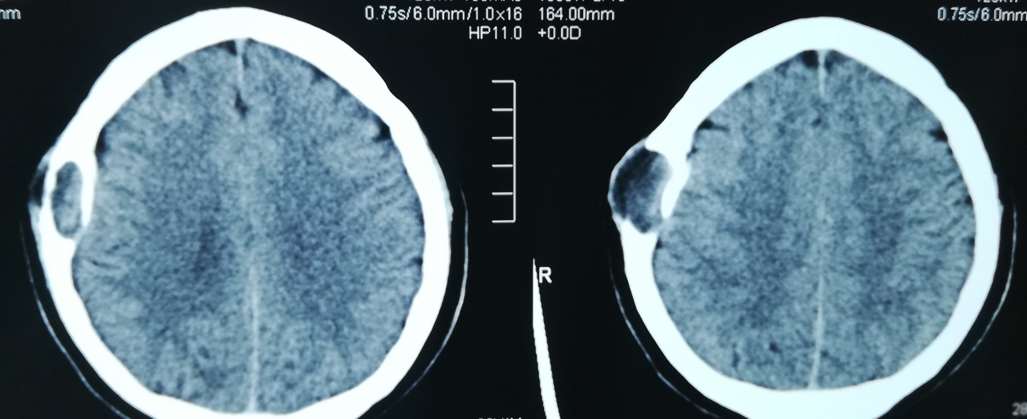
- Skull lesions arise during the first decade
Pathophysiology
- Traumatic embedding of squamous epithelium in the bone that forms a mass (Clin Orthop Relat Res 1970;68:84, J Bone Joint Surg Br 1982;64:456)
Clinical features
- Asymptomatic / painless lump
- Can become tender due to inflammation
Diagnosis
- Clinical, radiological and pathological correlation is adequate for diagnosis
Radiology description
- Xray
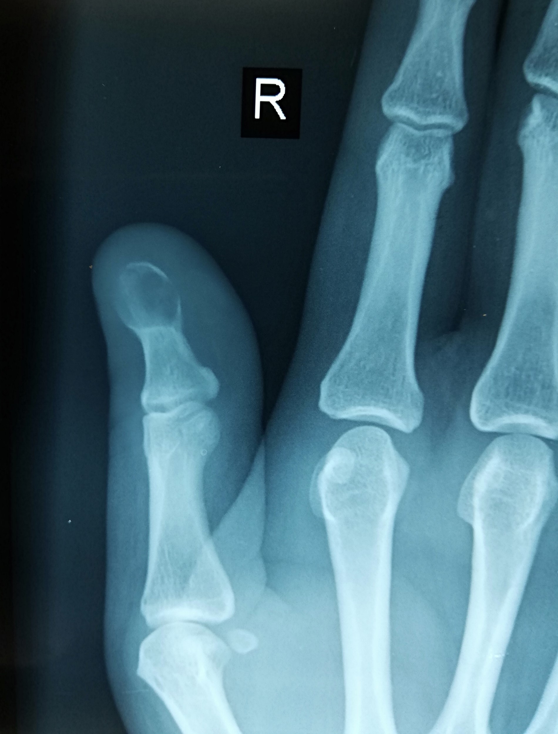
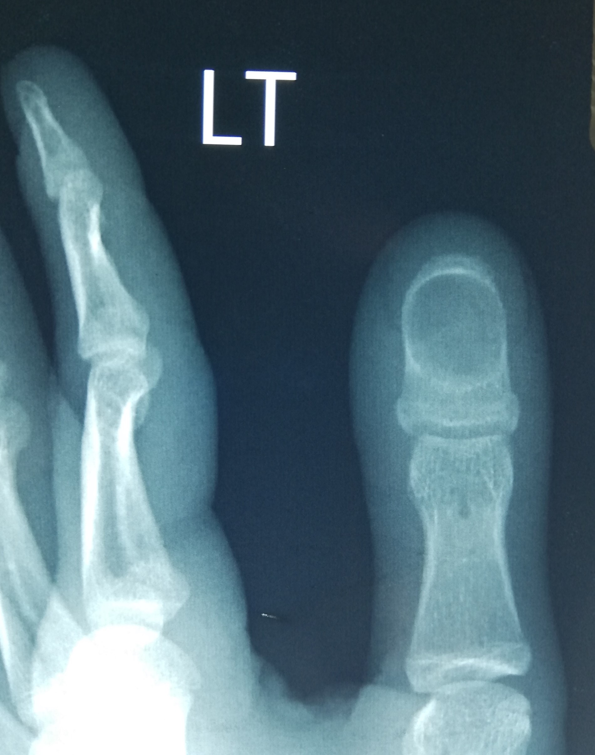
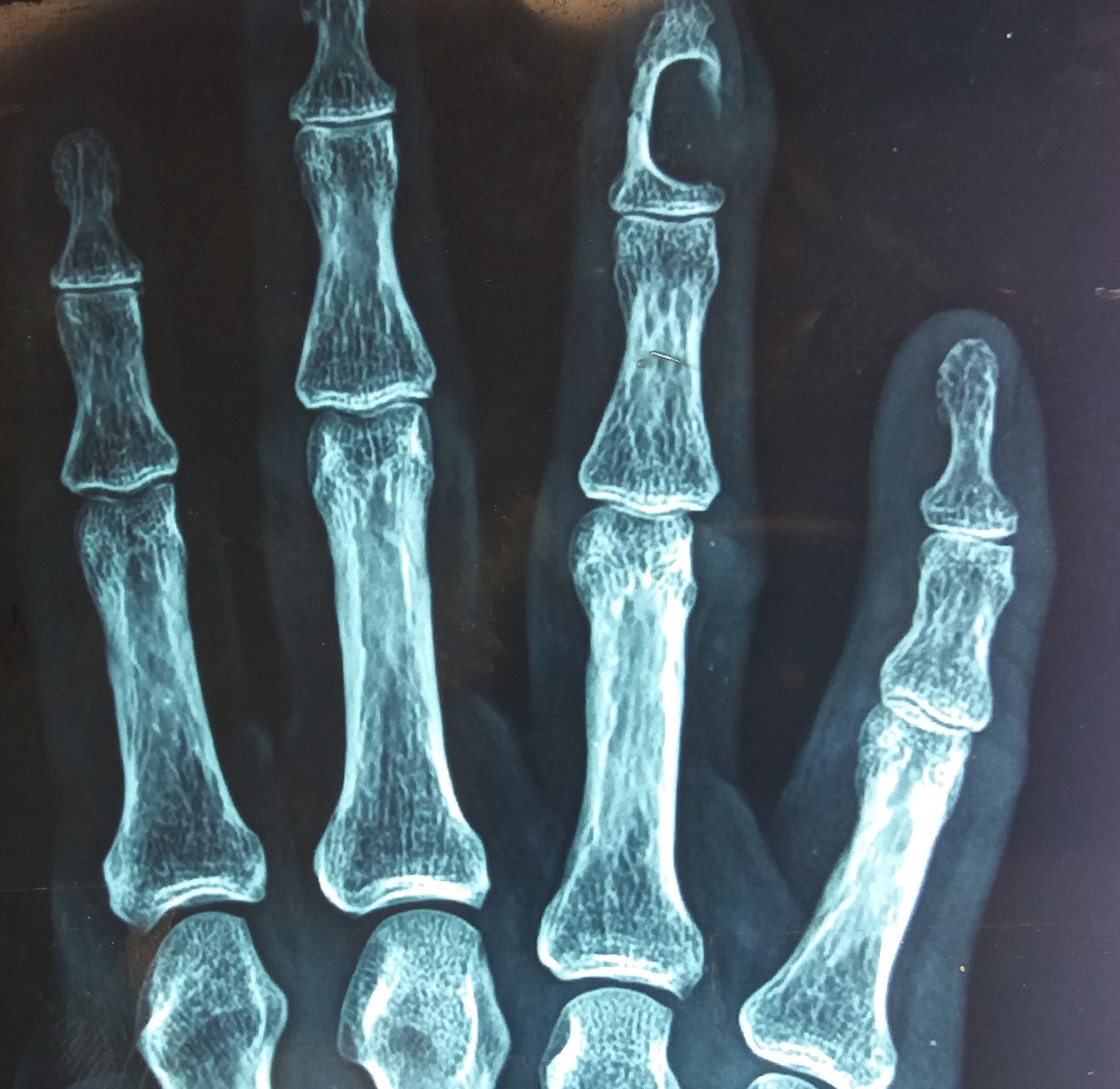
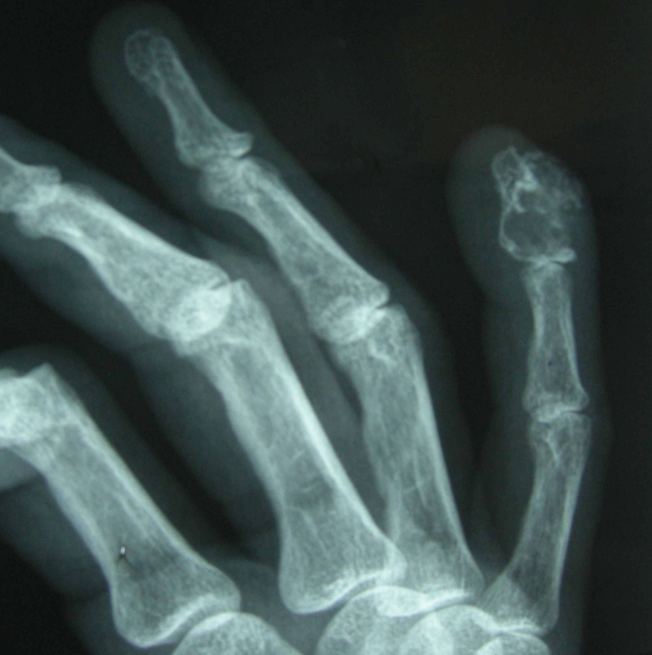
- Round osteolytic lesion with sharply demarcated sclerotic borders and thin cortex
- Expansion of bone
- Pathologic fracture is uncommon
- MRI (Clin Neurol Neurosurg 2021;200:106381):
- Hypointense on T1
- Hyperintense on T2
Radiology images
Prognostic factors
- Excellent prognosis
- Squamous cell carcinoma may very rarely arise in an epidermoid inclusion cyst; has been reported in the skull and finger (Neuroradiology 1999;41:570, Int J Surg Case Rep 2015;11:37)
Case reports
- 24 year old woman with an intraosseous epidermoid cyst of the skull (Dermatol Online J 2018;24:13030)
- 49 year old man with an epidermoid cyst of the anterior clinoid process (Clin Neurol Neurosurg 2021;200:106381)
- 63 year old woman with epidermal inclusion cyst of the fifth toe (Foot Ankle Spec 2017;10:470)
- 66 year old man with epidermal inclusion cyst of the knee (Eur J Orthop Surg Traumatol 2019;29:1355)
- 8 patients with intraosseous epidermoid cysts (J Hand Surg Eur Vol 2011;36:376)
Treatment
- Simple curettage or excision
Gross description
- Unilocular cyst filled with white to pale yellow, malodorous, cheesy material
Frozen section description
- Seldom required
Microscopic (histologic) description
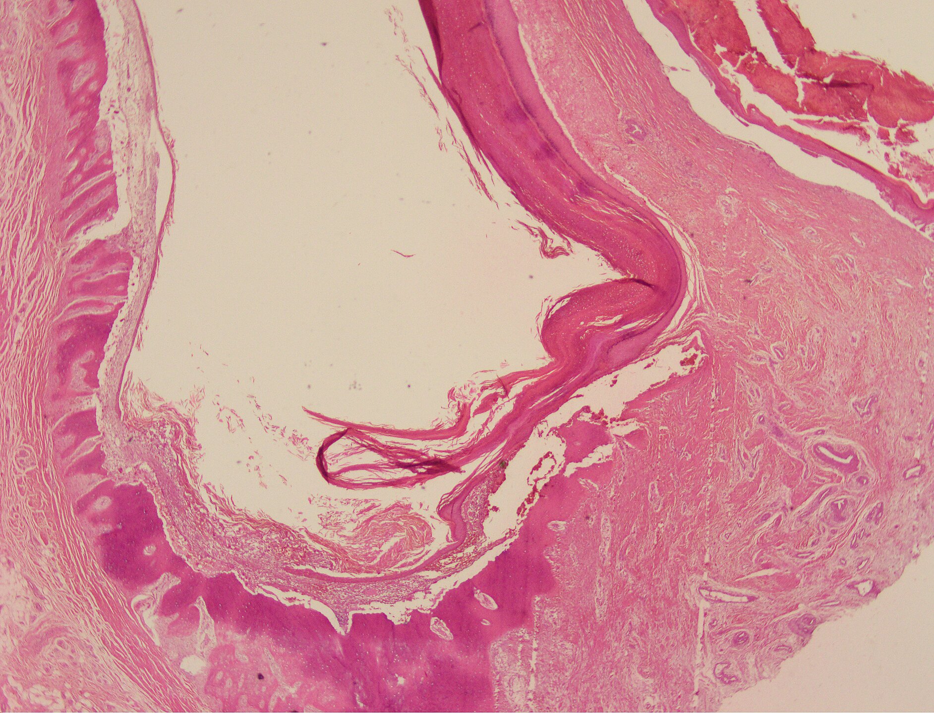
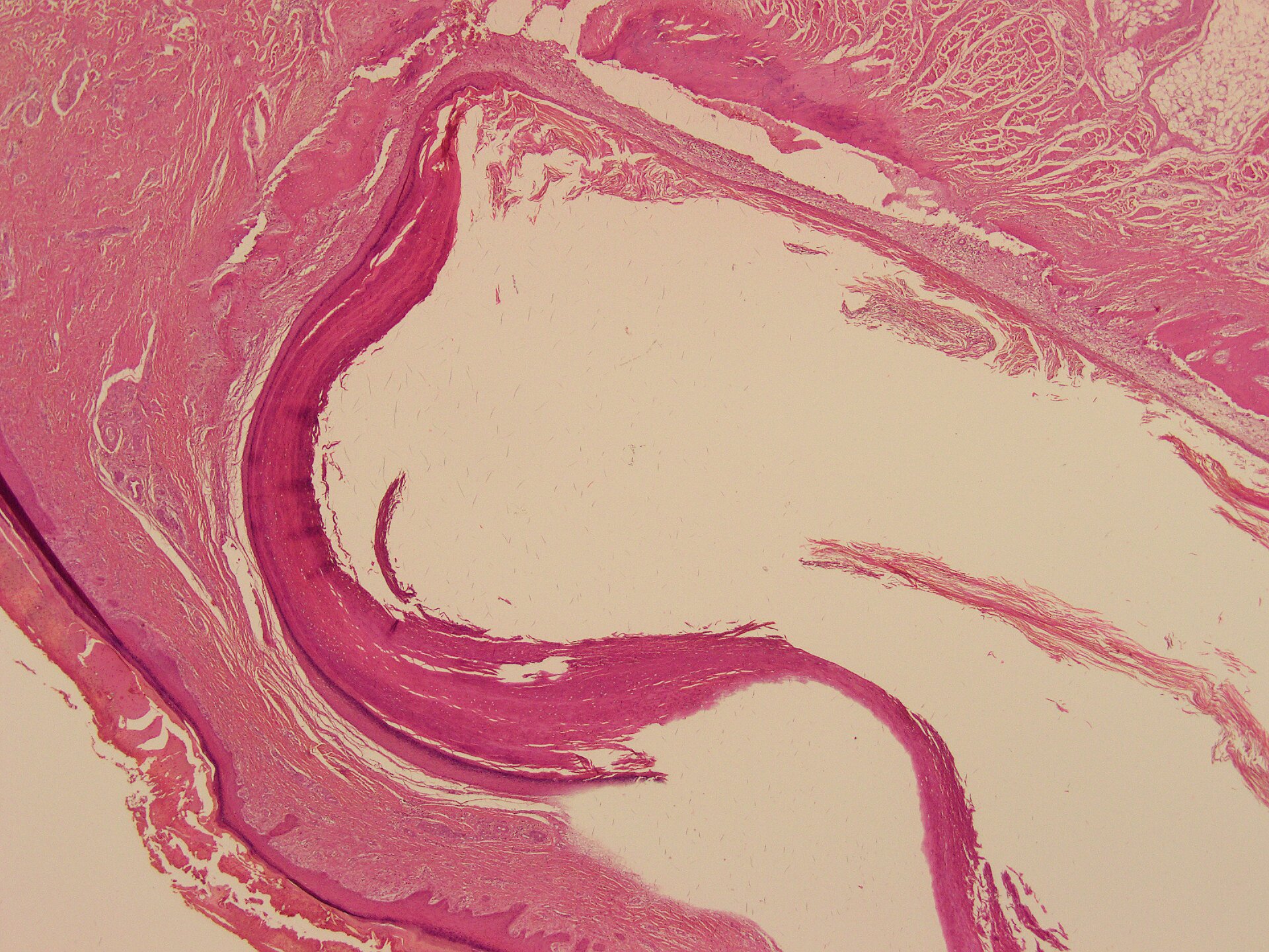
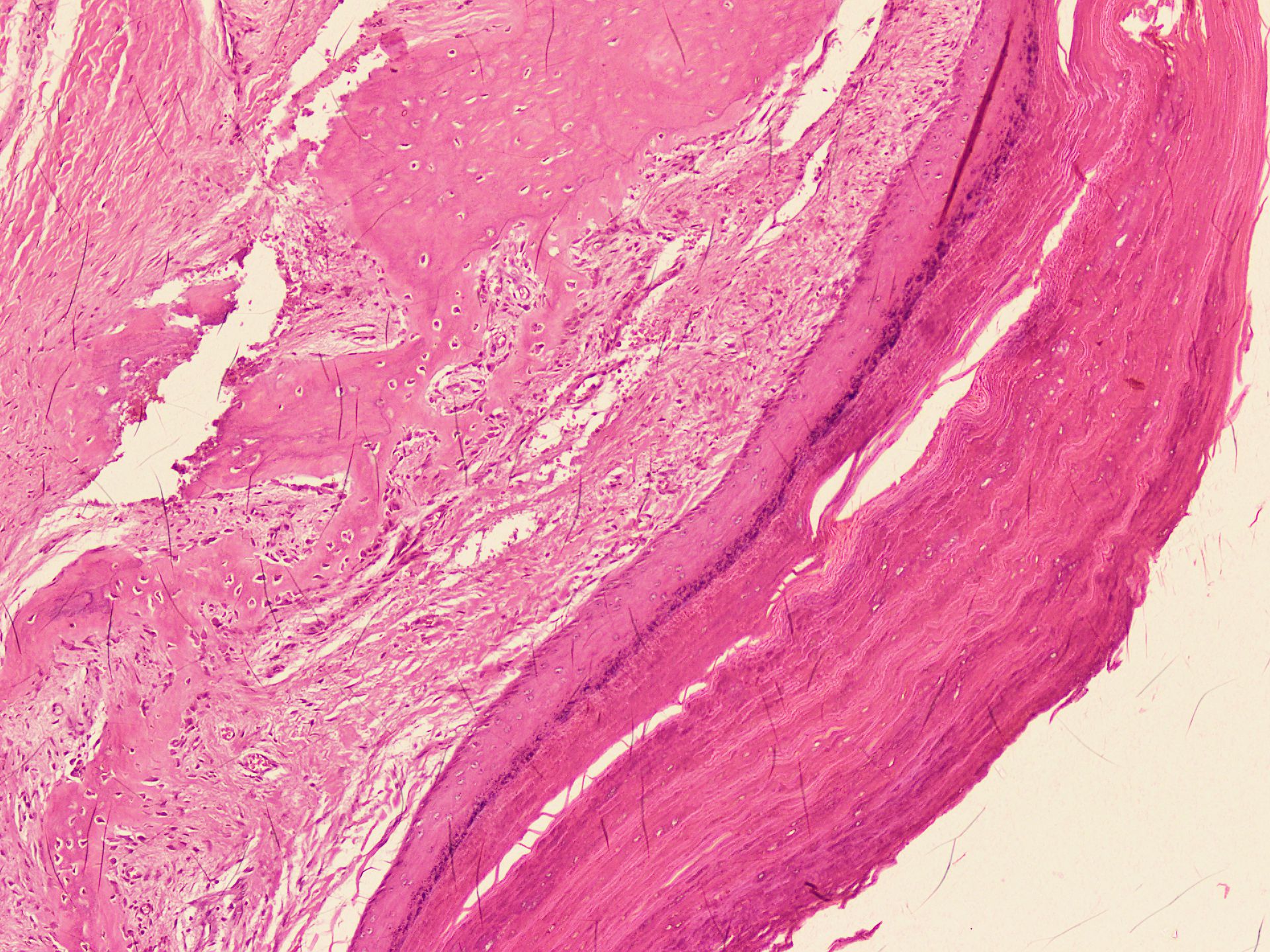
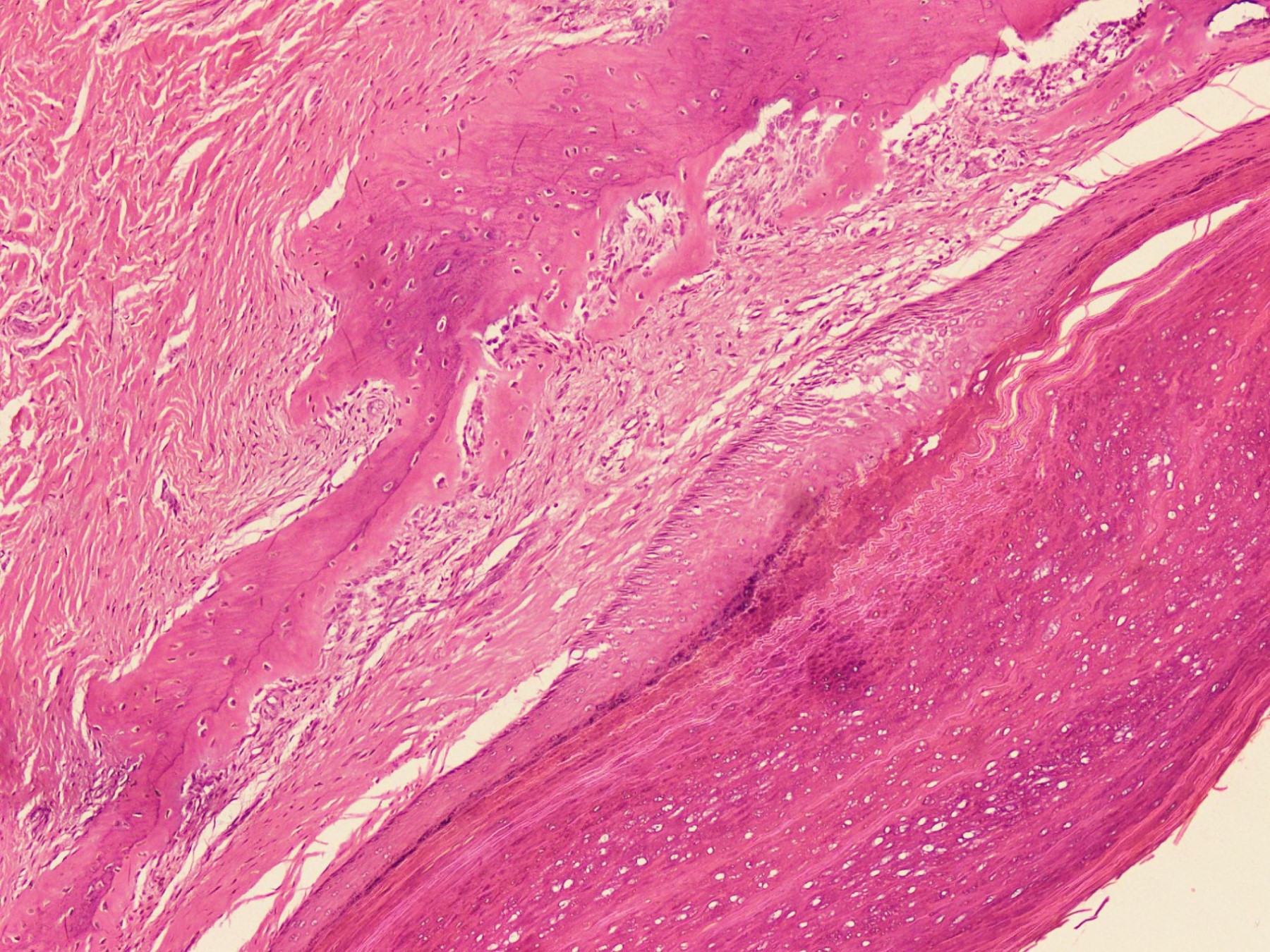
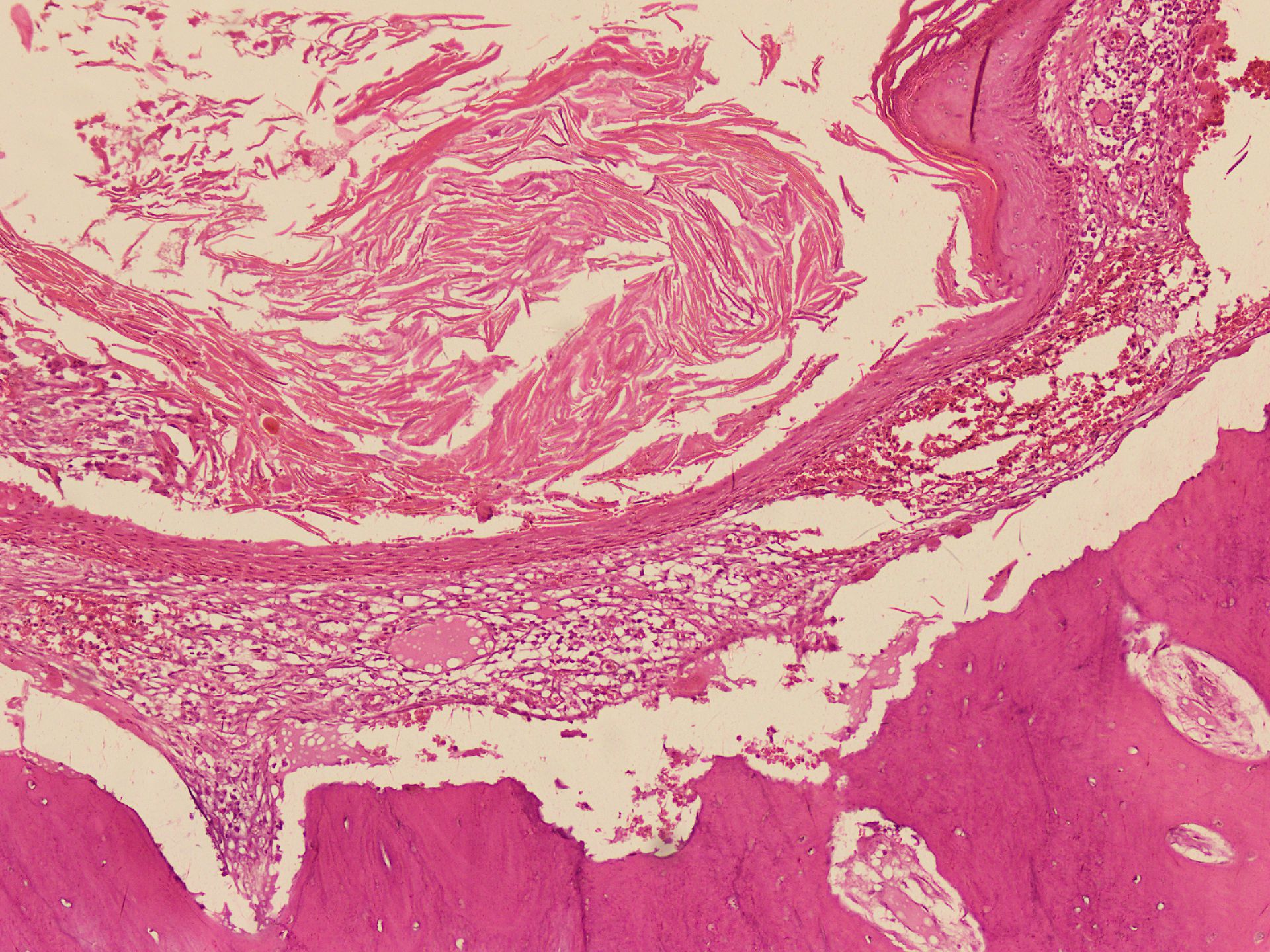
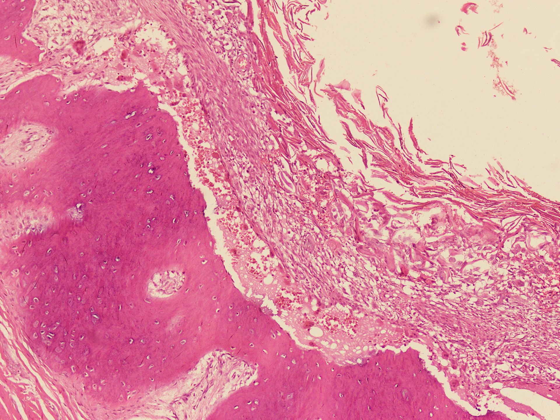
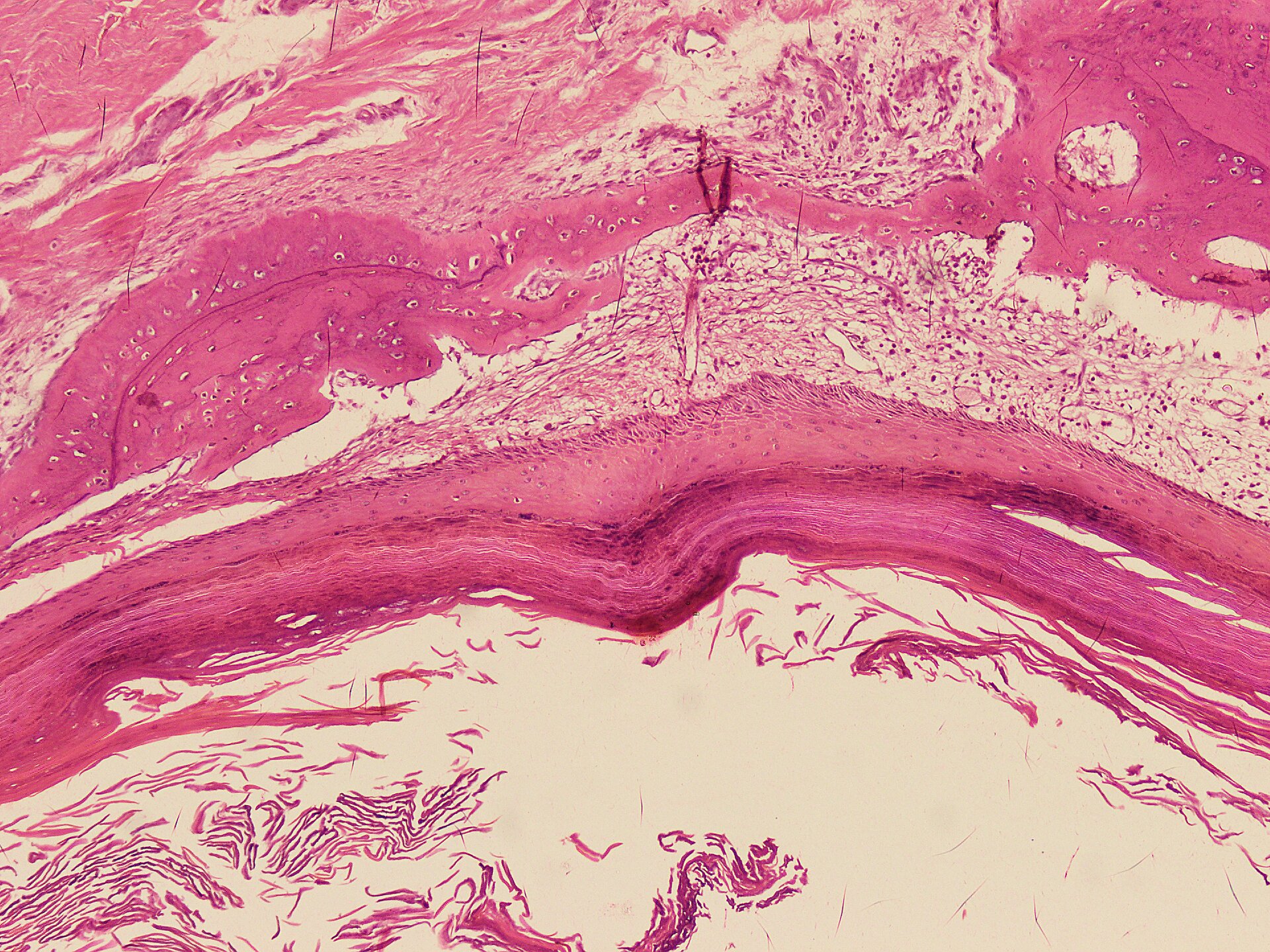
- Cyst is lined by squamous epithelium, including a granular layer
- Cyst wall is devoid of skin adnexal structures
- Cyst contents contain laminated keratin flakes
- Acute inflammation and foreign body type giant cell reaction may be present in the ruptured cyst
- Reference: Eur J Orthop Surg Traumatol 2019;29:1355
Microscopic (histologic) images
Positive stains
- H&E diagnosis
Sample pathology report
- Bone, right fifth distal phalanx, excision:
- Epidermoid inclusion cyst
Differential diagnosis
- Dermoid cyst:
- Presence of skin appendages in the cyst wall
- Compact keratin
- Enchondroma:
- Included in the radiological differential diagnosis
- Histologically composed of hypocellular cartilaginous nodules
- Glomus tumor:
- Rare in bone
- Included in the radiological differential diagnosis
- Histologically uniform small round cells with eosinophilic cytoplasm, distinct cell borders
- Osteomyelitis:
- Clinically mimics epidermal inclusion cyst
- Infiltration of bone by inflammatory cells including neutrophils, lymphocytes, and plasma cells
- Bone erosion and necrosis
- Reactive bone formation
- Squamous cell carcinoma:
- Associated precursor lesions, such as actinic keratosis or squamous cell carcinoma in situ are often present
- Invasion of dermis by tumor
- Tumor is composed of dysplastic squamous cells and may show lack of normal maturation
- Moderate and poorly differentiated carcinoma show focal or no keratinization
Additional references
Board review style question #1
Board review style answer #1
B. Cyst wall lined by benign squamous epithelium including a granular layer. The presence of a granular layer in the squamous epithelium and lamellated keratin are the key features that differentiate the epidermoid inclusion cyst from the trichilemmal (pilar) cyst.
Comment Here
Reference: Epidermoid inclusion cyst
Comment Here
Reference: Epidermoid inclusion cyst
Board review style question #2
Board review style answer #2
D. Presence of skin adnexal structures in the cyst wall. Dermoid cyst differs from epidermoid inclusion cyst by the presence of skin appendages.
Comment Here
Reference: Epidermoid inclusion cyst
Comment Here
Reference: Epidermoid inclusion cyst


