Table of Contents
Definition / general | Essential features | Terminology | ICD coding | Epidemiology | Sites | Pathophysiology | Etiology | Diagrams / tables | Clinical features | Diagnosis | Laboratory | Radiology description | Radiology images | Prognostic factors | Case reports | Treatment | Clinical images | Gross description | Gross images | Microscopic (histologic) description | Microscopic (histologic) images | Positive stains | Negative stains | Molecular / cytogenetics description | Sample pathology report | Differential diagnosis | Additional references | Board review style question #1 | Board review style answer #1 | Board review style question #2 | Board review style answer #2Cite this page: Zoshchuk B, Morisetti M, Beeter MC, Yeh YA. Urachus. PathologyOutlines.com website. https://www.pathologyoutlines.com/topic/bladderurachus.html. Accessed April 1st, 2025.
Definition / general
- The urachus, originated from remnants of allantois, is a fibrous cord connecting the umbilicus to the anterosuperior aspect of the bladder dome; usually obliterates at birth and becomes the median umbilical ligament (Kumar: Robbins & Cotran Pathologic Basis of Disease, 10th Edition, 2020)
- Urachal pathology results from failure of involution of the embryonic structures, resulting in the formation of a urachal cyst, umbilical urachal sinus, vesicourachal diverticulum or patent urachus (completely patent, tubular remnant communicating the developing bladder and the umbilical cord) (StatPearls: Patent Urachus [Accessed 29 December 2022])
Essential features
- The urachus progressively constricts at 12 weeks of gestation and becomes a fibrous cord connecting the umbilicus to the anterosuperior aspect of the bladder dome by the end of gestation (Moore: The Developing Human - Clinically Oriented Embryology, 11th Edition, 2019)
Terminology
- Patent urachus: completely patent tubular lesion communicating the bladder and the umbilical cord
- Umbilical urachal sinus: tubulocystic lesion with an opening in the umbilicus while the other end is closed (BMJ Case Rep 2016;2016:bcr2016215374)
- Vesicourachal diverticulum: tubulocystic lesion with an opening in the bladder while the other end is closed
- Urachal cyst: cystic structure with both the umbilical and bladder ends closed (BMJ Case Rep 2016;2016:bcr2016215374)
- Urachal remnants within bladder wall: tubulocystic lesion located in the bladder wall
ICD coding
Epidemiology
- Uncommon, 1.03% of pediatric population (J Urol 2015;193:632)
- Affected age range is from 1 to 20 years (Urology 1998;52:120)
- Average age of diagnosis: 4 years
- Rare in adults; urachal anomalies usually involute in early childhood (Urol Int 2022;106:195)
- Urachal cyst is the most common type of urachal anomaly (J Pediatr Surg 2013;48:2148)
- Patent urachus:
- 1 to 2 or 2.5 per 1,000,000 live births (Medicina (Kaunas) 2022;58:1621)
- M:F = 2:1
Sites
- Retzius space: prevesical or retropubic extraperitoneal space between the pubic symphysis and the urinary bladder
- Patent urachus: a tube-like structure between the umbilicus and bladder (diagram 1D) (BMJ Case Rep 2016;2016:bcr2016215374)
- Urachal sinus: a blind focal dilatation at the umbilical end (diagram 1E)
- Urachal cyst: a cystic structure along the urachus (diagram 1F)
- Vesicourachal diverticulum: an outpouching at the vesical end (diagram 1C) (BMJ Case Rep 2016;2016:bcr2016215374)
Pathophysiology
- At the second week of gestation, the allantois emerges from the posteroinferior yolk sac
- Allantois: an embryonic remnant with connection between the umbilicus and the cloaca (diagram 1A) (BMJ Case Rep 2016;2016:bcr2016215374)
- During the fourth to seventh weeks of gestation, the cloaca develops into urogenital sinus and the anal canal
- The superior part of the urogenital sinus continuous with the allantois develops into the urinary bladder (Differentiation 2018;103:66)
- During the fourth and fifth weeks of gestation, the urachal lumen (allantois) narrows to form a small tubular structure lined by urothelium (Moore: The Developing Human - Clinically Oriented Embryology, 8th Edition, 2007)
- By the twelfth week of gestation, the allantois progressively constricts, resulting in a fibromuscular strand structure known as urachus (Moore: The Developing Human - Clinically Oriented Embryology, 11th Edition, 2019)
- By the time of birth, the urachus becomes a thin fibrous cord that extends from the umbilicus to the urinary bladder dome (diagram 1B) (BMJ Case Rep 2016;2016:bcr2016215374)
- If the lumen of the allantois persists, it can develop into urachal anomalies:
- Urachal diverticulum (diagram 1C)
- Patent urachus (diagram 1D)
- Urachal sinus (diagram 1E)
- Urachal cyst (diagram 1F)
Etiology
- Developmental anomaly: defective obliteration of the lumen of allantois in the twelfth week of gestation (J Pediatr Surg 2013;48:2148)
- May be associated with other genitourinary malformations, especially posterior urethral valves (StatPearls: Patent Urachus [Accessed 10 January 2023])
Diagrams / tables
Clinical features
- Continuous or intermittent drainage of fluid from the umbilicus (Pediatr Emerg Care 2015;31:202)
- Additional manifestations include an enlarged or edematous umbilicus and delayed healing of the cord stump (J Paediatr Child Health 2017;53:1123)
- Urinary tract infection symptoms or calculi
- Abdominal mass (Clin Case Rep 2021;9:e04664)
- More often asymptomatic
Diagnosis
- Prenatal:
- Ultrasonography (US): increased thickness of the umbilicus observed as an extra-abdominal cystic mass (Medicina (Kaunas) 2022;58:1621)
- Early prenatal detection for appropriate counseling for postpartum corrective surgery
- Postnatal:
- Abnormally thick umbilical cord should prompt further investigation into the potential for a urachal anomaly (StatPearls: Patent Urachus [Accessed 10 January 2023])
- Demonstration of the fluid filled canal on longitudinal ultrasound or contrast filling on retrograde fistulography or voiding cystourography (VCUG)
- CT or MRI, usually depends on the bladder's filling status (Br J Radiol 2020;93:20190118)
- Surgical exploration (sometimes) (Urol Int 2022;106:195)
- Postsurgical histopathological examination
- Ultrasound: diagnostic for 82% of cysts, 100% of sinuses, 100% of patent urachus
- Voiding cystourethrogram: diagnostic for 100% of patent urachus
- CT scan: diagnostic for 71% of cysts (J Pediatr Urol 2007;3:500)
Laboratory
- Check creatinine level if urine in the umbilical drainage is suspected (StatPearls: Patent Urachus [Accessed 10 January 2023])
- Microorganisms cultured from the umbilical drainage often include Staphylococcus aureus, Escherichia coli, Enterococcus, Citrobacter and rarely, Proteus species
Radiology description
- Ultrasound:
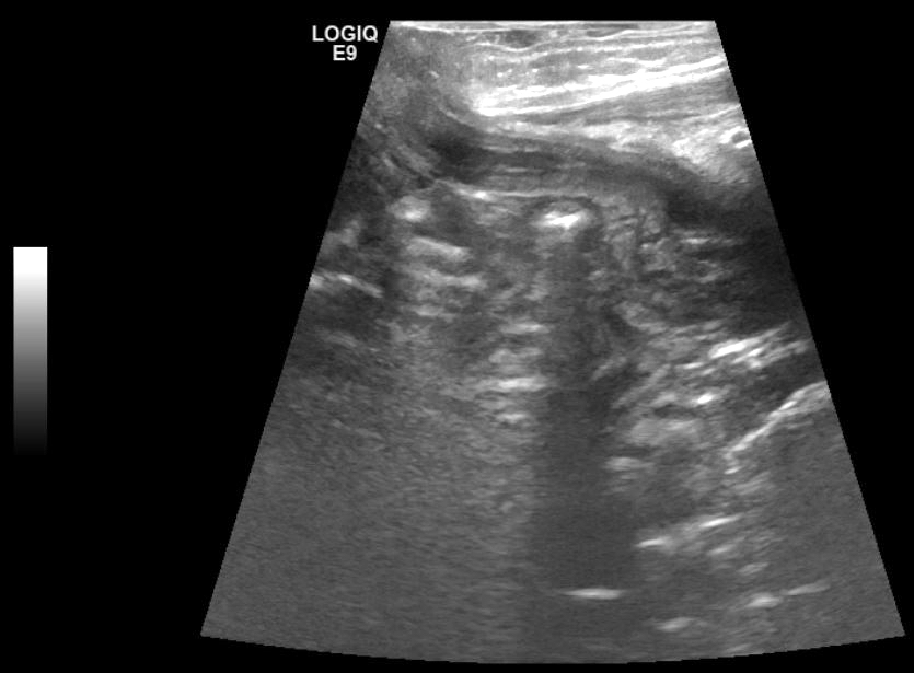
- Urachal cyst: ultrasound shows hypoechoic cystic mass under abdominal wall between the umbilicus and bladder dome (radiology image 1) (Medicine (Baltimore) 2020;99:e18884)
- Patent urachus: ultrasound shows tubular connection between the anterosuperior bladder and the umbilicus
- Umbilical urachal sinus: ultrasound shows thickened tubular structure along the midline below the umbilicus
- VCUG or fluoroscopic fistulogram with instilled contrast media identified the umbilical vesical tract (radiology image 2) (Medicine (Baltimore) 2022;101:e29187)
- Abdominal CT scan: tubular connection between the umbilicus and bladder (Pediatr Neonatol 2022;63:105)
- CT and MRI: air, fluid or calculi within the patent urachus (Br J Radiol 2020;93:20190118)
- Vesicourachal diverticulum: axial CT shows midline cystic lesion just above the bladder (Radiographics 2001;21:451)
Radiology images
Prognostic factors
- Prenatally, an increasing cystic mass may lead to compression on the umbilical vessels
- Complications of patent urachus: recurrent urinary infections and omphalitis (Semin Pediatr Surg 2007;16:41)
- Infection leads to bladder rupture (Urologe A 2003;42:834)
- Increased frequency in Prune belly syndrome (25 - 30% with patent urachus) (Pathol Annu 1977;12:17)
- Higher risk of urachal cancer (urachal adenocarcinoma) in adults, with an estimated incidence of 0.18 per 100,000 (Urol Int 2022;106:195, J Surg Case Rep 2018;2018:rjy056)
Case reports
- Newborn with prenatally ruptured patent urachus (Medicina (Kaunas) 2022;58:1621)
- Newborn boy with patent urachus and pyocele (Medicine (Baltimore) 2022;101:e29187)
- 3 month old girl with a persistently wet umbilicus since birth, local dermatitis and medical history of urinary tract infection (Pediatr Neonatol 2022;63:437)
- 4 year old boy with abdominal pain, vomiting and constipation (APSP J Case Rep 2017;8:8)
- 5 year old boy with severe acute abdominal pain (Medicine (Baltimore) 2020;99:e18884)
- 18 year old man and 20 year old woman with abdominal pain (Int J Surg Case Rep 2022;97:107394)
- 30 year old woman with dysuria and lower abdominal pain (Urol Ann 2018;10:219)
- 33 year old woman with pelvic pain and dysuria (Case Rep Obstet Gynecol 2015;2015:791408)
Treatment
- Surgical excision (open or laparoscopic) for patent urachus and other types of symptomatic urachal remnants (Semin Pediatr Surg 2007;16:41)
- Incision and drainage and antibiotics for infected umbilicus
- Spontaneous resolution in a subset of patients (J Pediatr Surg 2013;48:2148)
Clinical images
Gross description
- Urachal cyst: mucinous or fluid filled cyst with both umbilical and bladder ends closed and located between umbilicus and the bladder dome (Cheng: Urologic Surgical Pathology, 4th Edition, 2019)
- Patent urachus: completely persistent connection between the bladder and the umbilicus (J Pediatr Surg 2013;48:2148)
- Urachal diverticulum: outpouching of the bladder apex with umbilical end closed
- Urachal sinus: obliterated distal urachus and patent proximal urachus connected to the umbilicus (Urol Int 2022;106:195)
- Urachal remnants in bladder wall: deep in the bladder wall of the apex or in the muscularis propria of the bladder wall (Amin: Diagnostic Pathology - Genitourinary, 3rd Edition, 2022)
- Fibrous cord when the lumen is obliterated
Gross images
Microscopic (histologic) description
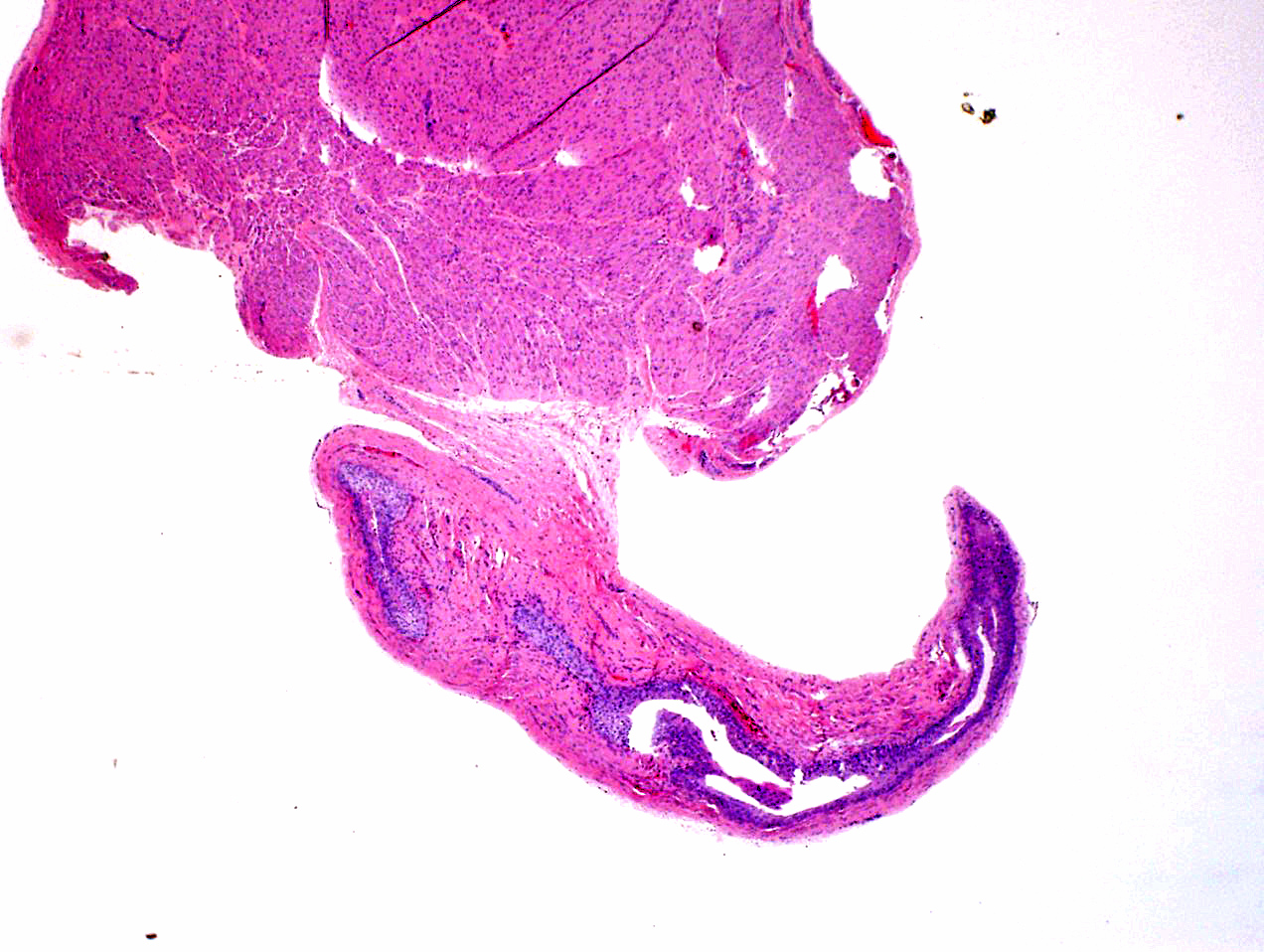
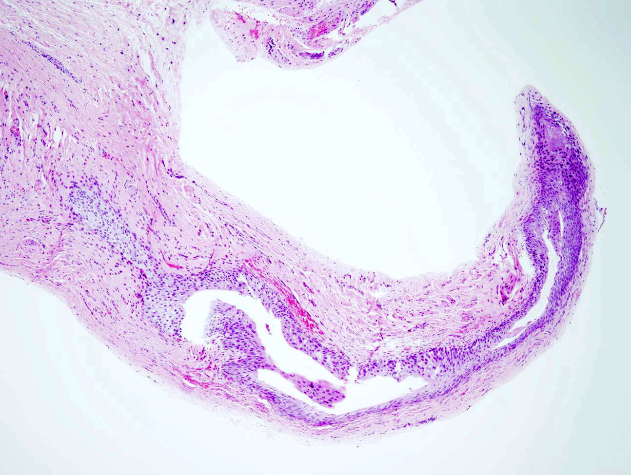
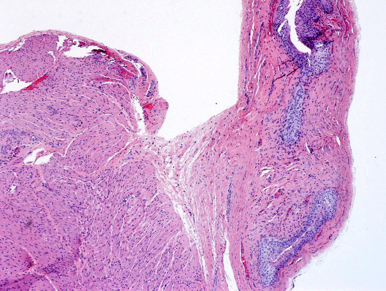
- Patent urachus, umbilical urachal sinus, vesicourachal diverticulum
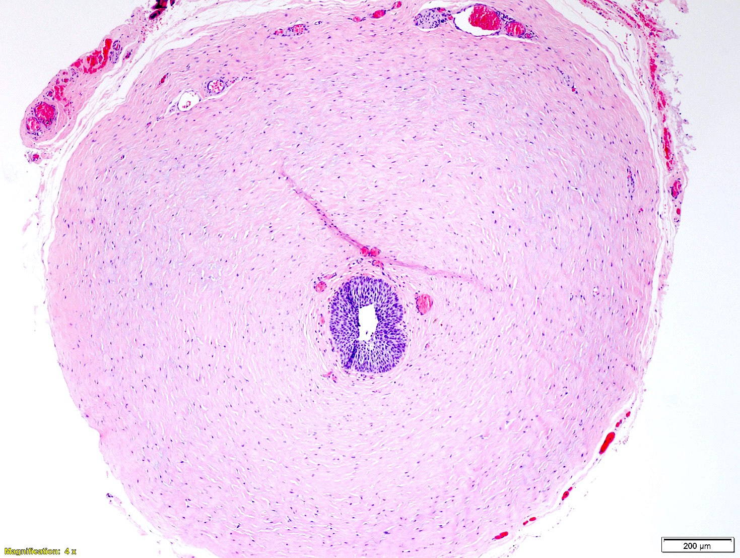
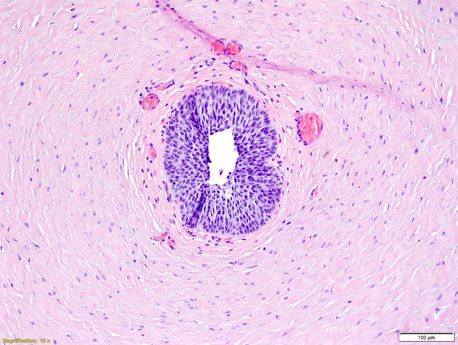
- Completely or incompletely opened structures lined by benign urothelium with or without squamous metaplasia
- Glandular metaplasia may present
- Urachal cyst
- Cystic remnants lined by benign cuboidal cells, flattened epithelial cells or urothelial cells
- Intestinal (columnar cells with or without goblet cells) metaplasia may present
- Urachal remnants in bladder wall (Cheng: Urologic Surgical Pathology, 4th Edition, 2019)
- Embryonic remnants in the bladder wall that are lined by benign urothelial cells
- Squamous or glandular (columnar cells with or without goblet cells) metaplasia may present
Microscopic (histologic) images
Positive stains
Negative stains
Molecular / cytogenetics description
- Microdeletion in 1q21.1q21.2 (1.82 Mb deletion) (Diagnostics (Basel) 2021;11:2332)
Sample pathology report
- Urinary bladder wall lesion, excision:
- Tubulocystic lesion lined by urothelial epithelium consistent with urachal remnants (see comment)
- Comment: Sections of the bladder wall lesion show a tubular structure lined by urothelium and attached to the muscularis propria. These findings are consistent with urachal remnants.
Differential diagnosis
- Patent omphalomesenteric (vitelline) duct:
- Cystic-like structure lined by intestinal epithelium and communication between the umbilicus and the ileum (Pediatr Pathol 1983;1:325, Hum Pathol 1989;20:458)
- Endometriosis (Arch Pathol Lab Med 2014;138:432):
- Endocervicosis (Arch Pathol Lab Med 2014;138:432):
- Endocervical-like glands with or without (CD10+) endometrial-like stroma
- Monolayer of columnar mucinous lining cells with abundant pale to clear cytoplasm
- Ciliated cells often present among mucinous cells
- Adenocarcinoma (primary or metastasis) (Am J Surg Pathol 2009;33:659):
- Primary urachal carcinoma (Am J Surg Pathol 2009;33:659):
- Metastatic adenocarcinoma:
- Infiltrating malignant glands with dysplastic / pleomorphic cells (Am J Surg Pathol 2001;25:1380)
- Invasive urothelial carcinoma (nested or glandular variant) (Mod Pathol 2003;16:1289):
Additional references
- BMJ Case Rep 2022;15:e247789, Clin Pract Cases Emerg Med 2022;6:186, Urology 2021;149:e1, Eur J Pediatr 2021;180:1987, BJR Case Rep 2016;2:20150226, Pediatr Emerg Care 2015;31:202, Intern Med 2014;53:1735, J Pediatr Surg 2007;42:e7, J Urol 2007;178:1615, Eur J Pediatr Surg 2004;14:206, J Urol 2003;169:1478, Int J Urol 1994;1:275, Am J Obstet Gynecol 1982;143:61
Board review style question #1
During bladder catheterization in a newborn boy, the tip of the catheter came out through his enlarged umbilical stump. What is the diagnosis?
- Patent omphalomesenteric duct
- Patent urachus
- Umbilical urachal sinus
- Urachal cyst
- Vesicourachal diverticulum
Board review style answer #1
B. Patent urachus. Patent urachus creates a tubular connection between the umbilicus and the anterosuperior wall of the bladder. Answer A is incorrect because patent omphalomesenteric duct has connection between umbilicus and small intestine. Answers C, D and E are incorrect because umbilical urachal sinus, urachal cyst and vesicourachal diverticulum cannot be probed through.
Comment Here
Reference: Urachus and patent urachus
Comment Here
Reference: Urachus and patent urachus
Board review style question #2
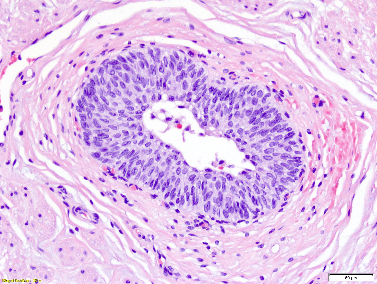
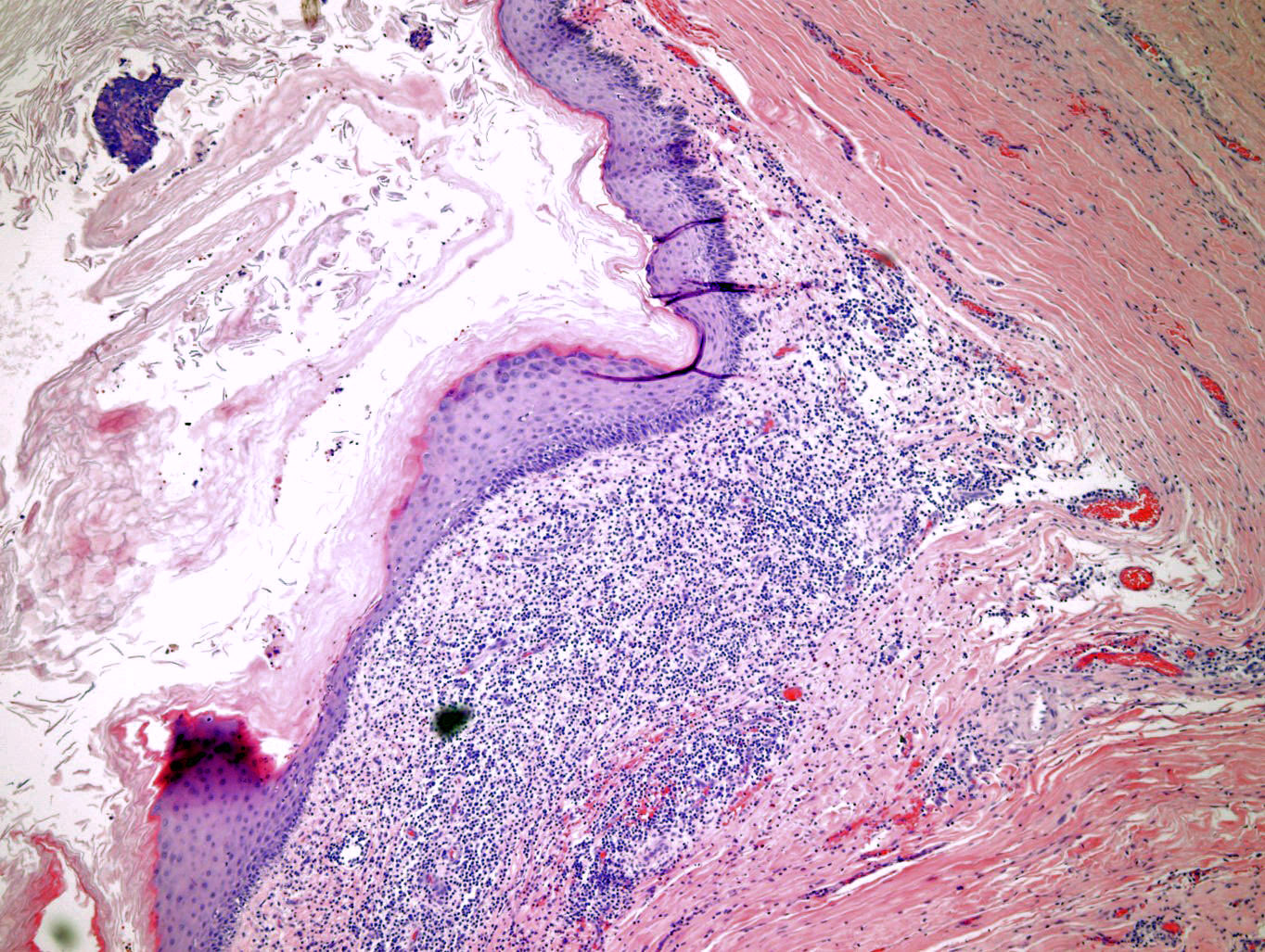
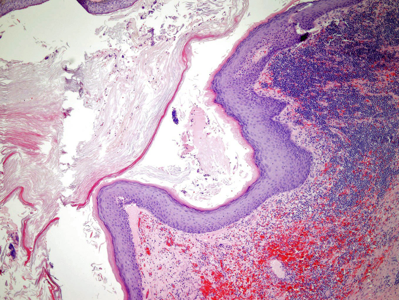
A 50 year old man complained of abdominal pain for several weeks. A noncontrast CT scan of the abdomen was performed. There was a cystic lesion in the anterosuperior bladder wall. No connection between the bladder and the umbilicus was noted. Surgical excision followed by pathological examination was performed. The photomicrograph is shown above. What is the diagnosis?
- Mesonephric remnant
- Omphalomesenteric duct remnant
- Urachal remnant in bladder wall
- Vitelline duct remnant
Board review style answer #2
C. Urachal remnant in bladder wall. Urachal remnants within the bladder wall are usually lined by urothelium but can show metaplastic changes, such as squamous metaplasia seen here. Mesonephric remnants are composed of small tubules lined by low columnar to cuboidal epithelial cells. The cells are immunoreactive to CK903, CD10 and vimentin. Omphalomesenteric duct remnant is the same as vitelline duct remnant, which is lined by gastrointestinal epithelium.
Comment Here
Reference: Urachus and patent urachus
Comment Here
Reference: Urachus and patent urachus


