Bladder & urothelial tract
General
Staging-renal pelvic carcinoma
Last author update: 9 September 2021
Last staff update: 1 December 2022
Copyright: 2003-2024, PathologyOutlines.com, Inc.
PubMed Search: Staging [title] renal pelvic tumors kidney
Page views in 2023: 3,904
Page views in 2024 to date: 3,188
Cite this page: Zynger DL. Staging-renal pelvic carcinoma. PathologyOutlines.com website. https://www.pathologyoutlines.com/topic/bladderstagingrenalpelvis.html. Accessed December 18th, 2024.
Definition / general
- All carcinomas of the renal pelvis are covered by this staging system
- These tumors are not covered: renal cell carcinoma, lymphoma and mesenchymal tumors
Essential features
- AJCC 7th edition staging was sunset on December 31, 2017; as of January 1, 2018, use of the 8th edition is mandatory
ICD coding
- ICD-10: C65.9 - renal pelvis
Primary tumor (pT)
- pTX: cannot be assessed
- pT0: no evidence of primary tumor
- pTa: noninvasive papillary carcinoma
- pTis: carcinoma in situ
- pT1: invades subepithelial connective tissue
- pT2: invades muscle
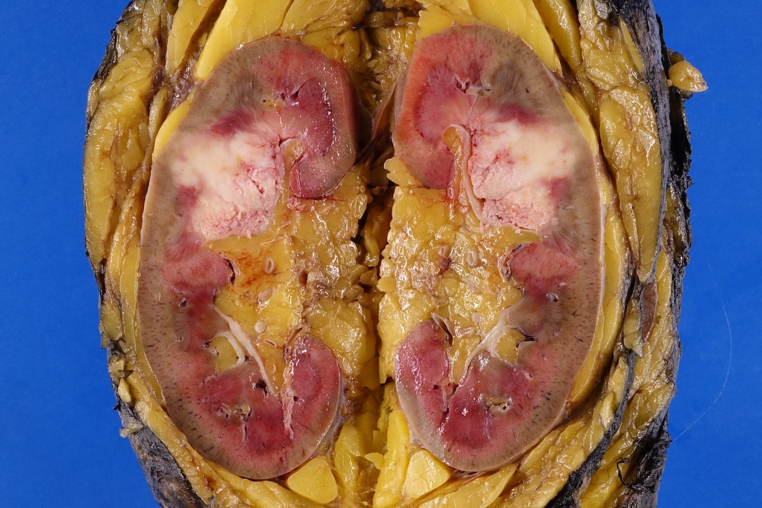
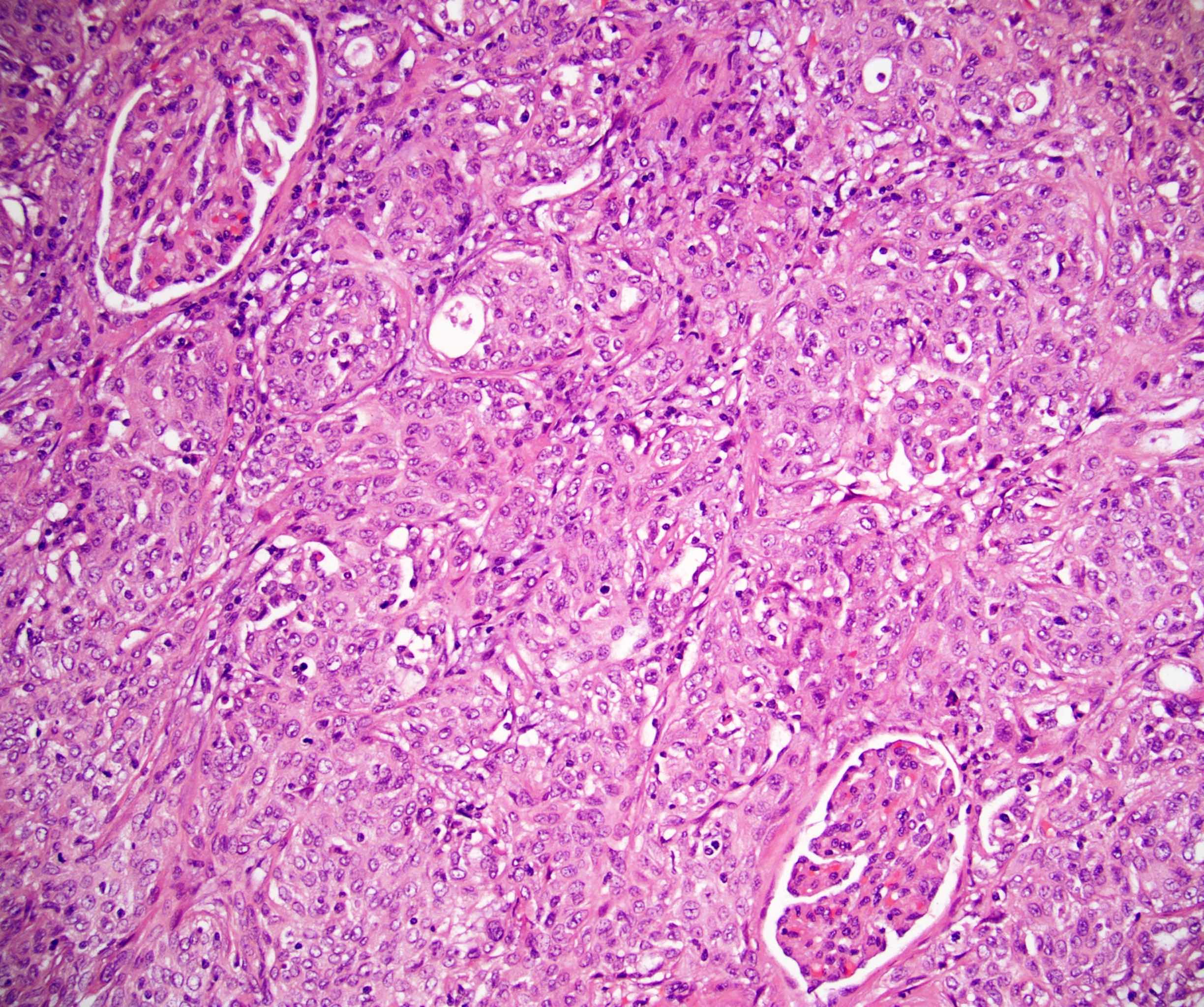
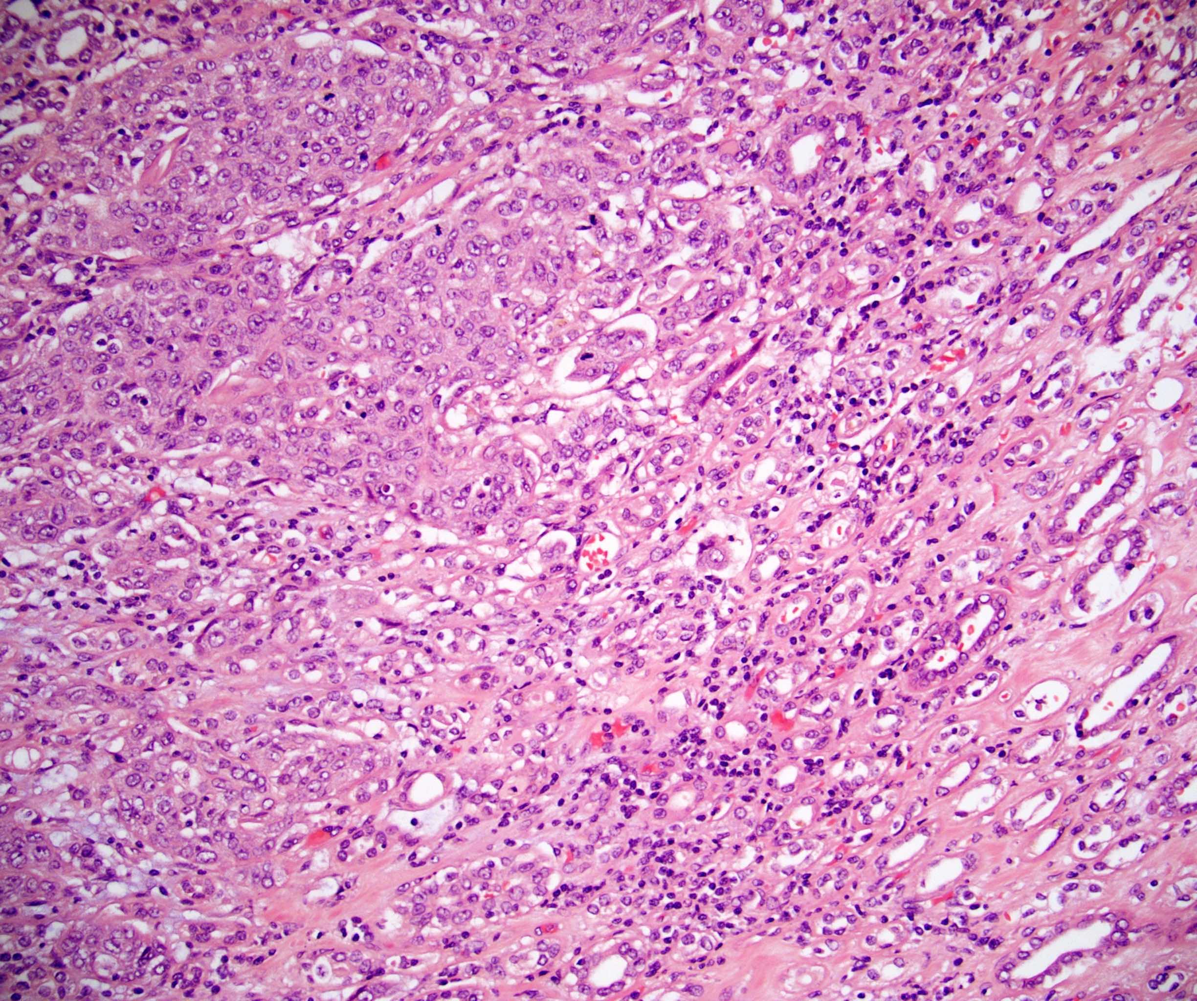
- pT3: invades peripelvic fat or renal parenchyma
- pT4: invades adjacent organs or perinephric fat
Regional lymph nodes (pN)
- pNX: cannot be assessed
- pN0: no regional lymph node metastasis
- pN1: 1 lymph node with tumor deposit ≤ 2 cm
- pN2: 1 lymph node with tumor deposit > 2 cm or metastases in multiple nodes
Notes:
- Regional lymph nodes include hilar, paracaval, aortic and retroperitoneal
Prefixes
- y: preoperative radiotherapy or chemotherapy
- r: recurrent tumor stage
AJCC prognostic stage groups
| Stage group 0a: | | Ta | | N0 | | M0
|
| Stage group 0is: | | Tis | | N0 | | M0
|
| Stage group I: | | T1 | | N0 | | M0
|
| Stage group II: | | T2 | | N0 | | M0
|
| Stage group III: | | T3 | | N0 | | M0
|
| Stage group IV: | | T4 | | NX - 2 | | M0 - 1
|
| | TX - 4 | | N1 - 2 | | M0 - 1
|
| | TX - 4 | | NX - 2 | | M1
|
Registry data collection variables
- Extranodal extension
- Size of largest tumor deposit within a lymph node
- Total number of lymph nodes
- Presence of carcinoma in situ
- Presence of noninvasive papillary carcinoma
- Lymphovascular invasion
- Urothelial carcinoma grade (high / low)
- Squamous cell / adenocarcinoma grade (1 - 3)
- Intratubular renal in situ spread
Histologic grade (G)
- Urothelial carcinoma
- LG: low grade
- HG: high grade
- Squamous cell / adenocarcinoma
- GX: cannot be assessed
- G1: well differentiated
- G2: moderately differentiated
- G3: poorly differentiated
Histopathologic type
- Noninvasive urothelial carcinoma
- Low grade papillary urothelial carcinoma
- High grade papillary urothelial carcinoma
- Urothelial carcinoma in situ
- Invasive urothelial carcinoma
- Conventional urothelial carcinoma
- Urothelial carcinoma variants
- Squamous cell carcinoma
- Adenocarcinoma
- Small cell carcinoma
Gross images
Contributed by Debra L. Zynger, M.D.

Renal pelvic and peripelvic fat invasion (pT3)
Microscopic (histologic) images
Contributed by Debra L. Zynger, M.D.


Renal parenchyma invasion (pT3)
Board review style question #1
A urothelial carcinoma of the renal pelvis at the deepest point of invasion involves the renal parenchyma. Which is the correct pT category?
- pT1
- pT2
- pT3
- pT4

Back to top


