Table of Contents
Definition / general | Essential features | Terminology | ICD coding | Epidemiology | Sites | Pathophysiology | Etiology | Clinical features | Diagnosis | Laboratory | Radiology description | Prognostic factors | Case reports | Treatment | Gross description | Gross images | Frozen section description | Microscopic (histologic) description | Microscopic (histologic) images | Positive stains | Negative stains | Molecular / cytogenetics description | Sample pathology report | Differential diagnosis | Additional references | Board review style question #1 | Board review style answer #1 | Board review style question #2 | Board review style answer #2Cite this page: Brown ML, Tretiakova M. Sarcomatoid variant. PathologyOutlines.com website. https://www.pathologyoutlines.com/topic/bladdersarcomatoidvariant.html. Accessed April 2nd, 2025.
Definition / general
- Variant of urothelial carcinoma; morphologically indistinguishable from sarcoma
- Heterologous elements may be present (Histopathology 2019;74:77)
Essential features
- Biphasic malignant neoplasm with morphologic and immunohistochemical evidence of both epithelial and mesenchymal differentiation
- Rare (0.3 - 0.6% of all urothelial carcinomas) and biologically aggressive variant
- Associated with radiation therapy or cyclophosphamide
- If present, heterologous elements should be mentioned in the report
Terminology
- WHO recommends the term sarcomatoid variant of urothelial carcinoma for these biphasic malignant neoplasms
- Terms carcinosarcoma, metaplastic carcinoma, spindle cell carcinoma and malignant mixed tumor have been used interchangeably but can lead to confusion and are therefore discouraged
ICD coding
- Location based ICD-10 coding:
- Renal pelvis, including pelviureteric junction and renal calyces
- Ureter, including ureteric orifice of bladder
- Bladder
- C67.0 - malignant neoplasm of trigone of bladder
- C67.1 - malignant neoplasm of dome of bladder
- C67.2 - malignant neoplasm of lateral wall of bladder
- C67.3 - malignant neoplasm of anterior wall of bladder
- C67.4 - malignant neoplasm of posterior wall of bladder
- C67.5 - malignant neoplasm of bladder neck
- C67.6 - malignant neoplasm of ureteric orifice
- C67.7 - malignant neoplasm of urachus
- C67.8 - malignant neoplasm of overlapping sites of bladder
- C67.9 - malignant neoplasm of bladder, unspecified
Epidemiology
- Estimated to represent 0.3 - 0.6% of all urothelial carcinomas (Mod Pathol 2009;22:S96, Eur Urol Focus 2020;6:653)
- Seen in up to 2.4% of all high grade urothelial carcinomas in renal pelvis
- Age 45 - 82 years (mean 71.6); M > F (2 - 3:1) (Oncol Lett 2014;8:1208, Pathol Res Pract 1996;192:1218)
- Although rare, sarcomatoid carcinoma is more common than primary sarcoma
Sites
- Kidney pelvis, proximal ureter and bladder
Pathophysiology
- TERT C228T promoter mutations in 35% of sarcomatoid carcinoma (Histopathology 2019;74:77, Eur Urol Focus 2020;6:653)
Etiology
- Probably has a common malignant clonal origin, with epithelial and mesenchymal differentiation (J Pathol 2007;211:420)
- Common final pathway of all forms of bladder tumors, supported by molecular and morphologic evidence (Histopathology 2019;74:77)
- Risk factors include previous exposure to radiotherapy and intravesicular cyclophosphamide (Eur Urol Focus 2020;6:653)
Clinical features
- Gross hematuria, flank pain, an abdominal mass and hydronephrosis
- Similar to those of conventional urothelial tumors
- Reference: ISRN Urol 2014;2014:794563
Diagnosis
- Cystoscopy
- CT scan
- MRI
- Reference: ISRN Urol 2014;2014:794563
Laboratory
- Hematuria
Radiology description
- Thickened bladder wall with or without intraluminal papillary or nodular mass; heterologous elements such as calcification in osteosarcoma may be demonstrated (Radiographics 2006;26:553)
Prognostic factors
- Poor prognosis, as frequently presents at an advanced stage and is associated with a worse overall survival when compared with pure urothelial carcinoma (Eur Urol Focus 2020;6:653)
- Similar to bladder, sarcomatoid urothelial carcinoma of renal pelvis has a worse prognosis on univariate analysis (ISRN Urol 2014;2014:794563, Oncol Lett 2014;8:1208, J Urol 2012;188:398)
- Associated with higher tumor stage, multifocality, tumor necrosis, frequent metastases at presentation; however, insufficient case numbers in kidney to confirm independent negative prognostic impact on multivariate analysis (J Urol 2012;188:398)
- May have heterologous elements, without definite prognostic significance (Mod Pathol 2009;22:S96)
Case reports
- 49 year old man with urothelial carcinoma arising in renal pelvis with exuberant chondrosarcomatous element (Indian J Pathol Microbiol 2014;57:284)
- 63 year old man with squamous cell and sarcomatoid variant urothelial carcinoma (BMC Surg 2021;21:96)
- 68 year old man with sarcomatoid carcinoma arising in renal pelvis (Open J Pathol 2013;3:96)
- 72 year old man with sarcomatoid urothelial carcinoma of the ureter with heterologous elements of chondrosarcoma and osteosarcoma and divergent squamous differentiation (Urol Case Rep 2020;34:101484)
- Sarcomatoid carcinoma of renal pelvis with giant cell tumor-like features (Int J Urol 2005;12:199)
Treatment
- Chemotherapy resistant (Eur Urol Focus 2020;6:653)
- No standard treatment due to rarity (Cancer Cell Int 2020;20:550)
Gross description
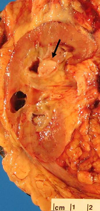
- Gray fleshy cut surface with infiltrative margins similar to sarcoma in appearance
Gross images
Frozen section description
- Not typically diagnosed on frozen section
Microscopic (histologic) description
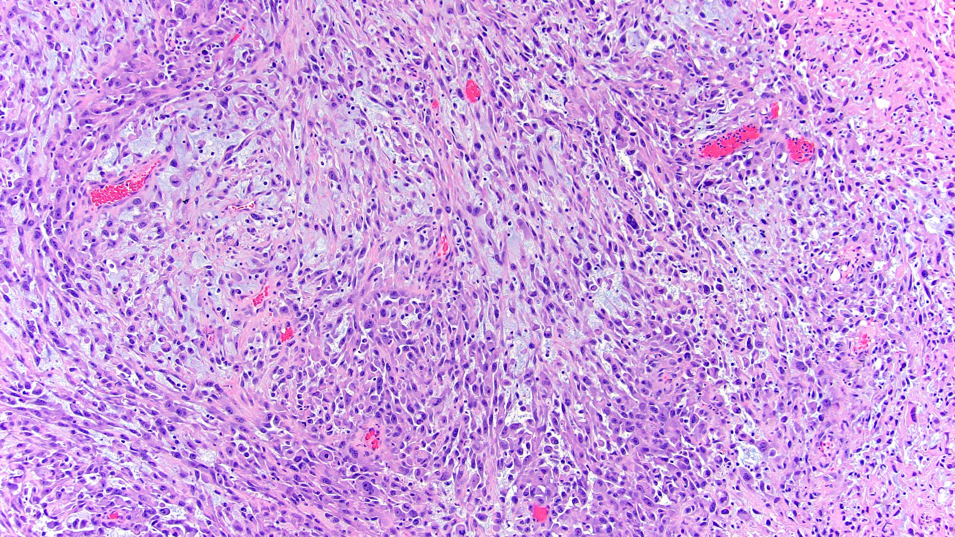
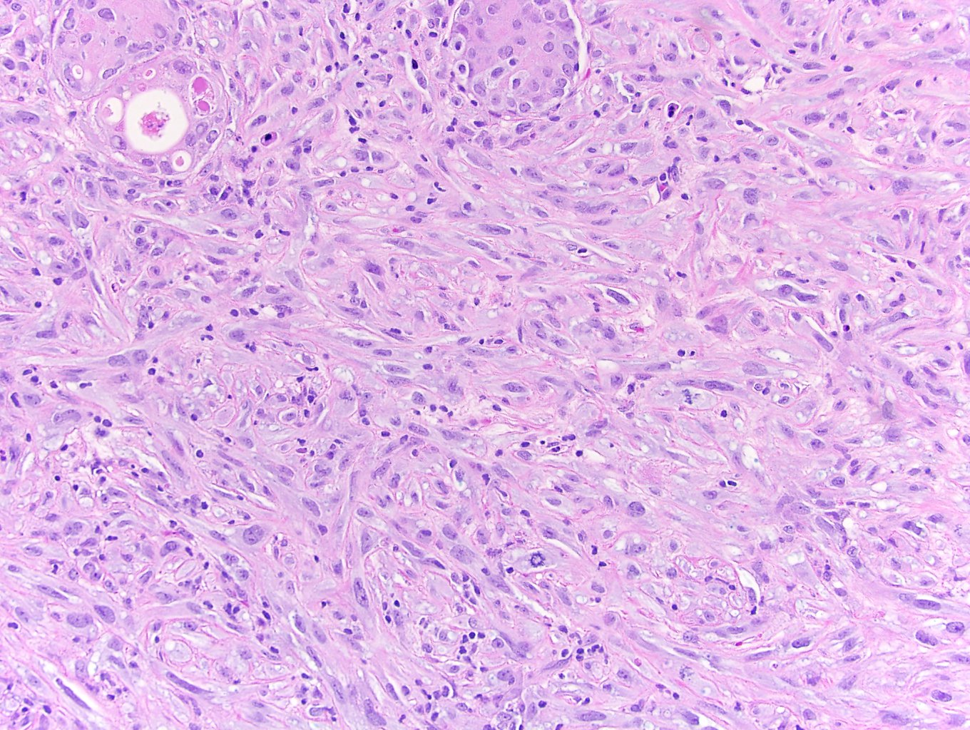
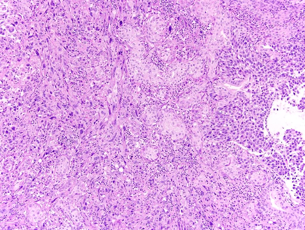
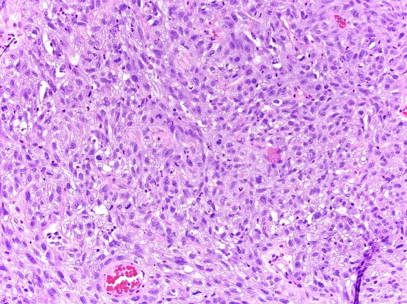
- Most common component is undifferentiated high grade spindle cell sarcoma (Histopathology 2019;74:77)
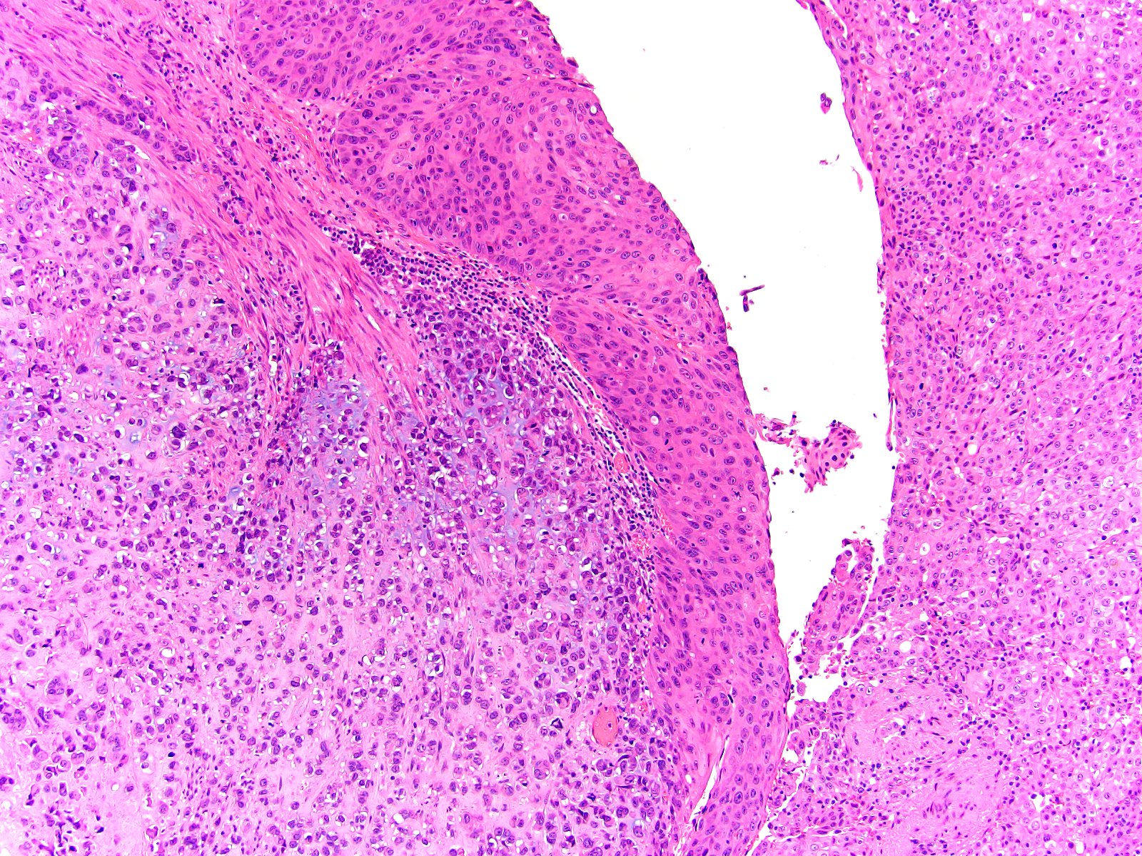
- Sarcomatoid areas admixed with conventional high grade urothelial carcinoma
- Spindle cell component may account for 10% to > 60% of tumor
- Epithelial component has urothelial differentiation or less commonly squamous or glandular differentiation
- Highly variable and can mimic nonepithelial neoplasms (J Biol Chem 2019;294:1579)
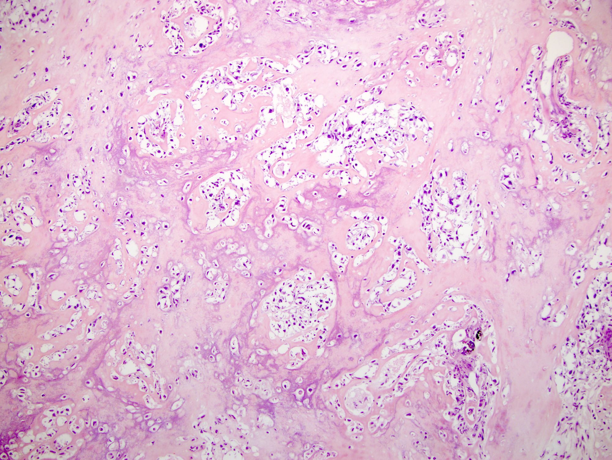
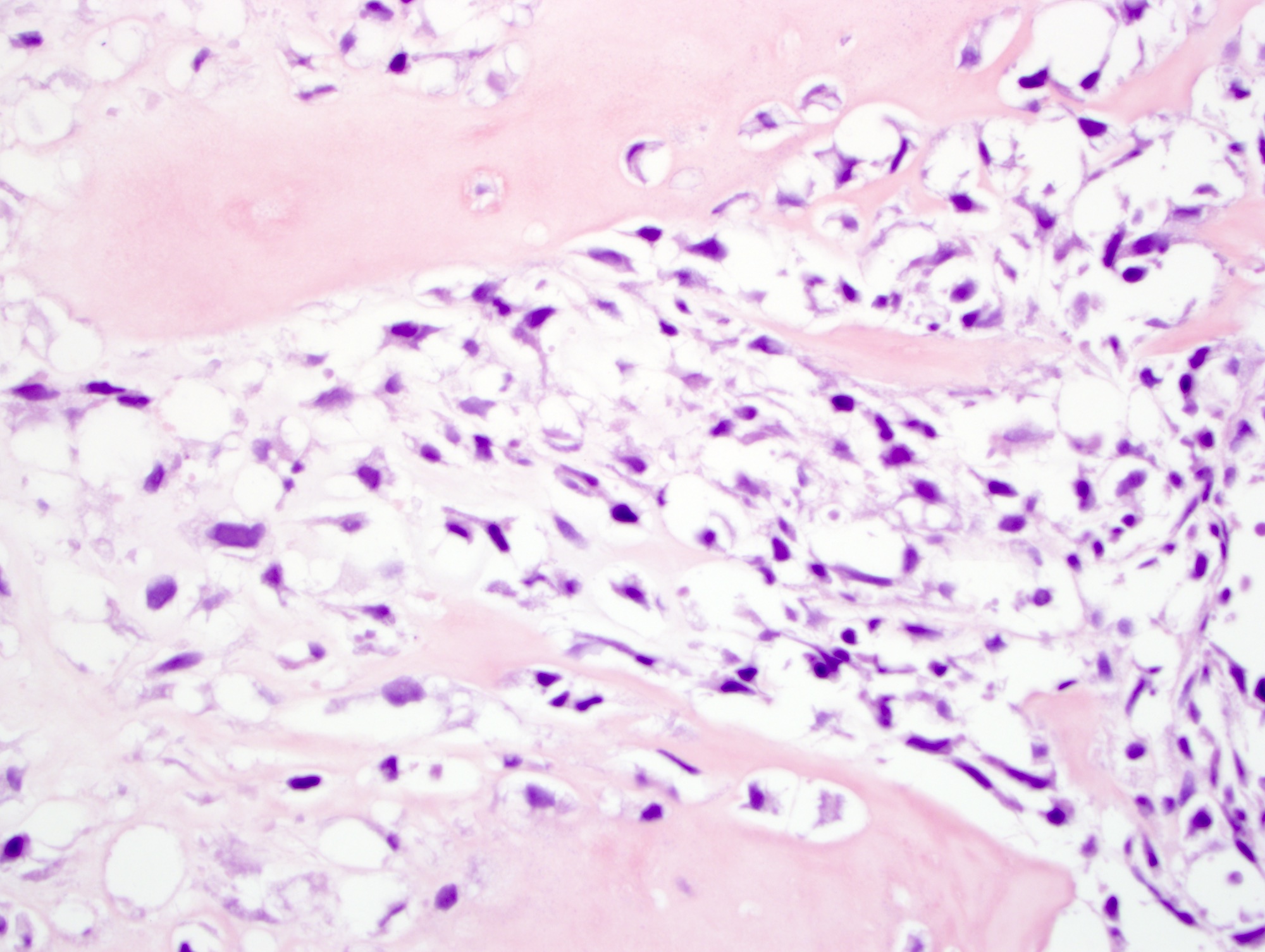
- Most common heterologous element is osteosarcoma, followed by chondrosarcoma, rhabdomyosarcoma, leiomyosarcoma, liposarcoma and angiosarcoma (Histopathology 2019;74:77)
Microscopic (histologic) images
Contributed by Megan L. Brown, M.D., Nicole K. Andeen, M.D., Maria Tretiakova, M.D., Ph.D. and Kenneth A. Iczkowski, M.D.
Positive stains
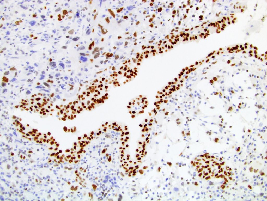
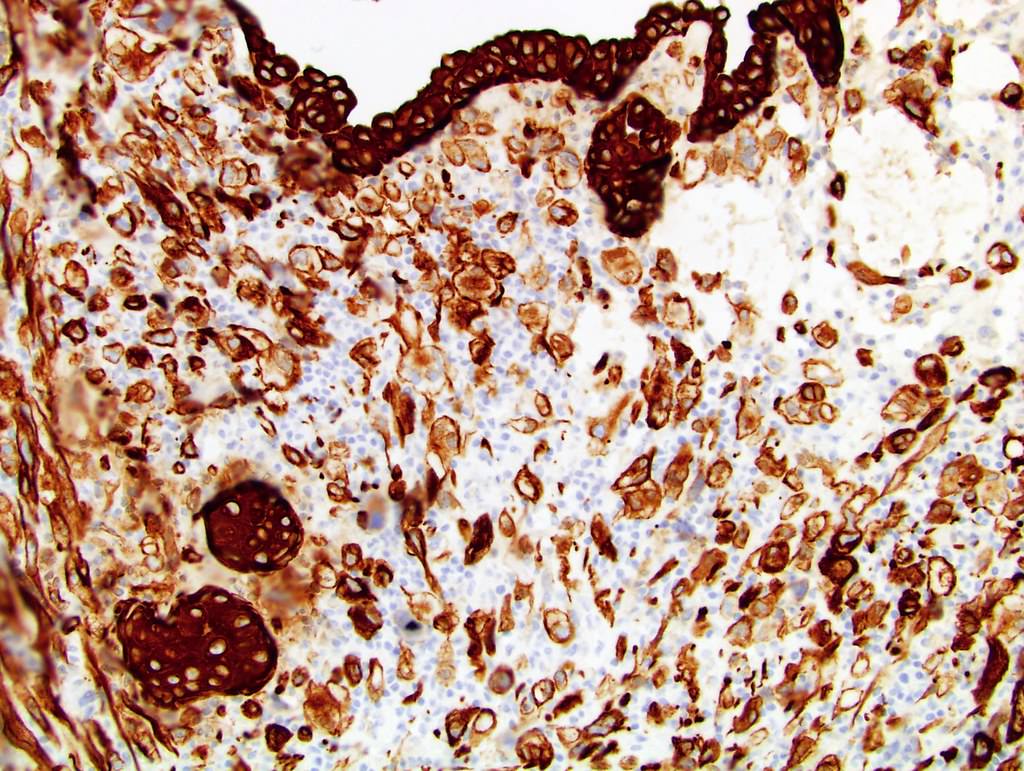
- Spindle cell component: vimentin (100%), EMA (100%), SMA (73%), AE1 / AE3 (70%), OSCAR cytokeratin (68%), p63 (50%), GATA3 and CK7 (Am J Surg Pathol 2009;33:99, Pathol Res Pract 1996;192:1218)
- PAX8 variable (Histopathology 2019;74:77)
Negative stains
- ALK1 (Am J Surg Pathol 2009;33:99)
- Desmin, calponin, h-caldesmon, myogenin (Histopathology 2019;74:77)
- CK5/6 (27% positive), 34βE12 (25% positive)
Molecular / cytogenetics description
- Overexpression of markers representative of epithelial - mesenchymal transition, including vimentin, FOXC2, SNAIL and ZEB1 (Histopathology 2019;74:77)
- TERT C228T promoter mutations in 35% of sarcomatoid carcinoma (Histopathology 2019;74:77, Eur Urol Focus 2020;6:653)
- Studies in urinary bladder showed:
- Similar patterns of allelic losses (J Pathol 2007;211:420)
- Similar TP53 point mutations between the carcinomatous and sarcomatous elements (Mod Pathol 2009;22:113)
- This suggests a common clonal origin with divergent differentiation (J Pathol 2007;211:420)
Sample pathology report
- Bladder, transurethral resection:
- High grade urothelial carcinoma with sarcomatoid differentiation (80%) (see comment)
- Comment: Invasive of muscularis propria (T2)
- Angiolymphatic invasion absent
- Muscularis propria present, involved by tumor
Differential diagnosis
- Inflammatory myofibroblastic tumor (IMT):
- IMT does not have associated malignant epithelial component
- Immunohistochemically, IMT expresses pankeratins but is usually negative for high molecular weight cytokeratins 34βE12 and CK5/6 and should express ALK (Histopathology 2009;55:491)
- Primary or metastatic sarcoma
- Sarcomatoid renal cell carcinoma:
- Identification and classification of associated in situ or invasive epithelial component is necessary; may require additional sampling and immunostaining
- Urothelial carcinoma with pseudosarcomatous stroma:
- Characterized by myxoid stroma, blood vessels and atypical cells
- Unlike sarcomatoid urothelial carcinoma, the stromal component lacks an expansile growth pattern, mitoses or expression of epithelial markers (Arch Pathol Lab Med 2007;131:1244, Mod Pathol 2009;22:S96)
Additional references
Board review style question #1
A 65 year old presented with bladder wall thickening on ultrasound. Transurethral resection of bladder tumor is performed and shows a lesion with features on H&E as seen in the image shown above. Which of the following is likely in the patient’s medical history?
- Asbestos exposure
- Family history of prostate adenocarcinoma
- High nitrate consumption in their diet
- Intravesical cyclophosphamide
- Woodworking hobby
Board review style answer #1
D. Intravesical cyclophosphamide has been associated with the development of sarcomatoid variant of urothelial carcinoma (Eur Urol Focus 2020;6:653)
Comment Here
Reference: Sarcomatoid variant
Comment Here
Reference: Sarcomatoid variant
Board review style question #2
Board review style answer #2
D. Osteosarcoma is the most commonly identified heterologous element in the sarcomatoid variant of urothelial carcinoma (Histopathology 2019;74:77)
Comment Here
Reference: Sarcomatoid variant
Comment Here
Reference: Sarcomatoid variant


