Superpage
Superpage Topics
Acanthamoeba
Acinetobacter (pending)
Actinomyces
Anaplasma
Angiostrongyliasis
Arthropod ectoparasites (pending)
Artifacts
Ascaris
Aspergillus
Babesia
Balamuthia
Biosafety for lab and pathology (pending)
Blastocystis
Blastomyces
Blood culture contamination (pending)
Borrelia burgdorferi / Lyme disease
Borrelia recurrentis / relapsing fever (pending)
Candida auris
Candida species (pending)
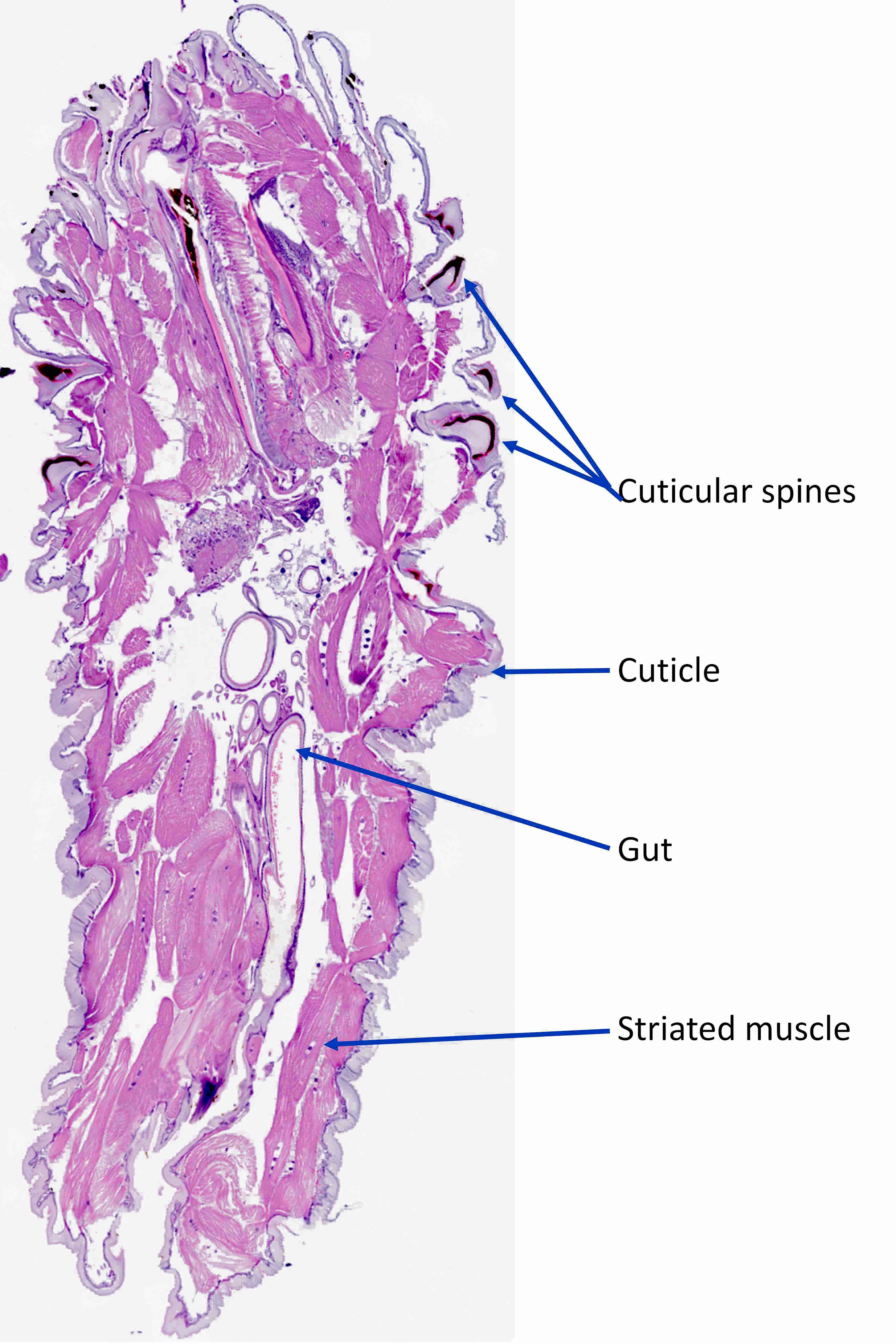
Cimex lectularius (bed bug)
Clostridioides difficile
Clostridium perfringens / C. septicum
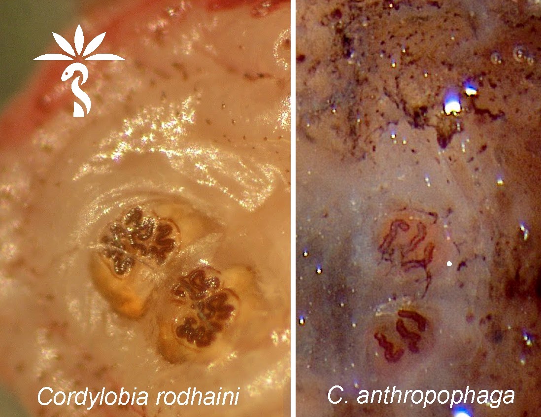
Cordylobia rodhaini (Lund fly)
Corynebacterium diphtheriae (pending)
COVID-19 (SARS-CoV-2) testing
Cryptococcus neoformans & gattii
Cutibacterium acnes
Cyclospora cayetanensis
Dematiaceous molds
Dengue fever
Dermatophagoides
Dermatophytes (pending)
Diphyllobothrium latum
Dipylidium caninum
Dirofilaria immitis (pending)
Dirofilaria repens
DNA Viruses
E. coli
Ebola (pending)
Echinococcal cyst
Ehrlichia
Enterococcus (pending)
Enteromonas hominis
Fasciola
Filariasis
Fusobacterium necrophorum
Haemophilus influenzae
Histoplasma capsulatum
HIV testing (pending)
Hookworm
HSV1 / HSV2
Hyaline molds
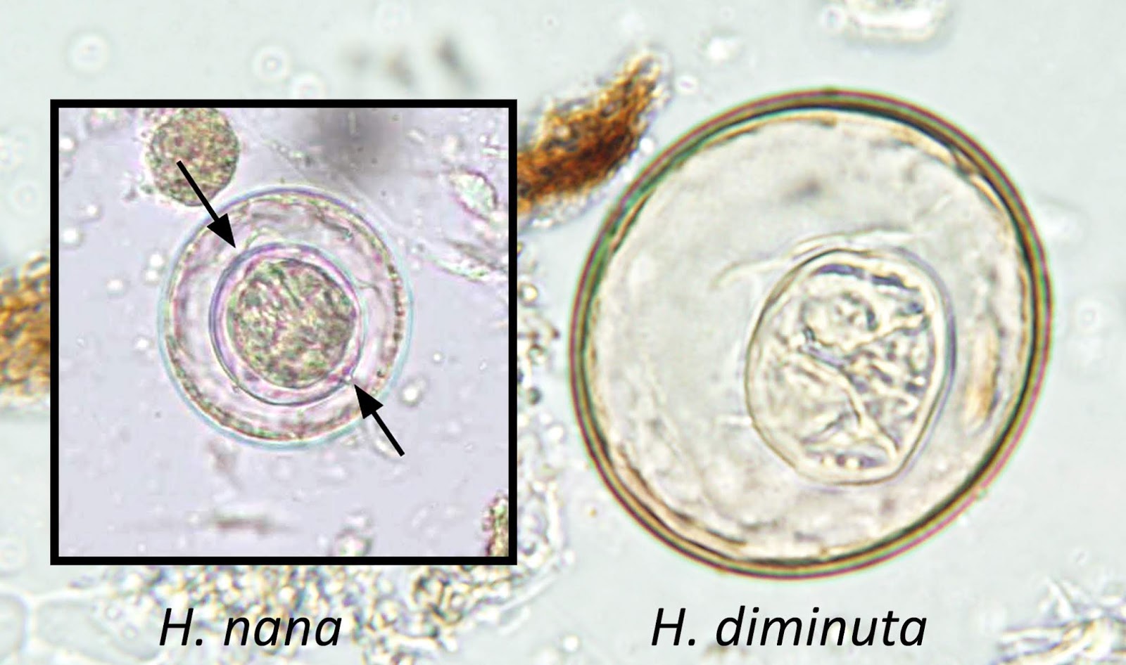
Hymenolepis diminuta
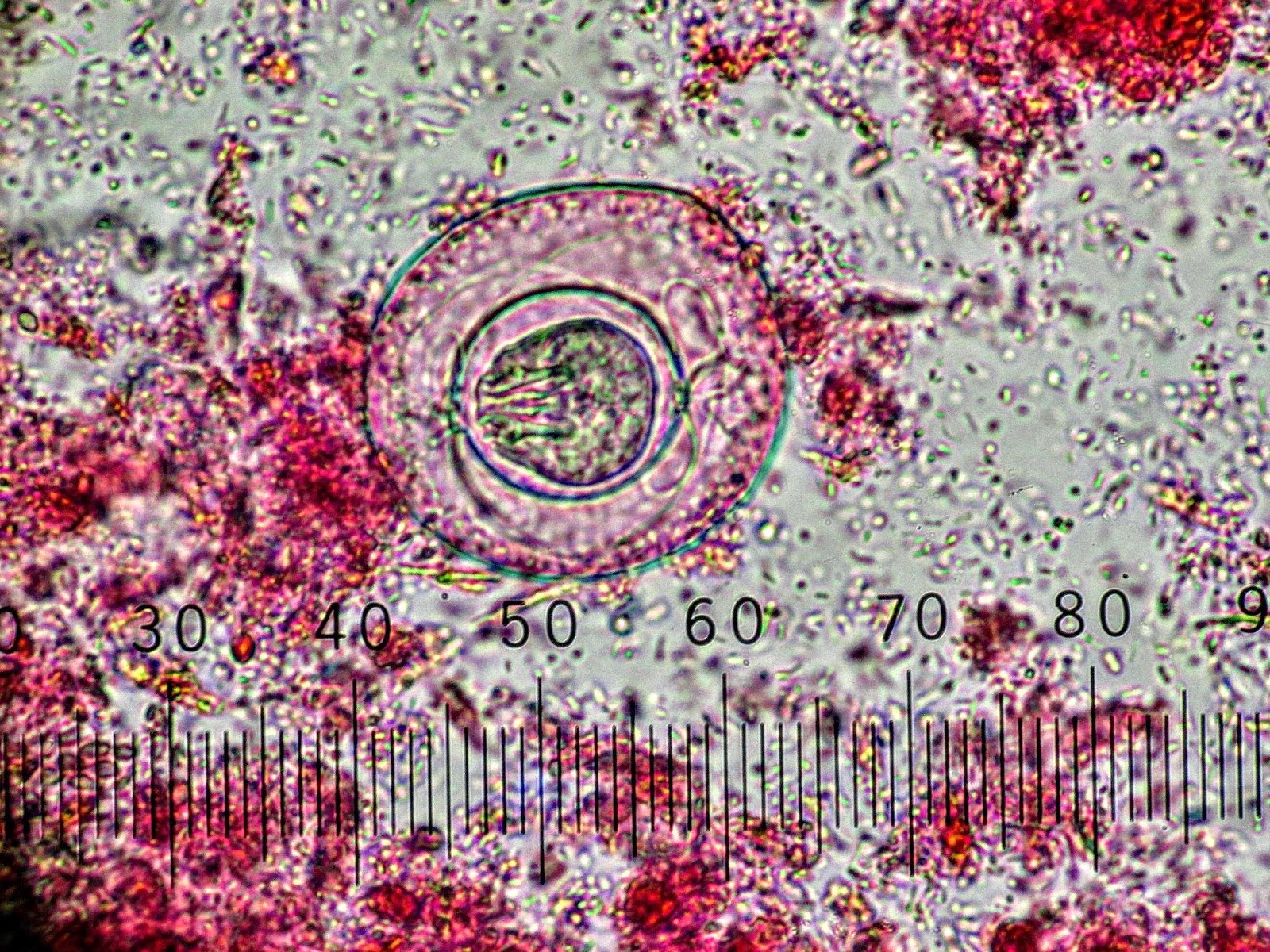
Hymenolepis nana
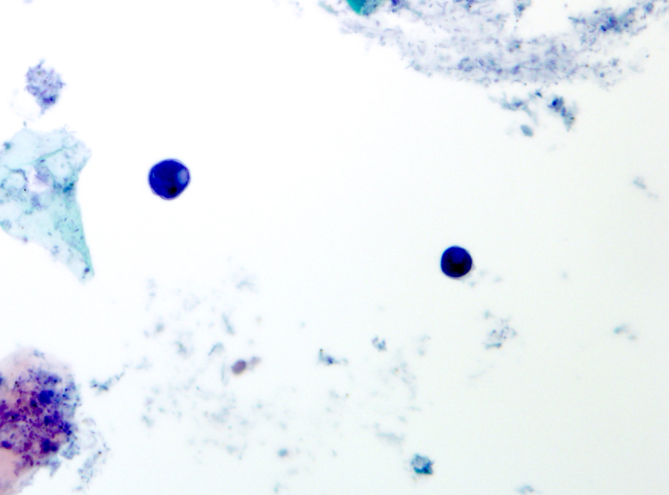
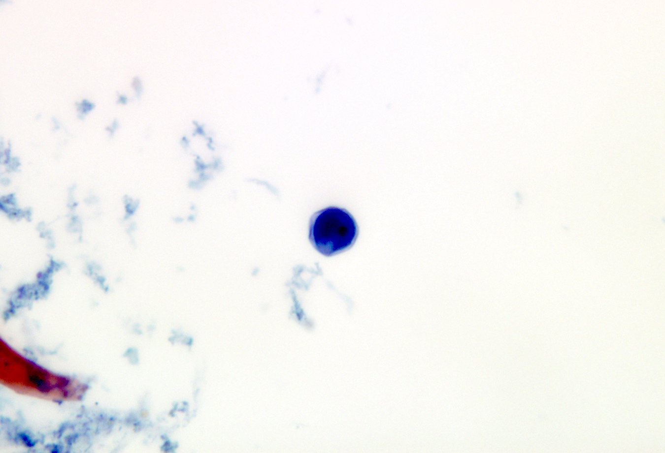
Iodamoeba bütschlii
Klebsiella oxytoca
Legionella
Leishmania
Leptospira
Listeria monocytogenes
Loa loa
M. genitalium
M. hominis / Ureaplasma spp.
M. pneumoniae
Macracanthorhynchus
MALDI-ToF MS (pending)
Microsporidia
Mpox / orthopoxvirus
Mycobacteria non-TB
Myiasis
Naegleria (pending)
Neisseria gonorrhoeae (pending)
Neisseria meningitidis
Nocardia
Norovirus (pending)
Orf
Paragonimus westermani (pending)
Parvovirus (erythrovirus) B19 (pending)
Pediculosis (lice)
Plasmodium falciparum
Plasmodium non-falciparum
Raillietina
Respiratory virus panel (PCR) (pending)
Sarcocystis
Schistosomiasis
Serratia species
Shigella
Staph aureus
Staph coagulase negative
Streptococcus pneumoniae
Streptococcus-other
Strongyloides
Syphilis (pending)
Taenia saginata
Taenia solium (neurocysticercosis)
Taenia solium (neurocysticercosis)
Taenia species
Talaromyces marneffei
Tick (Hyalomma)
Tick (Ixodes)
Total laboratory automation (pending)
Toxocara
Trichinella (pending)
Trichomonas (pending)
Trichostrongylus
Trypanosomes (pending)
Vibrio vulnificus (pending)
Whipworm
Yellow fever
ZygomycetesAcanthamoeba
Table of Contents
Definition / general | Case reports | Microscopic (histologic) description | Microscopic (histologic) imagesDefinition / general
- Rare but serious complication of contact lenses due to contamination of contact lens cleaning systems
- Organisms are ubiquitous protozoa in soil and fresh water
- Infections wax and wane, may infiltrate cornea along corneal nerve and cause pain
Case reports
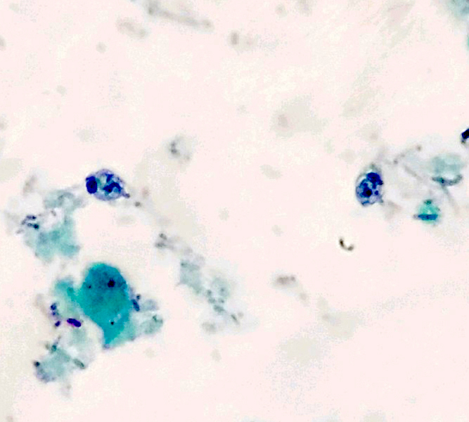
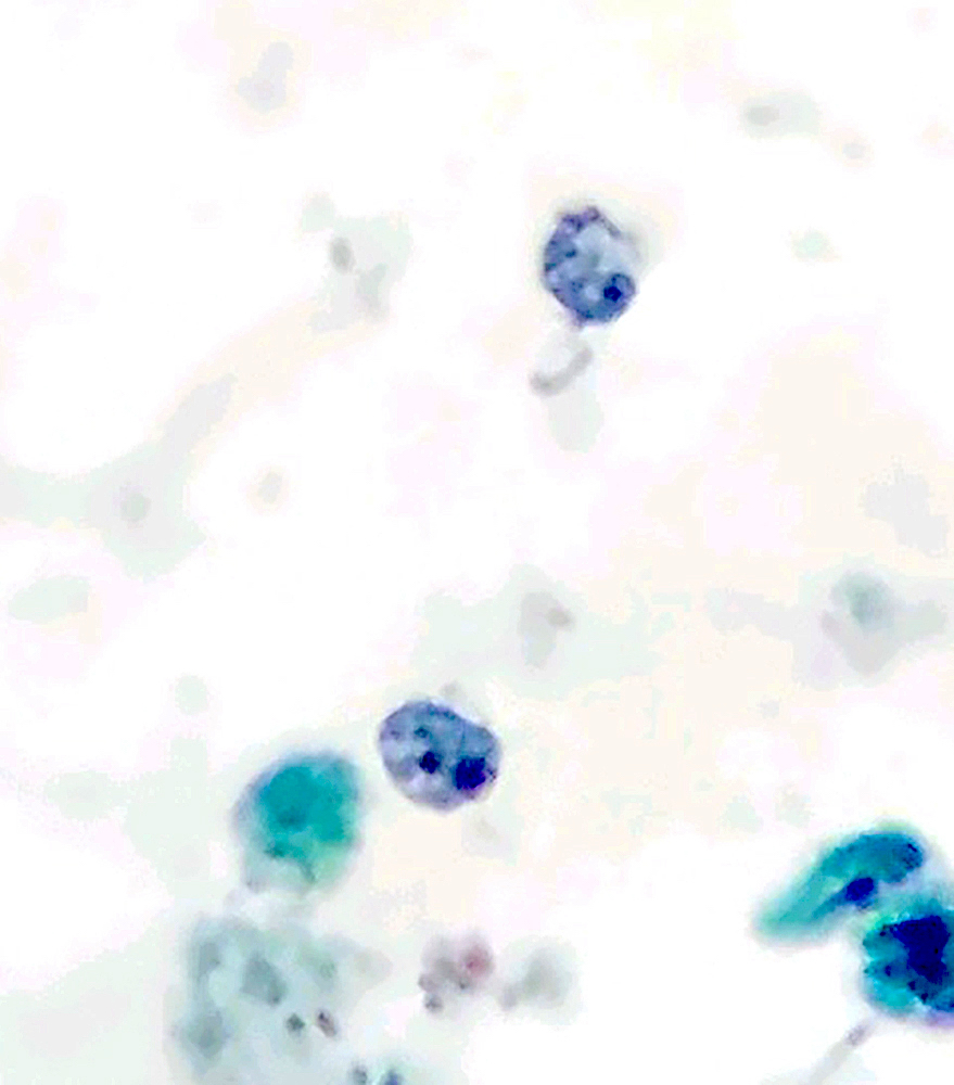
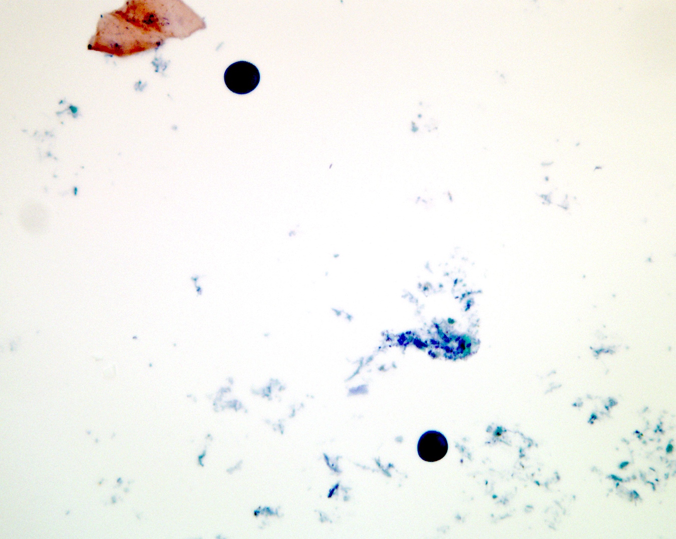
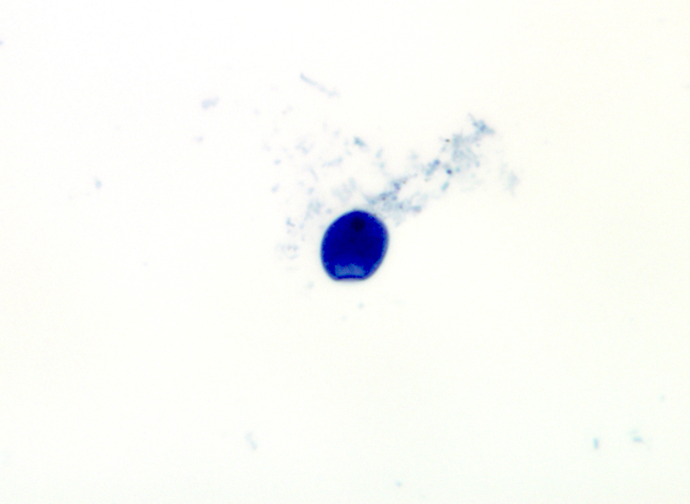
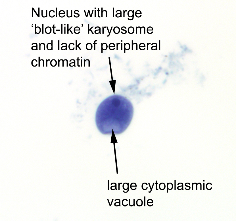
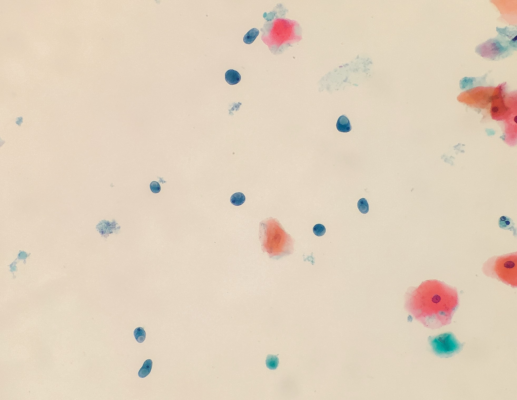
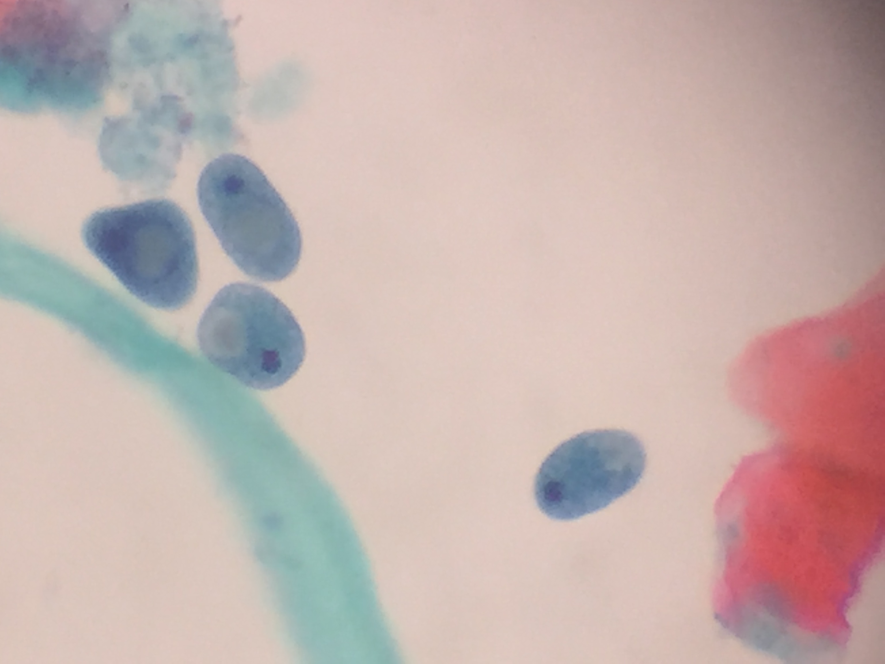
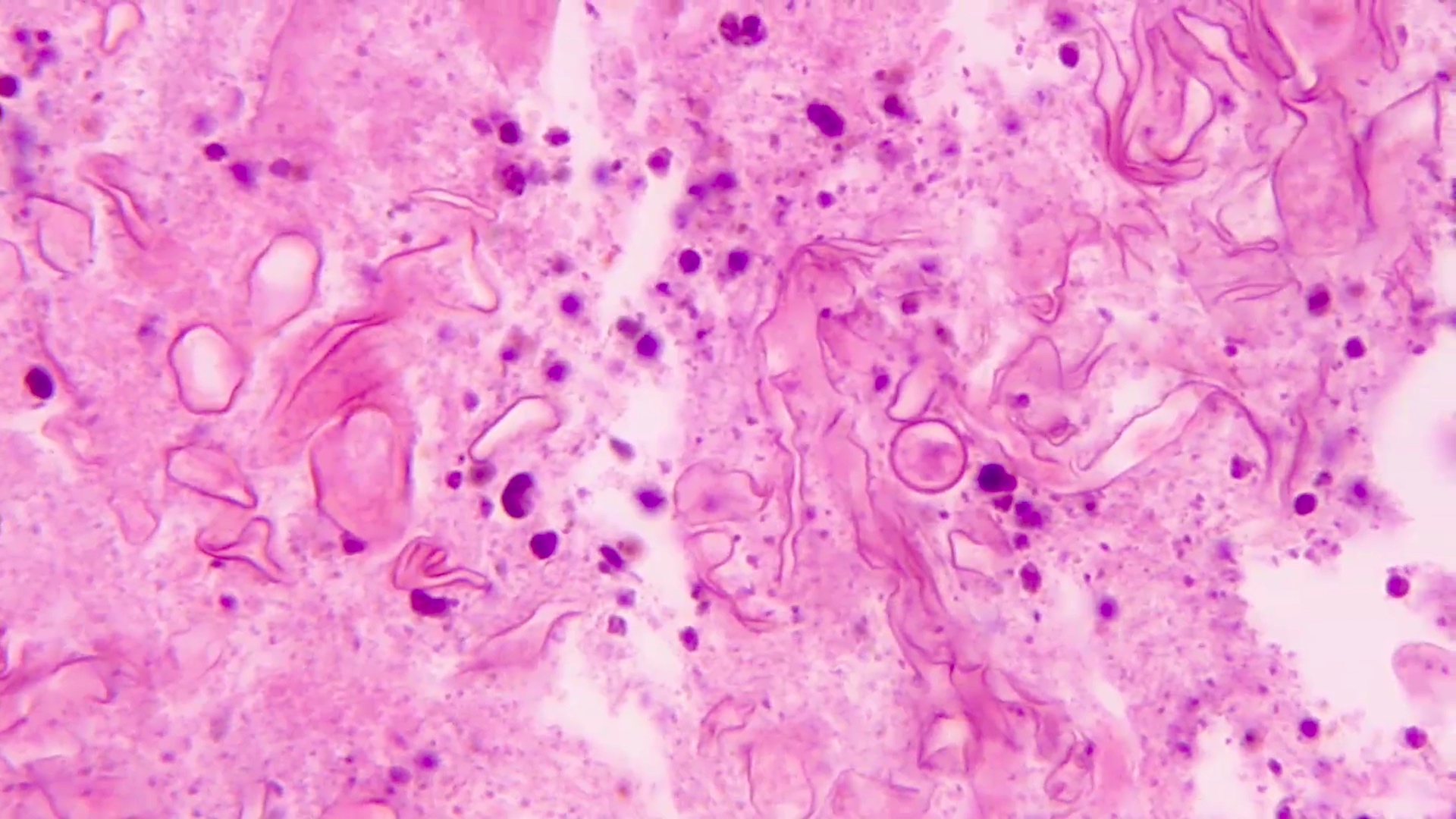
- Structures were seen on a non-nutrient agar culture that had been inoculated with corneal scrapings (Pritt: Creepy Dreadful Wonderful Parasites Blog - Case of the Week 542 [Accessed 25 June 2019])
Microscopic (histologic) description
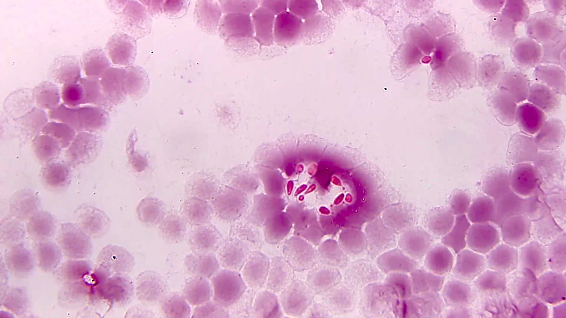
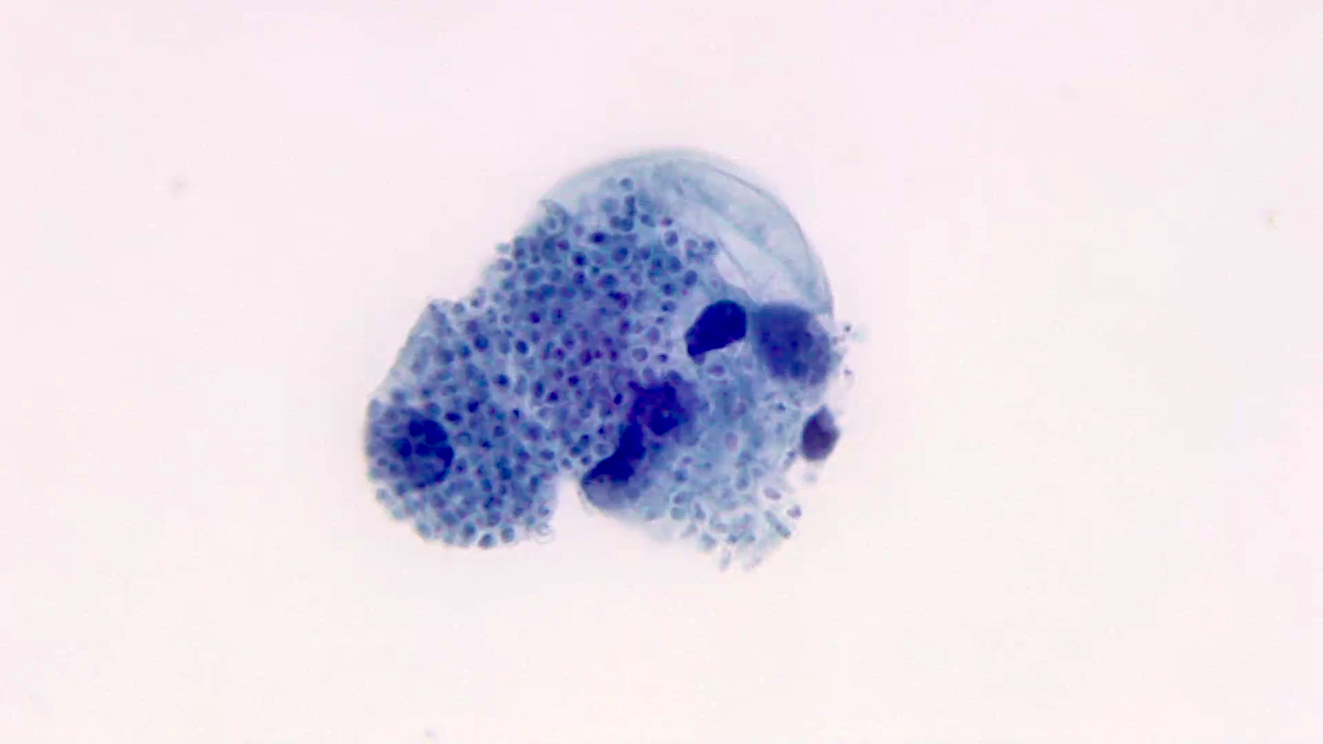
- Characteristic double walled cyst and trophozoite (Pritt: Creepy Dreadful Wonderful Parasites Blog - Answer to Case 542 [Accessed 25 June 2019])
Microscopic (histologic) images
Acinetobacter (pending)
[Pending]
Actinomyces
Table of Contents
Definition / general | Essential features | Epidemiology | Sites | Pathophysiology | Clinical features | Laboratory | Case reports | Treatment | Microscopic (histologic) description | Microscopic (histologic) images | Molecular / cytogenetics description | Differential diagnosis | Additional references | Board review style question #1 | Board review style answer #1 | Board review style question #2 | Board review style answer #2Definition / general
- Gram positive genus containing over 40 species
- Taxonomy: genera Actinobacteria, family Actinomycetaceae
- Common species:
- Actinomyces israelii
- Actinomyces bovis
- Actinomyces gerencseriae
- Actinomyces graevenitzii
- Actinomyces odontolyticus
- Actinomyces meyeri
- Actinomyces neuii
- Actinomyces turicensis
Essential features
- Gram positive anaerobic bacilli with filamentous branching (Clin Microbiol Rev 2015;28:419)
- Anaerobic; modified acid fast stain negative
- Grow in tissue as intertwined aggregates (termed sulfur granules) to exclude oxygen
- Normal flora in tonsils; pathogenic elsewhere
- Actinomycosis is characterized by pyogranulomatous inflammation
- Most common causes of actinomycosis include: A. israelii, A. gerencseriae and A. graevenitzii
- A. meyeri, A. neuii and A. turicensis are emerging pathogens
Epidemiology
- Reside on mucosal surfaces: oral cavity, tonsillar crypts, genitourinary tract
- Opportunistic infection in skin, jaw bones, chest and abdominal cavities, lungs, liver, uterus
- Common involvement in polymicrobial infections
- Affects immunocompetent and immunocompromised hosts
- More common in developing nations
- Associated with poor dentition, smoking, heavy alcohol consumption, bisphosphonate related osteonecrosis of the jaw, osteoradionecrosis, prolonged use of intrauterine contraceptive devices
- Reference: Clin Microbiol Rev 2015;28:419
Sites
- Orocervicofacial; > 50% of infections (Oral Surg 2017;123:586)
- Disseminated:
- Chest and abdominal cavities
- Lungs
- Liver
- CNS
- Uterus
Pathophysiology
- Infection initiated upon access to tissues via trauma, surgical procedures, foreign bodies
- Formation of dense intertwined filamentous bacterial aggregates called sulfur granules exclude oxygen, host cells and immune mediators (antibodies, etc.), thus enabling optimal growth
- Reference: Infect Drug Resist 2014;7:183
Clinical features
- Indolent, slowly progressing pyogranulomatous disease
- Orocervicofacial: abscess formation with sinus tracts, purulent discharge, fistulae, tissue fibrosis; lumpy jaw formation
- Pneumonia: chronic cough, dyspnea, fibrosis, cavitation (Clin Microbiol Rev 2015;28:419)
- Disseminated: low grade fever, weight loss; tissue specific symptoms
- Uterus: purulent vaginal discharge, foul odor, cramping
Laboratory
- Most pathogens will grow on chocolate blood agar at 37 °C within 5 days
- Some aerotolerant Actinomyces species will grow on aerobic culture within 2 days
- Most exhibit nondistinct tan-white diphtheroid-like colonies; gamma hemolysis
- A. israelli exhibits a pathognomonic molar tooth colony appearance
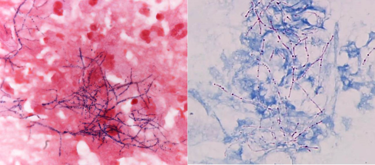
- Key feature: gram positive filamentous bacteria; staining negative with modified acid fast stain
- Acid fast stains include Kinyoun and Ziehl-Neelsen
- Actinomyces species lack high levels of mycolic acid in the cell wall and do not retain the carbol fuschin dye when using a weaker acid for decolorization (i.e., modified Kinyoun acid fast stain), resulting in a negative stain
- Nocardia, another gram positive filamentous bacteria, have abundant mycolic acid in the cell wall, can retain the carbol fuschin dye and stain positive with modified Kinyoun
- MALDI TOF mass spectrometry enables species level identification
- Reference: Infect Drug Resist 2014;7:183
Case reports
- 31 year old woman with Actinomyces bacteremia in association with tubo-ovarian abscesses and hysteroscopic sterilization (Obstet Gynecol 2014;124:451)
- 56 year old man with craniofacial Actinomyces osteomyelitis evolving from sinusitis (Radiol Case Rep 2017;13:104)
- 60 year old man with Actinomyces odontolyticus infection 3 months post robotic assisted laparoscopic prostatectomy (BMJ Case Rep 2019;12:e228184)
- 63 year old man with Actinomyces cavernous sinus infection (Pract Neurol 2018;18:373)
- 83 year old man with pyogenic granuloma associated with Actinomyces israelii (J Dent Sci 2018;13:285)
Treatment
- Susceptible to penicillin and most antibiotics used to treat gram positive bacteria
- Intrinsic resistance to metronidazole
- Surgical drainage recommended
- References: Microorganisms 2022;10:125, J Antimicrob Chemother 2005;56:407
Microscopic (histologic) description
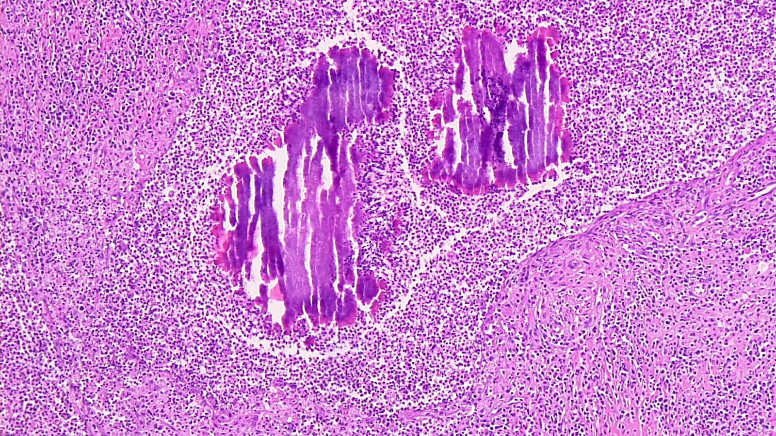
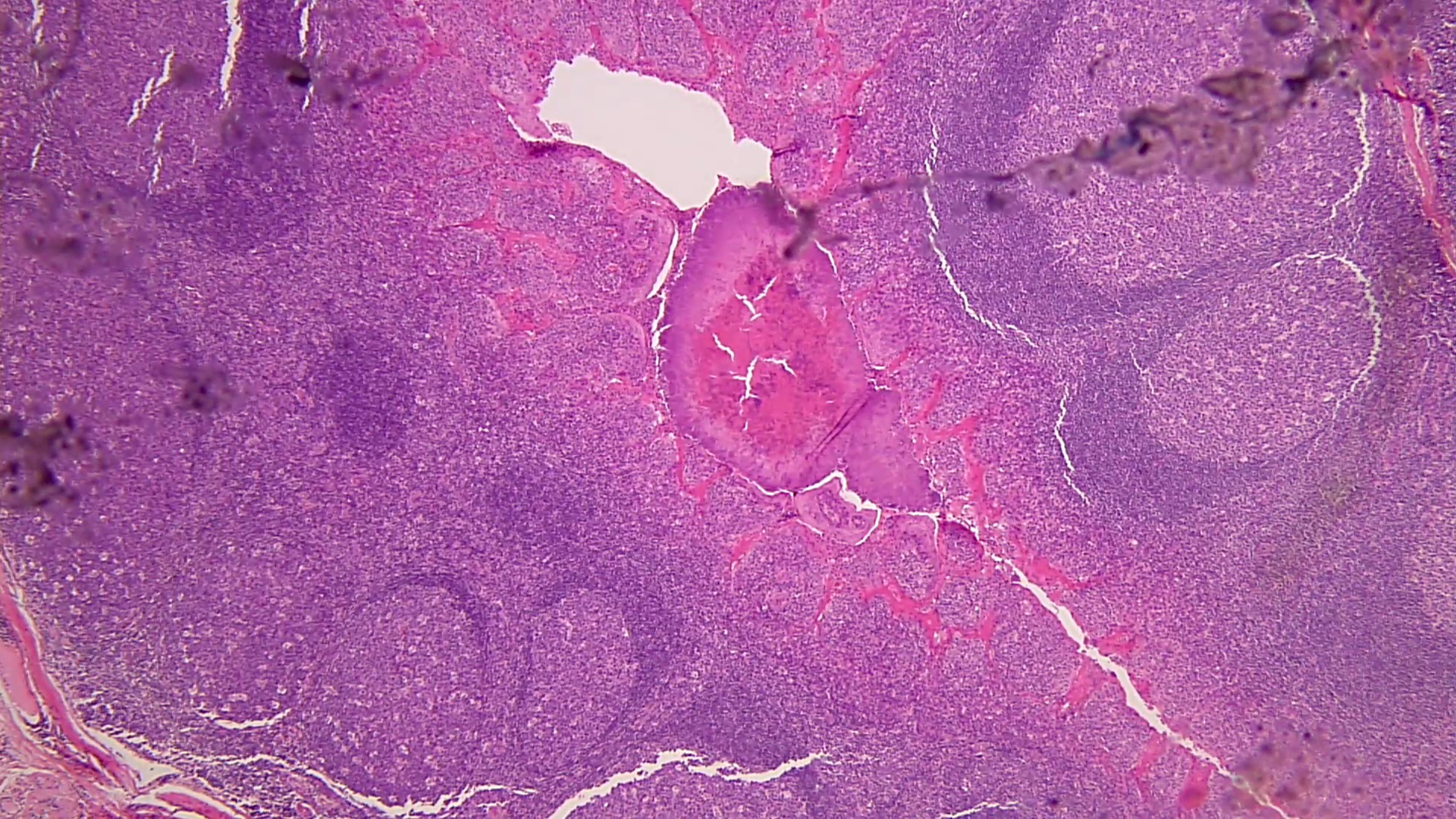
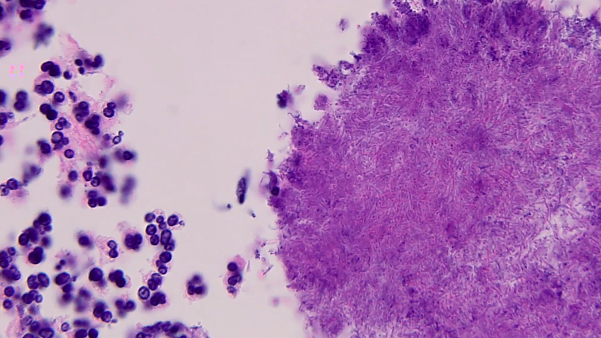
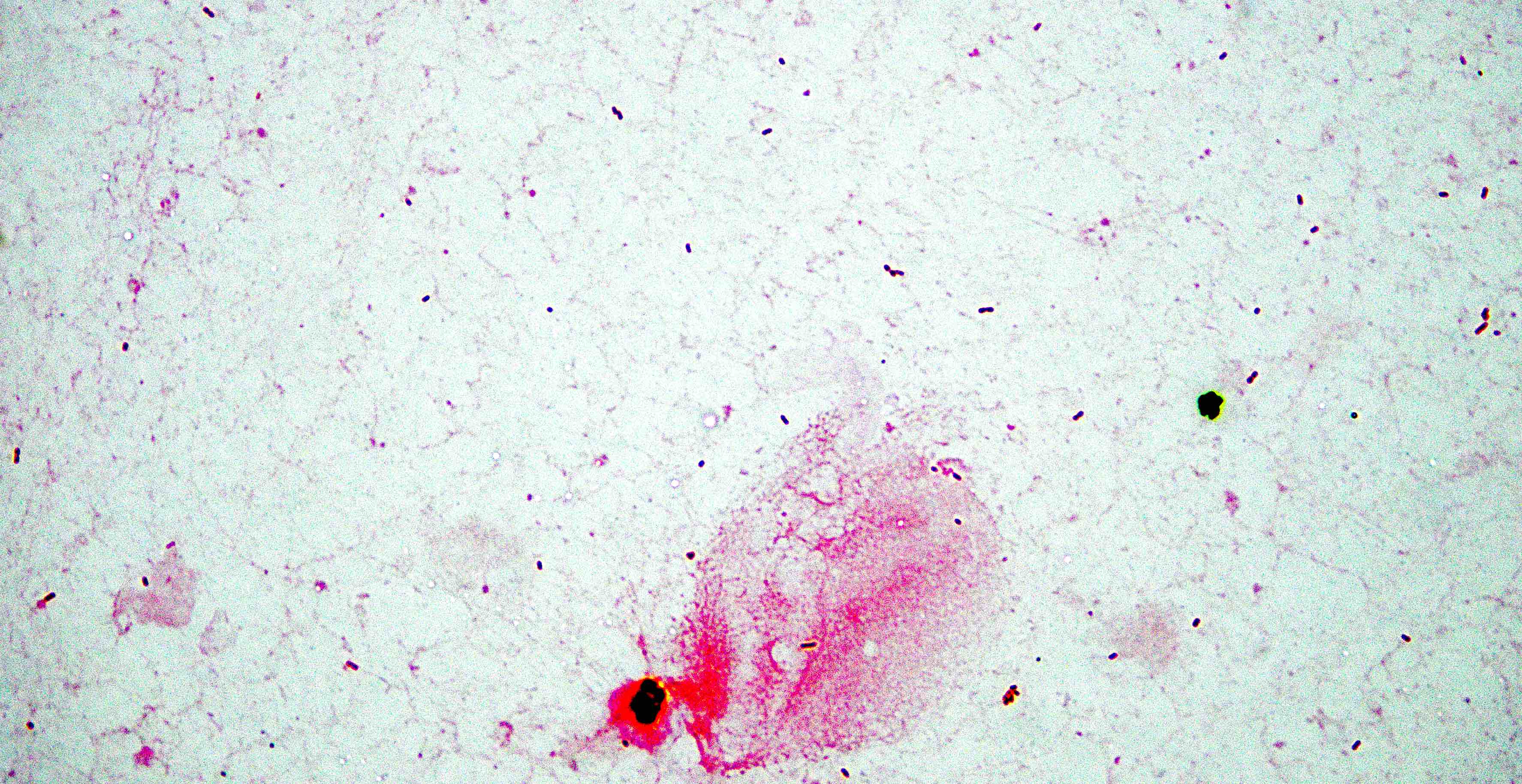
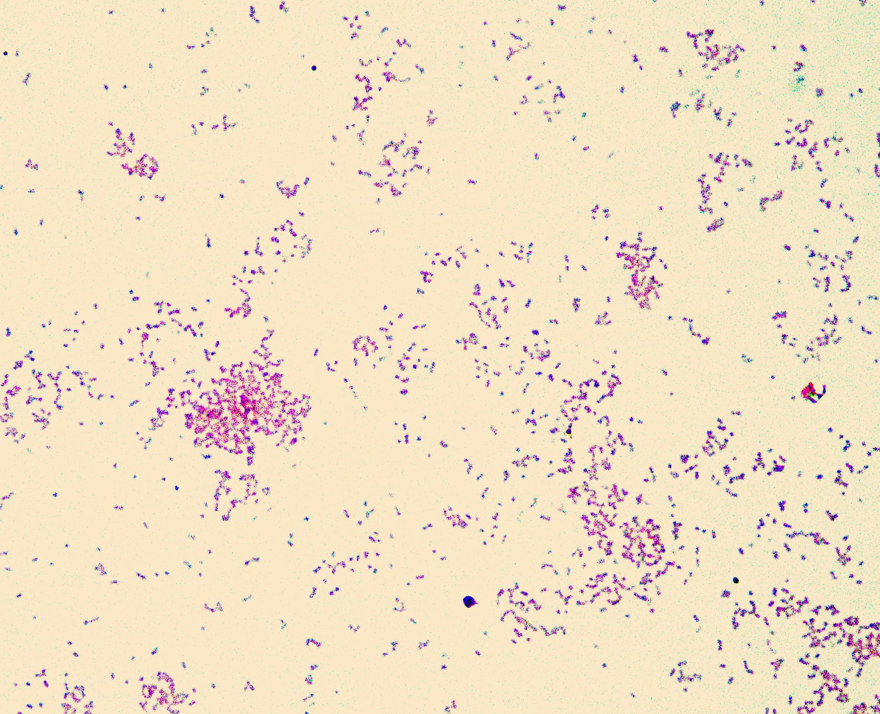
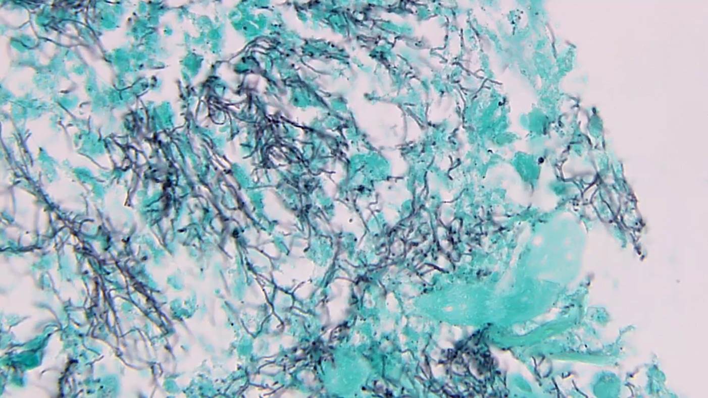
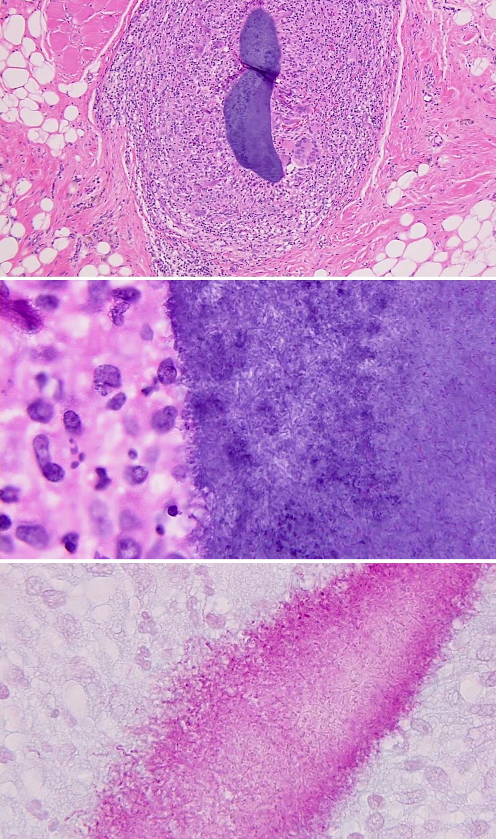
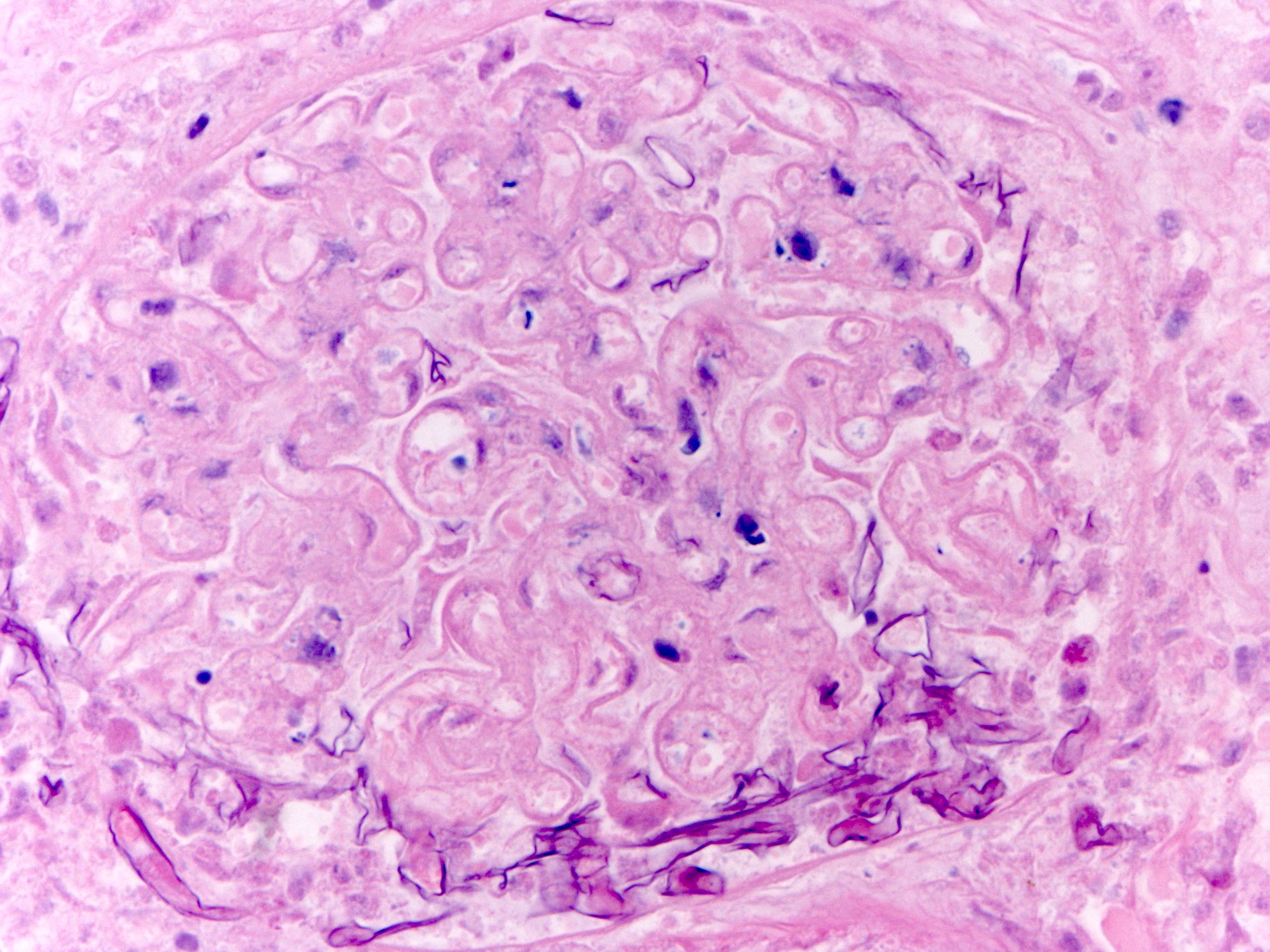
- Characteristic large basophilic filamentous bacterial aggregates (sulfur granules) that stain positive on GMS but negative with FITE stain and the traditional AFB stain
- Commensal organism frequently encountered histologically in tonsillar crypts, resembling starbursts
Microscopic (histologic) images
Molecular / cytogenetics description
- 16S rRNA sequencing is the current gold standard for species level identification in bacteria
Differential diagnosis
- Nocardiosis:
- Note aerobic growth and positive modified acid fast stain
- Botryomycosis:
- Note bacterial cocci (staphylococci) in tissue
- Tuberculosis:
- Note acid fast bacilli in tissue
- Mold infection:
- Note increased width (4 - 10 µm) of hyphae or yeast-like structures
- Malignancy:
- Note cellular atypia and absence of organisms
Additional references
Board review style question #1
A 43 year old woman presents to the emergency department with weight loss, dyspnea and a mild cough. A sputum culture Gram stain shows gram positive filamentous bacteria. Which stain is helpful for distinguishing the 2 major genera of filamentous bacteria?
- GMS
- Gram stain
- Modified Kinyoun
- Ziehl-Neelsen
Board review style answer #1
C. Modified Kinyoun. Actinomyces species, unlike Nocardia species, do not have high levels of mycolic acid in the cell wall and will not retain the carbol fuschin dye when using a weaker acid for decolorization (i.e., modified acid fast stain), resulting in a negative stain. Nocardia will retain the carbol fuschin dye and will stain positive. Both will stain gram positive, although Nocardia yield a beaded appearance due to the mycolic acid on the cell wall. Both will be positive by GMS performed on tissue and negative by Ziehl-Neelsen.
Comment Here
Reference: Actinomyces
Comment Here
Reference: Actinomyces
Board review style question #2
A homeless man with poor dentition presented with jaw pain with swelling, fistula formation and purulence. On exam, a small granule was expressed from this lesion and sent to microbiology. Half of the sample was crushed and stained as shown above and the other half was submitted for culture. A Gram stain of the slide revealed filamentous structures. Which culture conditions are optimal to isolate the likely etiologic agent?
- BCYE agar under aerobic conditions
- Blood agar under microaerophilic conditions
- CDC anaerobic agar under aerobic conditions
- Chocolate agar under anaerobic conditions
Board review style answer #2
D. Chocolate agar under anaerobic conditions. Actinomyces species grow optimally under anaerobic conditions. Some facultative anaerobes and aerotolerant species in this genus may grow in the presence of oxygen but extended culture may be required.
Comment Here
Reference: Actinomyces
Comment Here
Reference: Actinomyces
Anaplasma
Table of Contents
Definition / general | Clinical features | Case reports | Treatment | Microscopic (histologic) description | Peripheral smear images | Additional references | Board review style question #1 | Board review style answer #1Definition / general
- Anaplasma phagocytophilia: human granulocytotropic anaplasmosis (HGA, formally termed human granulocytic ehrlichiosis)
- Vector borne disease transmitted through bite of Ixodes ticks
- Bacteria is obligate intracellular pathogen that binds to P selectin glycoprotein ligand 1 (PSGL1 / CD162)
- Susceptibility also associated with expression of CD15s (J Clin Invest 1999;103:407)
- First described in USA in 1994
- Geographic distribution of A. phagocytophilia (HGA) reflects regions of US where their hard tick vectors reside: northeastern states, northwest Wisconsin, eastern Minnesota and Pacific northwest states
Clinical features
- Presents with fever, leukopenia, thrombocytopenia (70 - 90%) and elevated liver enzymes
- Mortality rate is 0.5 - 1% for HGA
- Particularly severe infections occur in elderly / immunocompromised
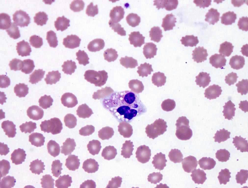
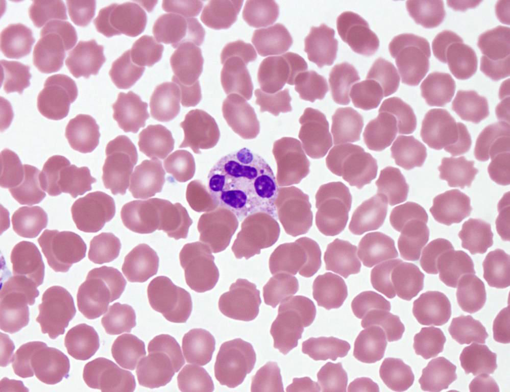
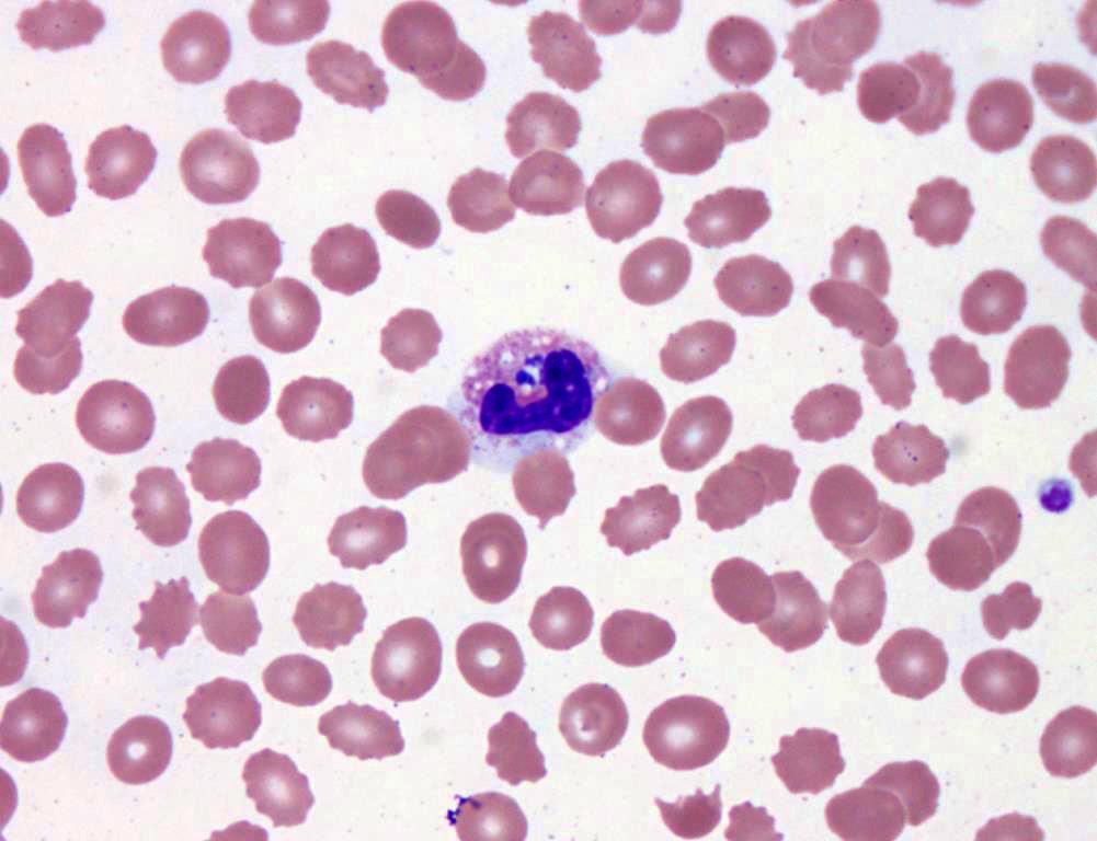
- Characteristic intracytoplasmic morulae (morula is Latin for mulberry): cytoplasmic membrane bound vacuoles with irregular edges containing hundreds to thousands of clustered gram negative bacteria
- Infected cells typically contain only 1 or 2 morulae although as many as 15 may be seen in immunosuppressed individuals
- Greatly variable percentage of peripheral blood films with detectable morulae in the literature (3 - 80%) with a higher number seen with HGA infection (50 - 80%) and in immunosuppressed individuals
Case reports
- 43 year old woman presented with fever, chills and muscle aches after a tick bite (Case of the Month #486)
- 78 year old man with Anaplasma phagocytophilum infection and CML (J Clin Pathol 2004;57:499)
- 3 pancreas transplant recipients with HGA / human granulocytic ehrlichiosis (Transpl Infect Dis 2001;3:34)
Treatment
- Most patients are seronegative during first few weeks of acute infection (60 - 97%), so therapeutic decisions must be based on clinical suspicion, peripheral blood findings and PCR (sensitivity is 60 - 85%, high degree of false positive results)
- Became a nationally reportable disease to US Centers for Disease Control in 1999
- Organisms are susceptible to tetracyclines and their derivatives, particularly doxycycline
Microscopic (histologic) description
- Peripheral blood: buffy coat examination may reveal intracytoplasmic inclusions (morulae - spherical structures with irregular edges) within neutrophils or monocytes
- Bone marrow: epithelioid granulomas; usually normo or hypercellular with intact trilineage maturation; rare hypoplasia; possible increased megakaryocytes
- Histopathologic bone marrow findings: inconsistent and likely to change during the course of the disease
- HGA organisms preferentially infect more mature rather than immature granulocytic cells in bone marrow
Peripheral smear images
Additional references
Board review style question #1
What of the following is true about anaplasmosis?
A. Anaplasmosis is typically transmitted by exposure to respiratory droplets.
B. Immunocompromised patients traveling to endemic regions should be vaccinated against Anaplasma.
C. A common presentation for patients with anaplasmosis includes relapsing fevers, neutrophilia and reactive thrombocytosis.
D. A diagnostic feature of anaplasmosis is the presence of neutrophilic morulae.
E. Anaplasma and malaria are common coinfections.
Board review style answer #1
D. Neutrophilic morulae are a characteristic feature of anaplasmosis on peripheral blood smear examination.
Comment Here
Reference: Anaplasmosis
Comment Here
Reference: Anaplasmosis
Angiostrongyliasis
Table of Contents
Definition / general | Essential features | Epidemiology | Sites | Pathophysiology | Clinical features | Diagnosis | Laboratory | Case reports | Treatment | Gross description | Gross images | Microscopic (histologic) description | Microscopic (histologic) images | Differential diagnosis | Board review style question #1 | Board review style answer #1 | Board review style question #2 | Board review style answer #2Definition / general
- Angiostrongylid nematodes are intra-arterial parasites from wild rodents that may infect humans, causing eosinophilic meningoencephalitis (Angiostrongylus cantonensis [ACa], also known as the rat lungworm), gastroenteritis and hepatitis (Angiostrongylus costaricensis [ACo])
Essential features
- Intense eosinophilic inflammation with granuloma and vasculitis, especially in the intestines, liver and meninges, calls attention to angiostrongyliasis
- Abdominal angiostrongyliasis may cause tumoral (inflammatory) intestinal lesions or necrotic congestive lesions and perforation
- Cerebrospinal fluid (CSF) eosinophilia is the main feature that leads to suspicion of cerebral angiostrongyliasis
- Ingestion of larvae developed in mollusks and freshwater crustaceans (ACa) leads to migration to the central nervous system (ACa, CNS) or to mesenteric blood vessels (ACo)
- Angiostrongyliasis is a food borne zoonotic disease
- Elimination of larvae in feces or CSF is hindered by the intense inflammatory reactions, for both ACa and ACo; it is not possible to detect larvae in stools
Epidemiology
- There is no clear age or gender susceptibility but like many zoonoses, focal and seasonal (spring / autumn, rainy periods) transmission is the rule (Rev Inst Med Trop Sao Paulo 1991;33:373)
- Some case series show a predominance of children and young adults
- Infective larvae do not invade skin but they may contaminate food, beverages or untreated water and they also may be ingested after consumption of raw mollusks or freshwater shrimps (ACa)
- Endemic areas are Southeast Asia and the Pacific Islands for ACa and the Americas (from southern U.S. to northern Argentina) for ACo (Lancet Infect Dis 2008;8:621, Parasit Vectors 2023;16:155)
Sites
- ACa larvae are located inside meningeal blood vessels or are less frequently seen moving freely inside eye chambers; they are usually not found in the CSF
- Late stage migration and maturation to adult worm inside pulmonary arteries (ACa) is extremely rare
- ACo adult worms live in the mesenteric artery branches of the ileocecal transition; less frequently, they also may develop inside the liver portal venous system
- Rare ectopic arterial location for ACo worms is possible (e.g., spermatic or lower limb arteries)
Pathophysiology
- Antigens secreted or eliminated intravascularly by parasites trigger intense eosinophilic inflammatory reactions
- ACo worms release eggs, followed by rapid embryogenesis and development of first stage larvae; in well adapted rodent hosts, these larvae are promptly eliminated with feces but in humans, they are trapped in tissues and are the focus for eosinophilic granulomatous reactions (Parasit Vectors 2023;16:155)
- Eosinophilic vasculitis and dead worm debris favor arterial mesenteric thrombosis, leading to focal necrosis and intestinal wall perforation
- Subacute slow antigen release may cause huge eosinophilic tissue infiltration and intestinal wall thickening
Clinical features
- Pain is the predominant symptom for both angiostrongyliasis infections
- Eosinophilic meningitis (ACa) is mainly manifested by severe headache and many other neurological signs and symptoms; from other less specific manifestations, migrating dysesthesias may indicate ACa infection
- Fever and neck stiffness are not always present and encephalitic syndromes are rare but are the main cause for poor outcomes
- Abdominal pain, either localized in right lower (ileocolitis) or upper (hepatitis) quadrants, may present as an acute abdominal syndrome, sometimes complicated by peritonitis (perforation) or intestinal obstruction (inflammatory tumoral lesions) caused by ACo infection (Rev Inst Med Trop Sao Paulo 1991;33:373)
Diagnosis
- Image examinations may disclose nonspecific tissue thickening; in the meninges for ACa and the intestinal wall for ACo
- Meningitis or painful abdominal syndromes, associated with blood / CSF eosinophilia, both provide strong evidence in favor of angiostrongyliasis (Pathogens 2023;12:624)
Laboratory
- Hallmark for angiostrongyliasis is eosinophilia detected in the blood (ACo) or CSF (ACa)
- Serology is available in reference laboratories which may lack extensive performance evaluation and their results should not be considered confirmatory
- DNA detection in serum, tissues (ACo) and CSF (ACa) confirms the etiology but it is also only available in reference centers (Parasit Vectors 2023;16:155, Clin Infect Dis 2021;73:e1594)
Case reports
- 2 year old boy with abdominal pain, bloody diarrhea, fever and palpable mass (Am J Trop Med Hyg 2022;106:1466)
- 24 year old man with fever, headache and lower limb paresthesia and weakness (Hawaii J Health Soc Welf 2021;80:40)
- 32 year old man with ileal perforation and 34 year old woman with pain in the right upper abdominal quadrant, hepatomegaly and hepatic nodular lesions with eosinophilic inflammatory lesions (Rev Inst Med Trop Sao Paulo 2008;50:339)
Treatment
- Anthelmintics are not recommended for ACo because they lack evidence of efficacy and the course of the infection is usually short and benign (Parasit Vectors 2023;16:155)
- Careful follow up is necessary for early diagnosis and surgical management of complications (i.e., intestinal obstruction and perforation)
- Corticosteroids are the mainstay of treatment for eosinophilic meningitis, especially focused on alleviating headache; although without clearly demonstrated efficacy, albendazole (15 mg/Kg/day, bid, 14 - 21 days) is recommended as an antihelminthic medication (Pathogens 2023;12:624)
Gross description
- Eosinophilic meningitis (ACa): according to autopsy studies, the brain and spinal cord are generally normal (Lancet Infect Dis 2008;8:621)
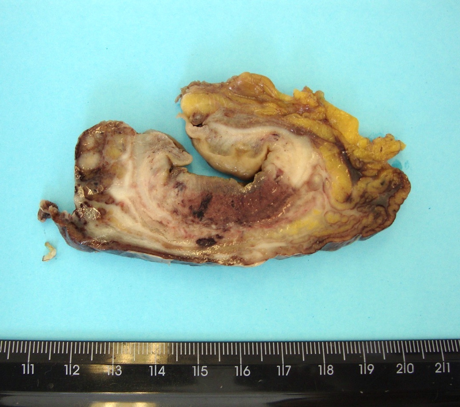
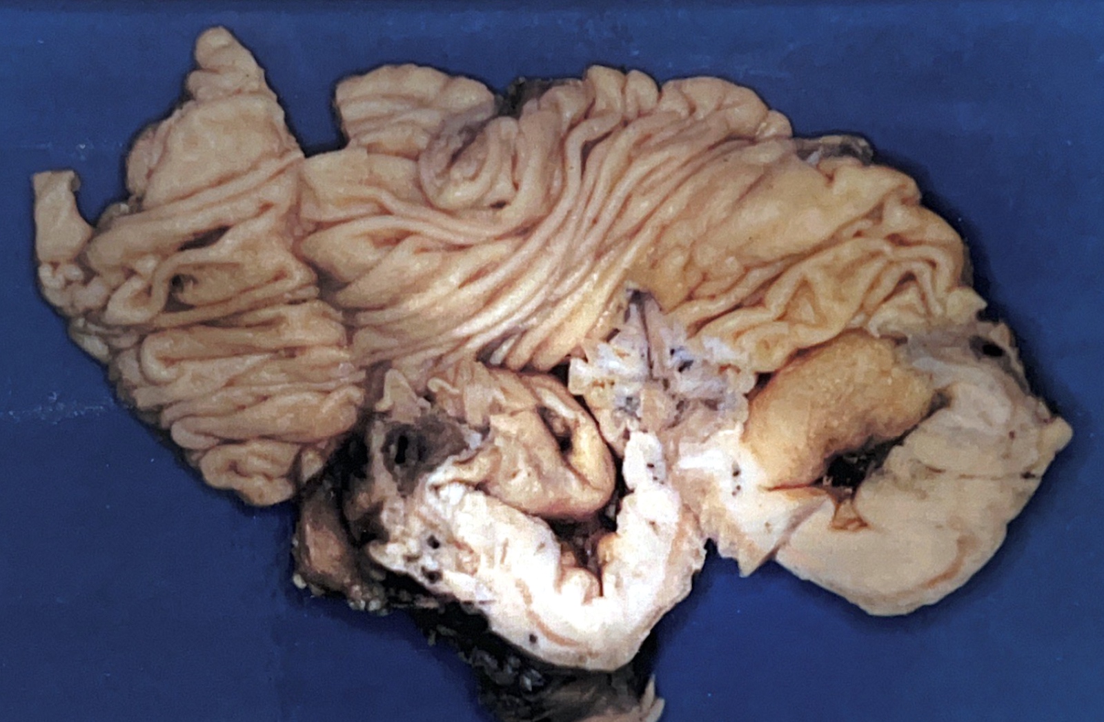
- Abdominal angiostrongyliasis (ACo): 2 types of intestinal lesions: i) infarction and ii) segmental thickening or tumoral (especially in the colon); multiple segmental small intestinal lesions may mimic Crohn's disease
- Macroscopy of the vermiform appendix is indistinct from bacterial acute appendicitis
- Multiple small white nodules may be seen on liver surfaces
- If lesions are suspicious, many sections from both the appendix and intestinal lesions should be examined in order to find parasitic structures
Gross images
Microscopic (histologic) description
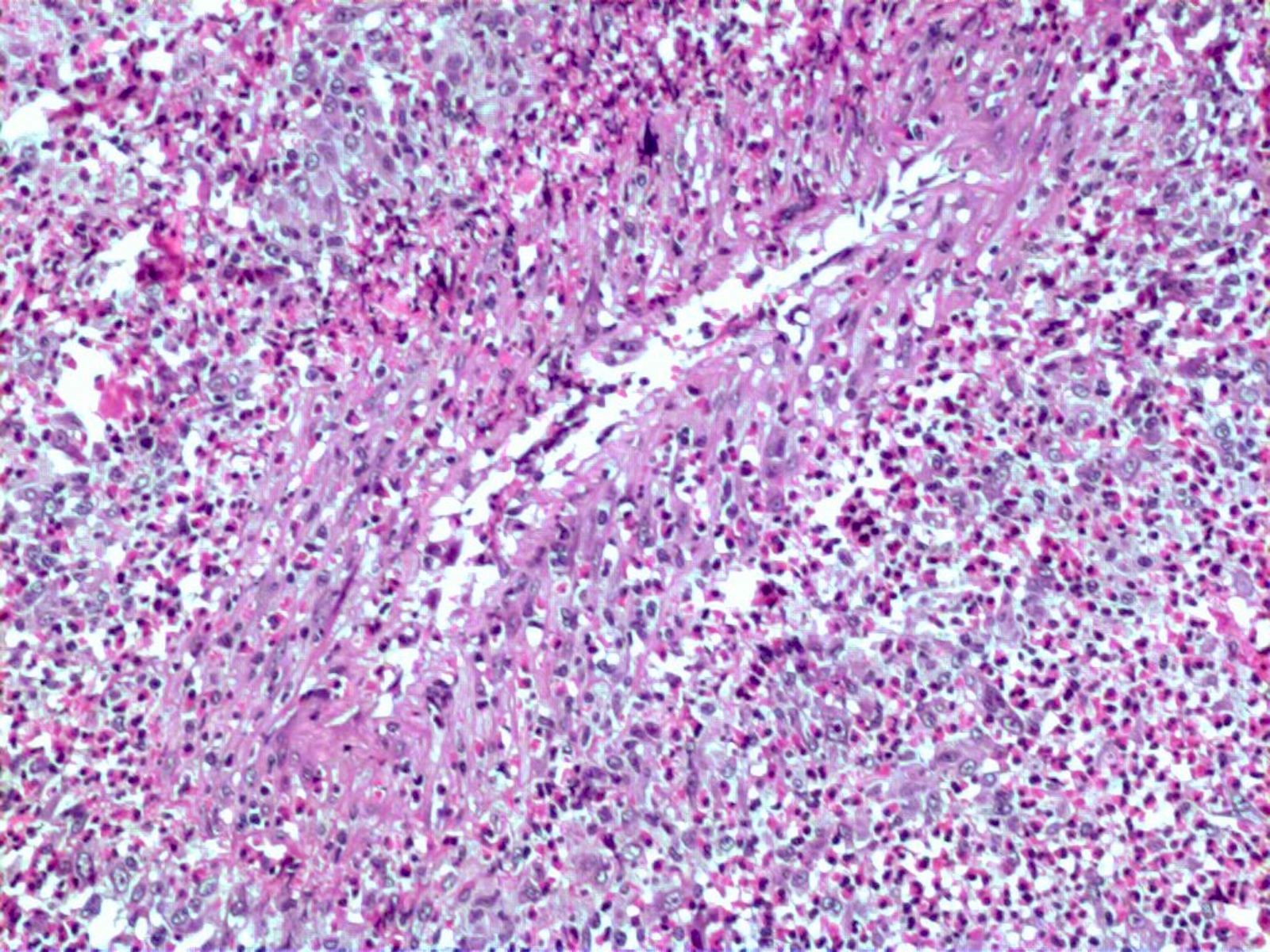
- Eosinophilic meningitis (ACa): meningeal eosinophilic infiltration, eosinophilic granulomas and brain with cell debris, thrombi and inflammatory cell (Acta Trop 2015;141:46)
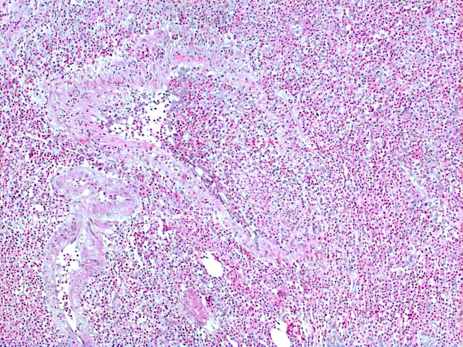
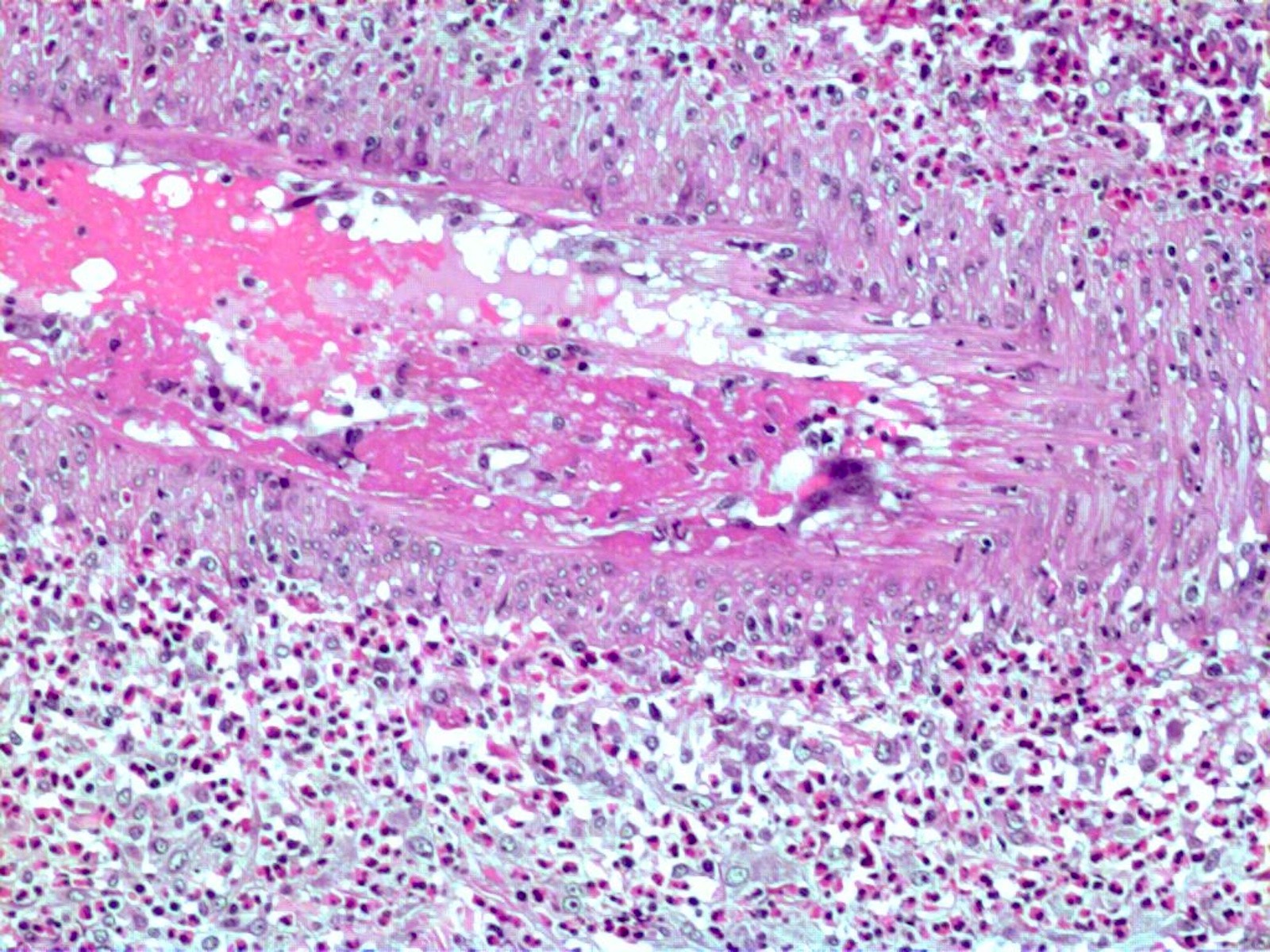
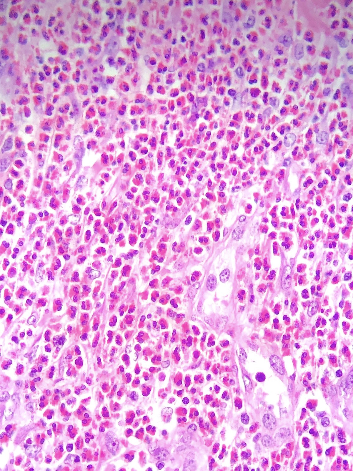
- Abdominal angiostrongyliasis (ACo): heavy eosinophilic infiltration around vessels of the submucosa and muscularis propria (Parasitol Res 1991;77:606)
- Granulomas with eosinophils may be found in the arterial wall or around capillaries and arterioles, sometimes associated with eggs or larvae
- Eosinophilic arteritis is a main histopathological feature
- Severe eosinophilic infiltration, granulomatous reaction and eosinophilic vasculitis, even in the absence of parasitic structures lead to a high probability of diagnosis of ACo infection
- Eggs and larvae are usually located inside capillaries and arterioles of submucosa and muscularis propria with severe granulomatous reaction
- Definitive diagnosis is made by finding parasitic structures (adult worms, eggs or larvae) inside vessels, mainly in the submucosa or mesenteric vessels; PCR in formalin fixed paraffin embedded (FFPE) specimens can be helpful (PLoS One 2014;9:e93658)
Microscopic (histologic) images
Contributed by Rubens Rodriguez, M.D., Ph.D.
Differential diagnosis
- Crohn's disease:
- Lesions are in the mesenteric side
- Granulomas are usually not associated with eosinophils and not adjacent to capillaries or arterioles
- Hepatitis caused by other agents:
- Without a heavy eosinophilic infiltrate
- Eosinophilic granulomas are not seen
Board review style question #1
Board review style answer #1
A. Abdominal angiostrongyliasis. Angiostrongylus worms live inside arteries causing a granulomatous reaction and eosinophilic infiltration. Answers B - D are incorrect because strongyloidiasis, ascariasis and schistosomiasis are not intra-arterial parasites and they are not associated with perivascular or intravascular granulomas or intense eosinophilia. Answer E is incorrect because there is a worm inside the arterial lumen.
Comment Here
Reference: Angiostrongyliasis
Comment Here
Reference: Angiostrongyliasis
Board review style question #2
A young adult presents with eosinophilic meningoencephalitis and a small meningeal tumor. What main finding would establish the presumptive diagnosis of neuroangiostrongyliasis?
- Abscess with intense neutrophilic inflammation
- Hemorrhage
- Histiocytosis and necrotic granulomas
- Mononuclear cellular infiltration
- Perivascular intense eosinophilic infiltration
Board review style answer #2
E. Perivascular intense eosinophilic infiltration. Presence of a large number of eosinophils in tissues (especially perivascular) is the single most common feature of angiostrongyliasis (cerebral or abdominal). The other answers are not common. Answer C is incorrect because granulomas are usually not necrotic and are always associated with eosinophilia and surrounding parasite structures. Answer A is incorrect because neutrophilic inflammation is not seen in neuroangiostrongyliasis and bacterially complicated secondary infections are not common (as they are with abdominal angiostrongyliasis). Answer B is incorrect because the hemorrhage is not related to neuroangiostrongyliasis. Answer D is incorrect because the most important inflammatory cells are eosinophils.
Comment Here
Reference: Angiostrongyliasis
Comment Here
Reference: Angiostrongyliasis
Arthropod ectoparasites (pending)
Table of Contents
Definition / generalDefinition / general
(pending)
Artifacts
Definition / general
- Artifacts include
- Stool:
- Epithelial and white blood cells
- Yeast and fungal elements
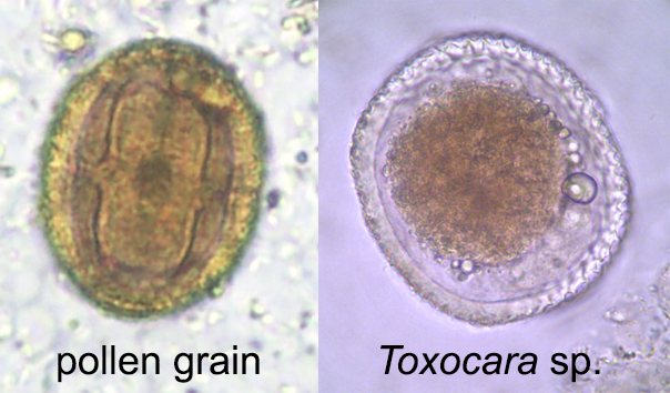
- Pollen grains
- Plant material and plant hairs
- Blood:
- Platelets
- Nucleated red blood cells
- Fungi
- Tissue:
- Yeast
- Seeds
- Other:
- Nonparasitic worms and larvae
- Stool:
Microscopic (histologic) images
Ascaris
Table of Contents
Case reports | Gross description | Gross images | Microscopic (histologic) description | Microscopic (histologic) images | Videos | Differential diagnosisCase reports
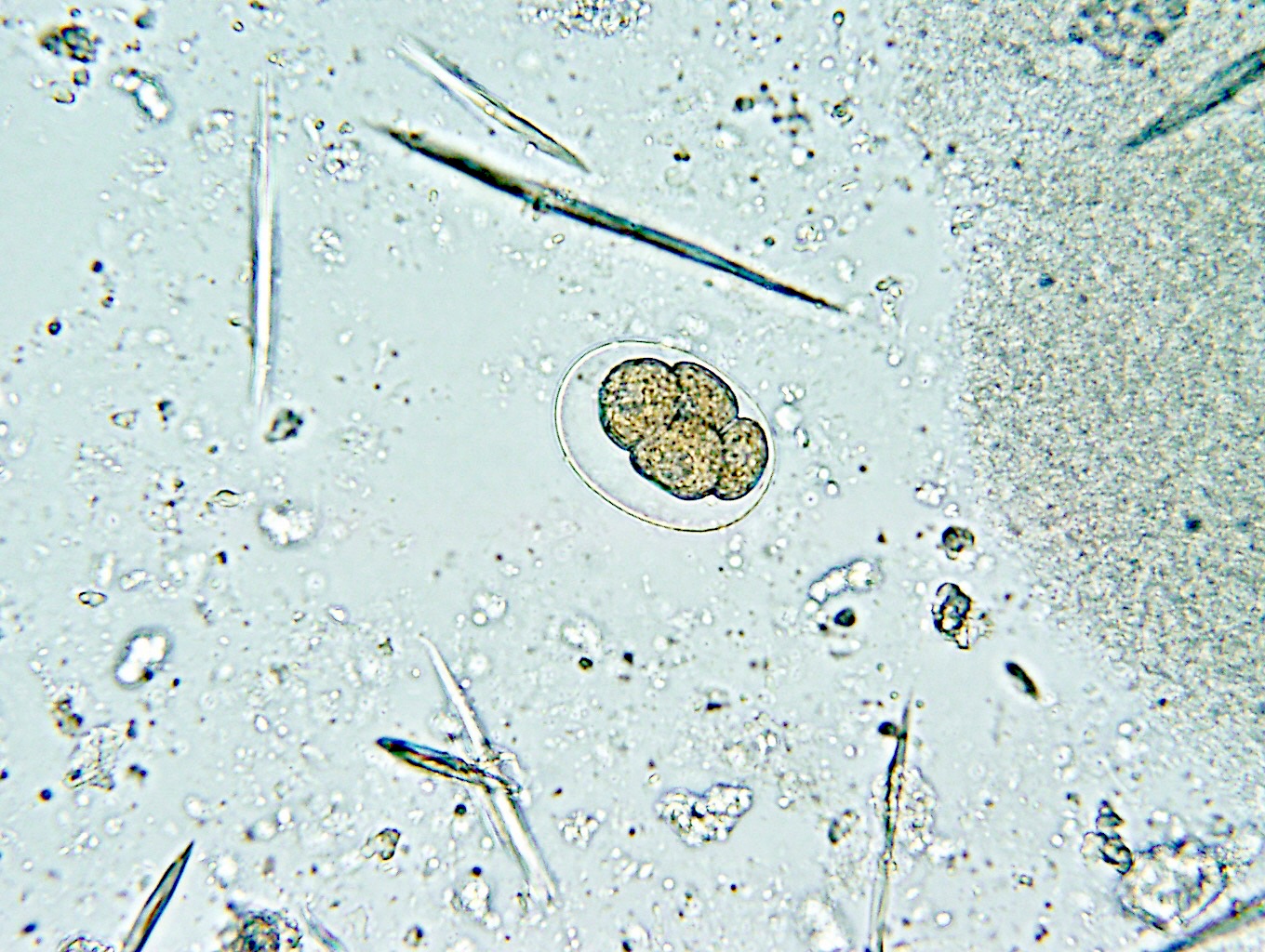
- Objects were seen in a concentrated wet prep of a stool specimen from an international adoptee from Ethiopia (Pritt: Creepy Dreadful Wonderful Parasites Blog - Case of the Week 550 [Accessed 7 August 2019])
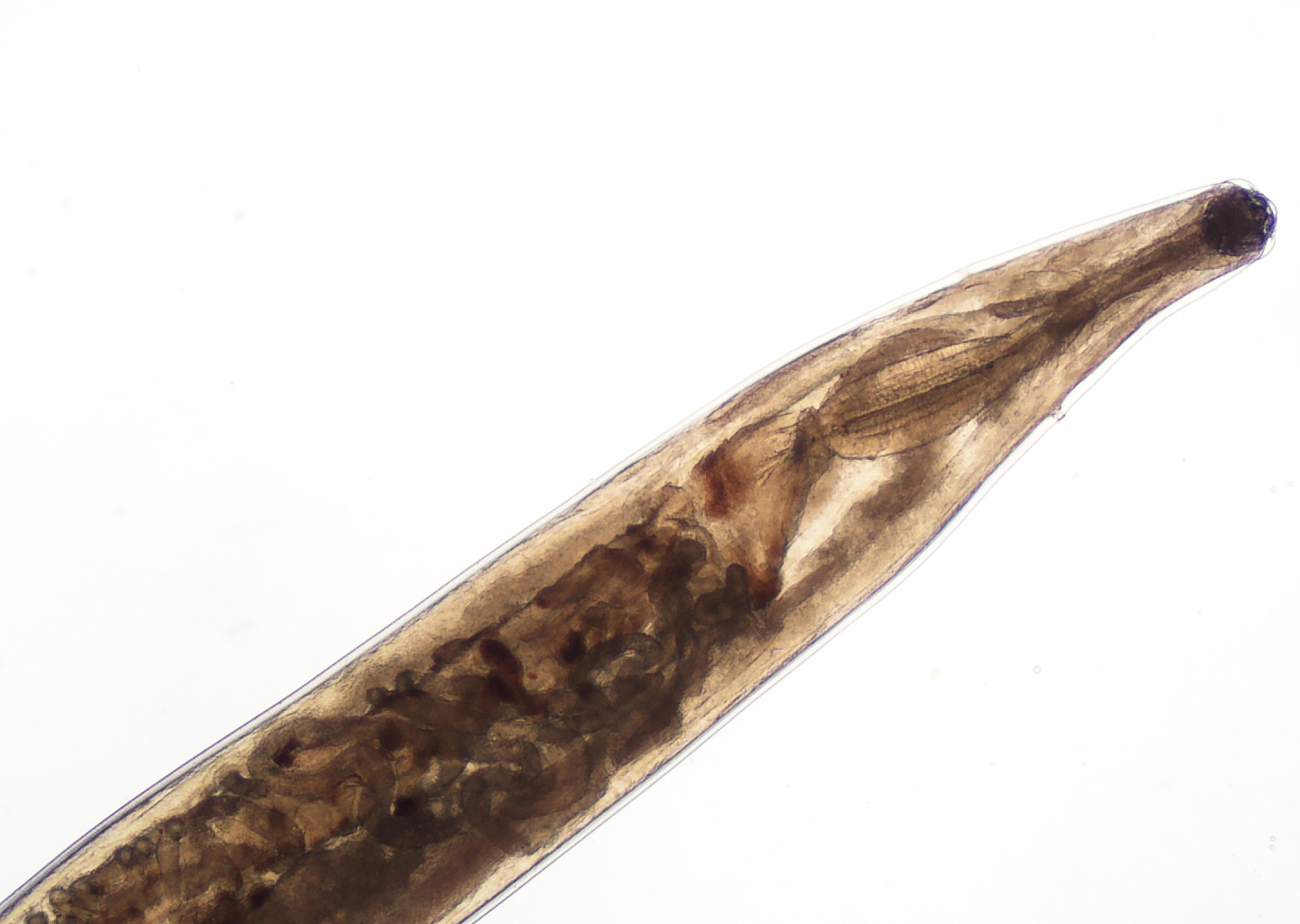
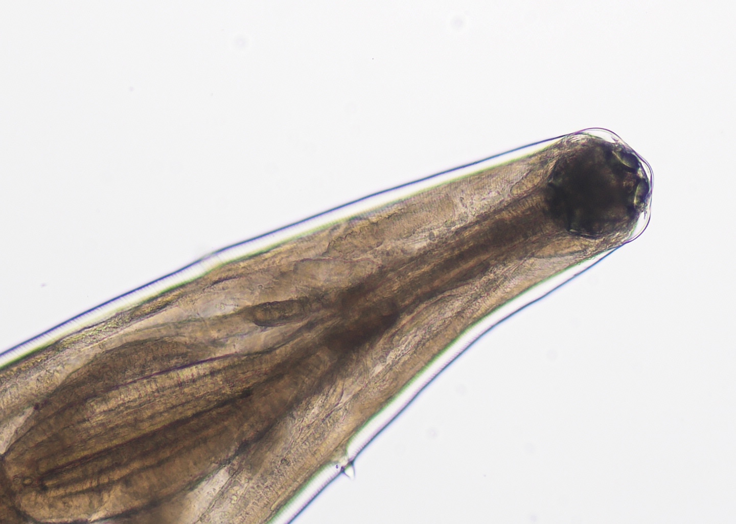
- Nematode in fresh (unfixed) stool specimen (Pritt: Creepy Dreadful Wonderful Parasites Blog - Case of the Week 553 [Accessed 9 August 2019])
Gross description
- Anterior end has characteristic 3 fleshy lips common to all ascarids (Pritt: Creepy Dreadful Wonderful Parasites Blog - Answer to Case 553 [Accessed 9 August 2019])
Gross images
Microscopic (histologic) description
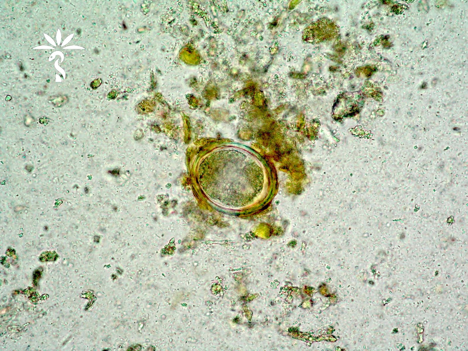
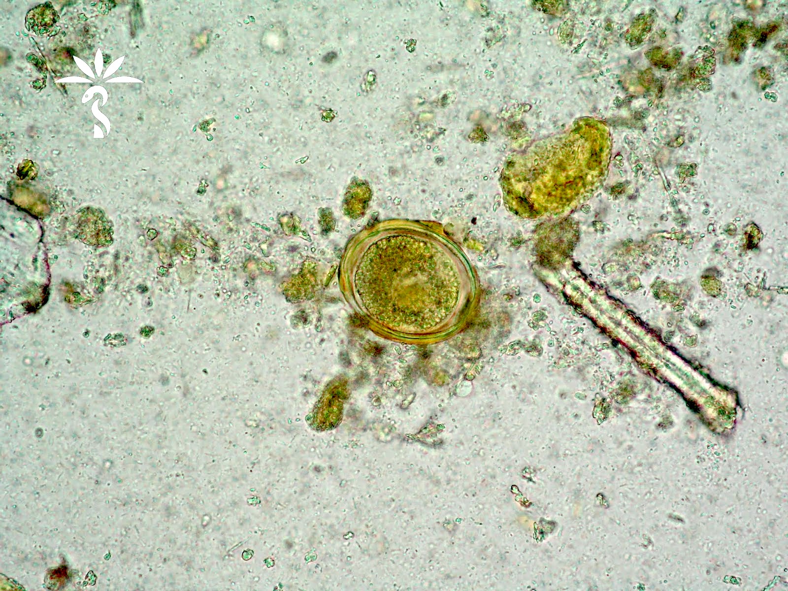
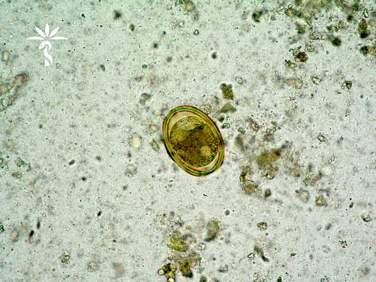
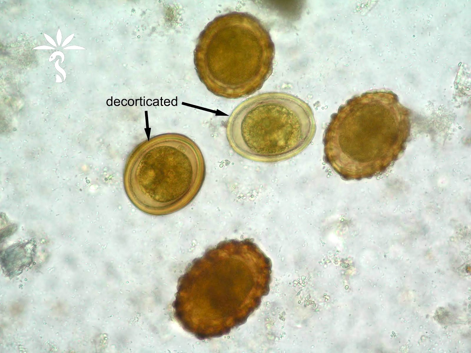
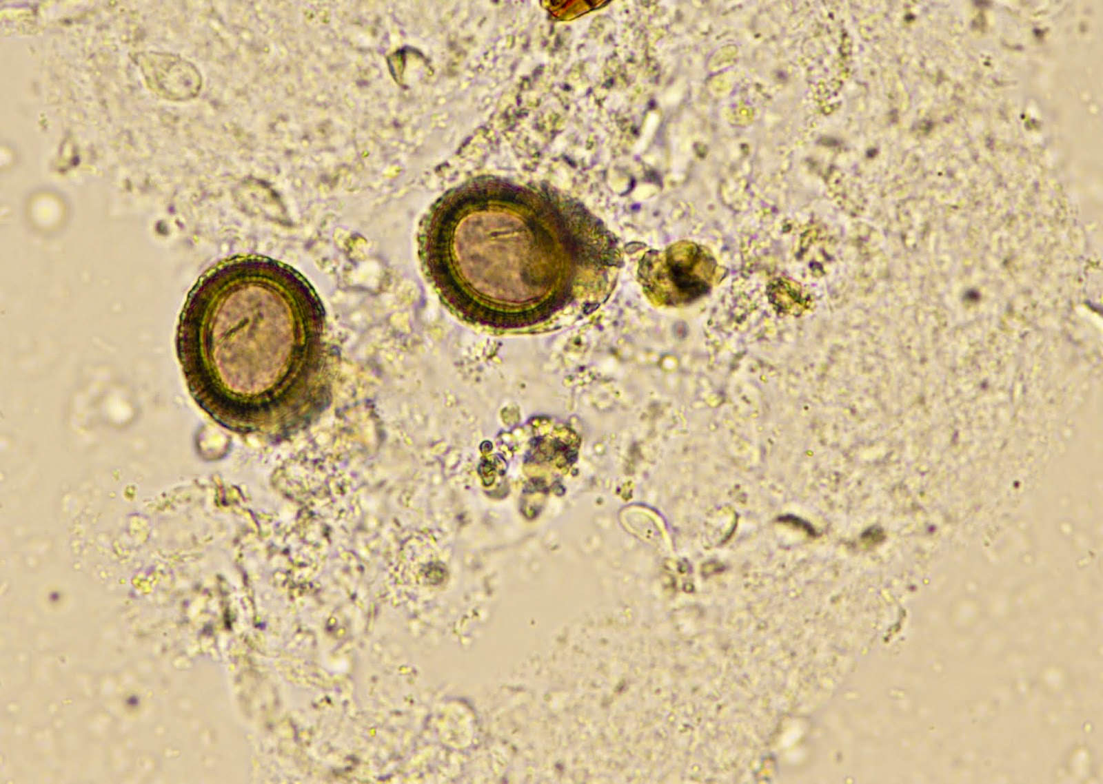
- Ascaris lumbricoides can have both mammillated and decorticated eggs (Pritt: Creepy Dreadful Wonderful Parasites Blog - Answer to Case 550 [Accessed 7 August 2019])
- Mammillated: having relatively small protrusions from the exterior, most commonly the surface
- Decorticated: to remove the bark, rind or husk from, i.e. to remove the outer mammillated layer
Microscopic (histologic) images
Videos
Creepy Dreadful Wonderful Parasites Case
Differential diagnosis
- Other helminth eggs such as hookworm and Schistosoma japonicum (Pritt: Creepy Dreadful Wonderful Parasites Blog - Answer to Case 550 [Accessed 7 August 2019]):
- When only decorticated eggs are seen, they can be confused for other helminth eggs
- Size and thick shell can be used to reliably differentiate them
Aspergillus
Babesia
Table of Contents
Definition / general | Essential features | Epidemiology | Sites | Pathophysiology | Clinical features | Diagnosis | Case reports | Treatment | Peripheral smear description | Peripheral smear images | Molecular / cytogenetics description | Differential diagnosis | Board review style question #1 | Board review style answer #1 | Board review style question #2 | Board review style answer #2Definition / general
- Babesia spp. are protozoan parasites that infect red blood cells
- Taxonomy:
- Phylum: Apicomplexa
- Order: Piroplasmida
- Family: Babesiidae
Essential features
- Babesiosis is caused by Babesia spp., transmitted via tick bites (most commonly, Ixodes) (Pathogens 2021;10:1447)
- Infection of erythrocytes leads to hemolytic anemia and cytokine production causing fever, jaundice, hepatosplenomegaly and, in severe cases, multiorgan failure (Pathogens 2022;11:399)
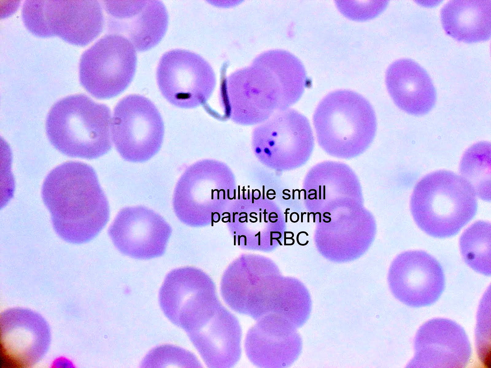
- Diagnosis is made upon seeing multiple infected red cells with extracellular ring forms on blood smear
- Classical blood smear finding is a tetrad of intracellular ring forms (Maltese cross) with extracellular ring forms
- Mild infections resolve spontaneously
- Moderate and severe manifestations may require treatment (moderate: atovaquone and azithromycin; severe: clindamycin, quinine, exchange transfusion)
Epidemiology
- More than 70 species exist worldwide but most cases of babesiosis in the United States are due to B. microti
- In Europe, B. divergens is associated with more serious clinical syndrome (Pathogens 2021;10:1165)
- In the Northeastern U.S., Ixodes scapularis, commonly known as deer tick or black legged tick, is the main species of tick that transmits B. microti (Pathogens 2021;10:1447)
- Splenectomy, HIV infection, immunosuppression and advanced age increase the likelihood of severe infection (Pathogens 2022;11:399)
- Humans are incidental hosts; natural hosts include small rodents (voles, field mice, etc.) (Pathogens 2021;10:1447)
- Other modes of transmission include transfusion, organ transplantation and transplacental (Pathogens 2022;11:399)
- Co-infection with Anaplasma and Borrelia can occur, as both diseases are vectored by Ixodes scapularis (Pathogens 2021;10:1447)
Sites
- Parasite found in the blood
Pathophysiology
- Babesia parasites are maintained in animal tick cycles, where ticks have transovarian and stage to stage transmission (Trop Parasitol 2015;5:94)
- Cycle of human infection (Pathogens 2021;10:1447):
- A Babesia infected tick will inject sporozoites via saliva into the bloodstream in the form of pyriform bodies
- Trophozoites infect erythrocytes and asexually reproduce via binary fission
- Erythrocyte lysis releases merozoites that infect other erythrocytes or are taken up by feeding ticks
- Lysis of red cells leads to a cascade of inflammatory responses resulting in fever, malaise with more severe disease (i.e., disseminated intravascular coagulation [DIC], renal failure, shock) in patients with splenectomy, immunosuppression and advanced age (Pathogens 2022;11:399)
Clinical features
- Symptoms include fever without periodicity, malaise, headache, chills, fatigue, weakness
- Signs include hemolytic anemia, hepatosplenomegaly
Diagnosis
- Diagnosis is made on thick and thin blood smear with Giemsa stain (gold standard)
- When parasitemia is low and in cases of screening (particularly for blood products), serological assays and antigen capture assays can be performed (Pathogens 2022;11:399)
- During acute disease, polymerase chain reaction (PCR) may be used for diagnosis in the form of organism specific Babesia spp. PCR or as part of larger tick borne disease PCR panels that include anaplasmosis, ehrlichiosis and babesiosis
Case reports
- 37 year old man in Singapore who acquired Babesia microti infection in the U.S. (Emerg Infect Dis 2020;26:826)
- 66 year old man in the South Bronx presenting with febrile illness (Clin Pract Cases Emerg Med 2018;2:61)
- 70 year old woman presenting with asplenic sepsis (Turk J Haematol 2019;36:284)
- 81 year old man from upstate New York with fevers, malaise, vague abdominal pain and confusion (Proc (Bayl Univ Med Cent) 2020;34:97)
Treatment
- Mild: resolves spontaneously
- Moderate: combination atovaquone and azithromycin
- Severe: can be treated with clindamycin, quinine, sometimes exchange transfusion
Peripheral smear description
- Thick smear Giemsa stain:
- Red / purple chromatin dot with pale blue cytoplasm forming a ring
- Thin smear Giemsa stain (Pathogens 2022;11:399):
- Intracellular and extracellular ring forms
- Usually multiple forms within each infected red cell
- Maltese cross = classical finding of tetrad of intracellular ring forms
Peripheral smear images
Molecular / cytogenetics description
- PCR tests for Babesia DNA exist and can be used in cases of low parasitemia and blood product screening
Differential diagnosis
- Plasmodium falciparum (Pathogens 2021;10:1165):
- Up to 2 intracellular ring forms
- No extracellular ring forms
- Can see banana shaped gametocytes
- Clinical history of travel to endemic area
- May exhibit periodicity in fever
- Other Plasmodium spp. (Pathogens 2021;10:1165):
- Red cells with normally 1 ring form, occasionally 2
- Will commonly see other forms: schizonts, gametocytes, etc.
- No extracellular ring forms
- Clinical history of travel to endemic area
- May exhibit periodicity in fever
- If there has been a known Ixodes tick exposure and general symptoms of fever and malaise, differential diagnoses should include the following (Trends Parasitol 2018;34:295):
- Lyme disease:
- No visible parasites on blood smear
- Serology and PCR studies positive for Borrelia burgdorferi, a spirochete bacterium
- Commonly co-infects with Babesia
- Tick borne relapsing fever:
- No visible parasites on blood smear
- Serology and PCR studies positive for Borrelia miyamotoi
- Anaplasmosis:
- No visible intraerythrocytic or extracellular trophozoites
- May have morula observed in granulocytes
- Serology and PCR studies positive for Anaplasma phagocytophilum, a tick borne bacterium
- Ehrlichiosis:
- No visible intraerythrocytic or extracellular trophozoites
- May have morula observed in monocytes or granulocytes
- Serology and PCR studies positive for Ehrlichia spp.
- Powassan virus disease:
- No visible parasites on blood smear
- Laboratory diagnosis by testing serum or cerebrospinal fluid for virus specific antibodies
- Lyme disease:
Board review style question #1
Board review style answer #1
A. It causes more severe disease in asplenic patients. Babesiosis is less severe in those with an intact spleen, as the diseased red cells can be removed. The primary treatment of babesiosis is with atovaquone and amoxicillin. Babesiosis can co-infect with Lyme disease, Anaplasma and other tick borne diseases. Babesia is endemic to U.S. (most characteristically Mid-Atlantic to Northeastern U.S.) and Europe, not to sub-Saharan Africa (unlike Plasmodium spp.).
Comment Here
Reference: Babesia
Comment Here
Reference: Babesia
Board review style question #2
The clinical microbiology lab received a blood specimen from a 10 year old boy presenting with fever, malaise and chills. Significant history includes recent camping trip to Connecticut. Seen on thin smear are intraerythrocytic and extraerythrocytic ring forms and some red cells with up to 6 ring forms. What is the most likely diagnosis?
- Babesia microti
- Borrelia burgdorferi
- Plasmodium falciparum
- Plasmodium malariae
Board review style answer #2
A. Babesia microti. Babesia spp. are characterized by multiple intracellular ring forms and extracellular forms on blood smear. Babesiosis is common in the Northeastern U.S. due to prevalence of Ixodes (deer) tick. Without travel to malaria endemic regions, Plasmodium can be eliminated as a potential diagnosis. While the Ixodes scapularis tick can also vector Borrelia burgdorferi, the causative agent of Lyme disease, B. burgdorferi is a spirochete that is unlikely to be observed on blood smears.
Comment Here
Reference: Babesia
Comment Here
Reference: Babesia
Balamuthia
Table of Contents
Definition / general | Essential features | Epidemiology | Sites | Pathophysiology | Clinical features | Diagnosis | Laboratory | Case reports | Treatment | Microscopic (histologic) description | Microscopic (histologic) images | Differential diagnosis | Additional references | Board review style question #1 | Board review style answer #1 | Board review style question #2 | Board review style answer #2Definition / general
- Taxonomy: genera Balamuthia, family Balamuthiidae
- Single celled, free living amoeba discovered from brain tissue fragments of a mandrill baboon (old world monkey) that died from a neurological condition at San Diego Zoo Wild Animal Park in California in 1986 (Clin Microbiol Rev 2008;21:435)
- Rare cause of chronic granulomatous amebic encephalitis, disseminated disease or skin lesions in immunocompetent and immunocompromised individuals
- Enters nasal passages or ulcerated broken skin
- Hematogenous dissemination to the central nervous system (CNS)
- Similar disease spectrum as Acanthamoeba
Essential features
- 2 stage life cycle (Trop Parasitol 2015;5:15)
- Trophozoite: 12 - 60 μm in diameter, infective, pleomorphic, uninucleated / binucleated, with 1 - 3 nucleoli (helps differentiate from Acanthamoeba) and long, slender pseudopodia
- Cyst: 6 - 30 μm, dormant round cells that form under harsh conditions; double walled with wrinkled fibrous outer wall (exocyst); 1 or 2 nuclei
Epidemiology
- Isolated from soil, dust, fresh water
- Human exposure is common but infection is rare
- > 100 confirmed cases in the U.S.; > 200 worldwide (CDC: Parasites - Balamuthia mandrillaris - Granulomatous Amebic Encephalitis (GAE) - General Information [Accessed 15 December 2022])
- Given the difficulty of diagnosing infection, many undiagnosed infections are suspected
- Cases in the Southwestern U.S. and Latin America (Peru), with limited cases in Asia, Australia and Europe, have involved the development of facial skin lesions
- Increased risk in Hispanic ethnicity and immunocompromised conditions: diabetes, HIV / AIDS, solid organ transplant, liver cirrhosis, renal failure and cancer (Clin Infect Dis 2019;68:1815)
Sites
- Skin, brain, spinal cord
Pathophysiology
- Entry via nasal passages or broken skin; rarely organ donation with dissemination to the CNS (Trop Parasitol 2015;5:15)
- It is transmitted on contact with skin wounds and cuts or when dust containing the parasite is breathed (e.g., during gardening)
- Organisms are difficult to eradicate with tropism to blood vessels
- Initial acute inflammatory response develops into chronic granulomatous inflammation with persistent perivascular ameba involving CD4 and CD8 T cells, epithelioid histiocytes and multinucleated giant cells
- In immunocompromised patients, granulomas may or may not be present
Clinical features
- Skin:
- Involvement of face, palate, extremities and trunk (Clin Infect Dis 2019;68:1815)
- Single or disseminated chronic plaque-like ulcerated skin lesion; may be mistaken for chronic fungal or mycobacterial infection and progress to CNS disease
- CNS:
- Granulomatous amebic encephalitis:
- Indolent progression initially over weeks to months
- Usually fatal with 90% mortality
- Hematogenous spread from skin or nasal passages (Clin Infect Dis 2019;68:1815)
- Meningoencephalitis: fever, headache, stiff neck, nausea, vomiting, lethargy with neurological deficits including ataxia, impaired speech, focal deficit and seizures
- Granulomatous amebic encephalitis:
Diagnosis
- Identification of cysts or trophozoites in brain or skin biopsy
Laboratory
- Skin:
- Biopsy and microscopic evaluation of formalin fixed paraffin embedded (FFPE) tissue
- Direct microscopic examination with Calcofluor white dye
- RT-PCR on fresh tissue is required for definitive diagnosis
- CNS:
- Cerebrospinal fluid (BMC Microbiol 2008;8:210)
- High lymphocytes, low glucose, high protein
- Extremely rare to identify amebae on cerebrospinal fluid
- Biopsy and microscopic evaluation of FFPE tissue
- RT-PCR on fresh tissue or cerebrospinal fluid required for definitive diagnosis
- In the U.S., the CDC offers RT-PCR for B. mandrillaris and morphologic mimics Naegleria fowleri and Acanthamoeba species
- Negative cerebrospinal fluid RT-PCR result does not rule out intraparenchymal brain infection
- Cerebrospinal fluid (BMC Microbiol 2008;8:210)
- CDC ID pathology branch offers immunohistochemical staining
- Antibodies in serum
- Indirect immunofluorescence assay (CDC: Parasites - Balamuthia mandrillaris - Granulomatous Amebic Encephalitis (GAE) - Diagnosis & Detection [Accessed 15 December 2022])
- Culture requires special media and is not available in clinical labs
Case reports
- 3 year old girl with B. madrillaris granulomatous amebic encephalitis (Am J Trop Med Hyg 2021;104:1836)
- 52 year old woman diagnosed with B. mandrillaris showing unusual lab and radiological findings (Arch Neurol 2000;57:1210)
- 72 year old immunocompetent woman with Balamuthia encephalitis (Arch Pathol Lab Med 2004;128:466)
Treatment
- Miltefosine in combination with drugs such as flucytosine, pentamidine, fluconazole, sulfadiazine and either azithromycin / clarithromycin (Clin Infect Dis 2010;51:e7)
Microscopic (histologic) description
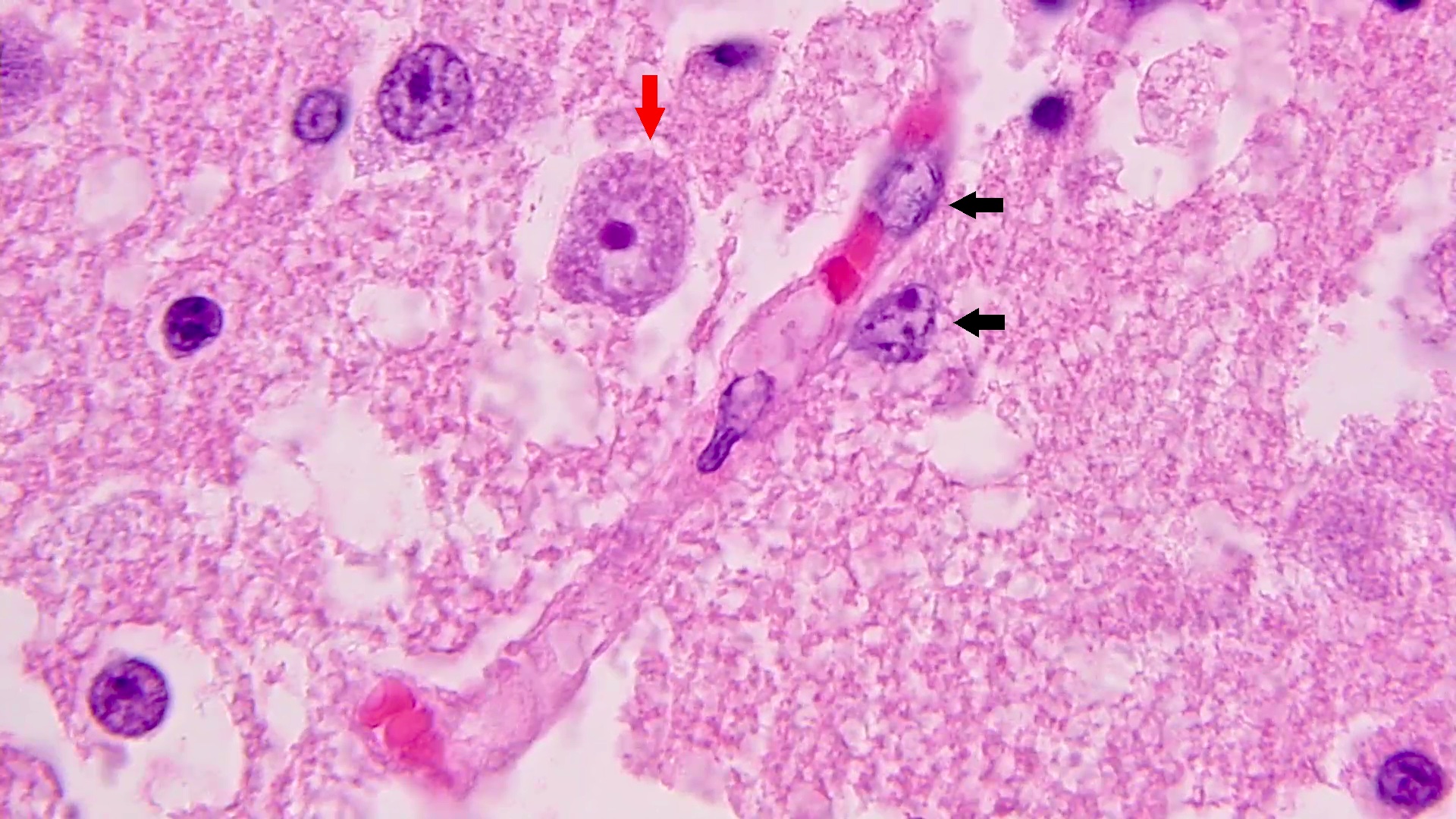
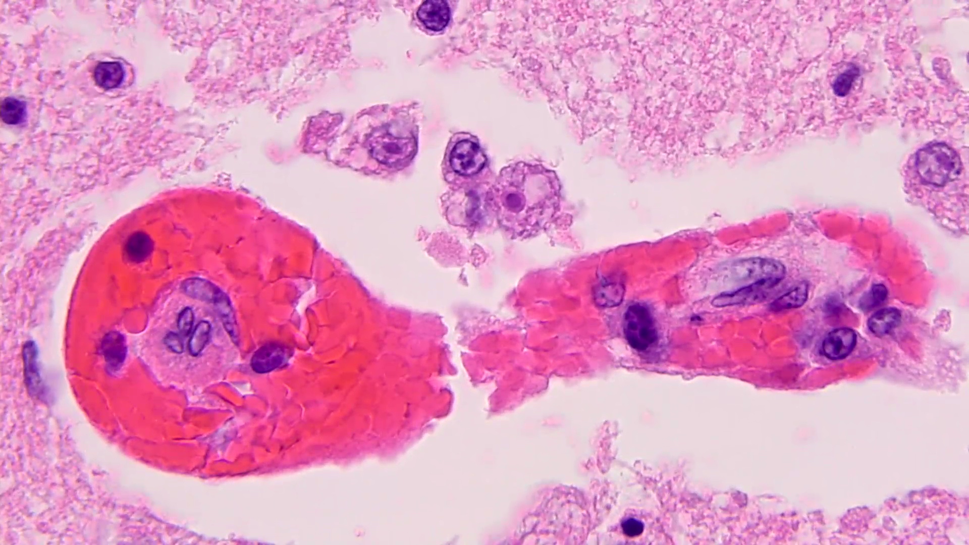
- Chronic granulomatous inflammation with cysts or trophozoites surrounding blood vessels
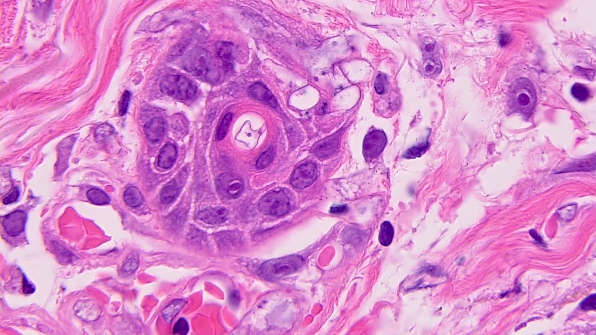
- Cysts have 2 walls: wrinkled fibrous outer exocyst and inner endocyst and appear hexagonal, spherical or star shaped
- Refractive granules may be present in the inner wall with no pores
- May be uninucleate / binucleate
- Trophozoites are pleomorphic with long pseudopodia
- May be uninucleate / binucleate with multiple nucleoli in infected tissue
- Reference: Trop Parasitol 2015;5:15
Microscopic (histologic) images
Differential diagnosis
- Tuberculosis:
- Acid fast bacilli within granulomatous inflammation
- Acanthamoeba:
- Inner wall (endocyst) with hexagonal, spherical, star shaped or polygonal morphology
- Molecular identification often necessary for definitive distinction
- Naegleria:
- Acute illness and inflammation are predominant
- Cysts do not form in human tissues
- Granular cytoplasm with many vacuoles, single large nucleus and dense karyosome with no margination
- Sappinia pedata:
- Binucleated trophozoites and cysts
- Only 1 reported case in Texas (JAMA 2001;285:2450)
- Neurocysticercosis:
- Cysticerci present
- Pathognomonic calcaneous corpuscles within cestode tissue
- Histoplasma:
- Intracellular, uniformly sized, oval to round budding yeasts
- Mold infection:
- Infiltrating hyphae
- Aspergillus and neurotropic dematiaceous molds
- Reference: Emerg Microbes Infect 2020;9:1379
Additional references
Board review style question #1
A 13 year old girl presented with a 1 year history of a cutaneous skin lesion and recent onset of dizziness, blurry vision, diplopia and worsening headaches. Brain CT showed hypodense regions in the left lateral ventricle and a brain biopsy revealed granulomatous inflammation and the perivascular organisms shown above. What microscopic features can be used to distinguish this organism from Acanthamoeba species?
- Endocyst structure
- Exocyst structure
- Multiple nucleoli
- Vacuolated cytoplasm
Board review style answer #1
A. Endocyst structure. Balamuthia mandrillaris resembles Acanthamoeba and may require molecular identification for definitive identification in granulomatous CNS lesions. However, the inner wall (endocyst) of Acanthamoeba may exhibit hexagonal, spherical, star shaped or polygonal morphology that distinguishes it from the more uniform endocyst of Balamuthia species.
Comment Here
Reference: Balamuthia
Comment Here
Reference: Balamuthia
Board review style question #2
A 58 year old man presented with 2 month history of sporadic headaches that have increased in frequency. Fundoscopy showed bilateral papilledema and retinal hemorrhages. Brain CT showed a hypodense lesion in right temporal lobe. It was suspected to be brain tumor and resected. Amebic trophozoites were identified. What test can confirm the diagnosis?
- Cerebrospinal fluid microscopy
- Culture on agar
- Real time (RT) PCR
- Serology
Board review style answer #2
C. Real time PCR can detect up to a single organism per reaction and distinguish between Acanthamoeba, Balamuthia and Naegleria. PCR on fresh tissue is optimal. PCR on cerebrospinal fluid can be helpful to render the diagnosis but does not rule out intraparenchymal infection if negative.
Comment Here
Reference: Balamuthia
Comment Here
Reference: Balamuthia
Biosafety for lab and pathology (pending)
[Pending]
Blastocystis
Table of Contents
Definition / general | Diagrams / tables | Case reports | Microscopic (histologic) description | Microscopic (histologic) images | Additional referencesDefinition / general
- Single celled parasite that inhabits the gastrointestinal tracts of humans and animals (Wikipedia: Blastocystis [Accessed 28 June 2019])
Case reports
- Objects were seen in a trichrome stained stool specimen (Pritt: Creepy Dreadful Wonderful Parasites Blog - Case of the Week 546 [Accessed 28 June 2019])
Microscopic (histologic) description
- 4 common forms: vacuolar, granular, amoeboid and cyst (Wikipedia: Blastocystis [Accessed 28 June 2019])
Microscopic (histologic) images
Additional references
Blastomyces
Table of Contents
Definition / general | Essential features | Epidemiology | Sites | Pathophysiology | Clinical features | Laboratory | Case reports | Treatment | Microscopic (histologic) description | Microscopic (histologic) images | Positive stains | Molecular / cytogenetics description | Differential diagnosis | Additional references | Board review style question #1 | Board review style answer #1 | Board review style question #2 | Board review style answer #2Definition / general
- Taxonomy:
- Class: Eurotiomycetes; order: Onygenales; family: Ajellomycetaceae
- Common species:
- Blastomyces dermatitidis: most common cause of blastomycosis
- Blastomyces gilchristi: northern U.S. and Canada
- Blastomyces helicus: western U.S
- Blastomyces percursus: Africa
Essential features
- Dimorphic mold
- Body temperature, uniformly sized, extracellular round yeasts with a thick, refractile, double contoured cell wall and broad based budding
- Immunocompetent: a chronic granulomatous and suppurative disease of the lung or skin
- Immunocompromised: disseminated disease with poor prognosis
Epidemiology
- Cause of blastomycosis, a chronic pyogranulomatous inflammatory disease
- Soil organisms, associated with moist areas near riverbeds
- Midwestern, south central and southeastern U.S., particularly in areas surrounding the Ohio and Mississippi River valleys, the Great Lakes and the Saint Lawrence River
- Present in Canada, with a small number of documented cases in Africa and India
- References: J Clin Aesthet Dermatol 2009;2:22, Semin Respir Crit Care Med 2020;41:31
Sites
- Pulmonary blastomycosis: can be asymptomatic or range from self limited pulmonary infection to life threatening (eMedicine: Acute Respiratory Distress Syndrome (ARDS) [Accessed 26 January 2022])
- Extrapulmonary blastomycosis:
- Cutaneous: development of verrucous (wart-like) or ulcerative skin lesions
- Osseous: osteomyelitis along with contagious tissue abscesses and draining sinuses
- Genitourinary (usually male): involvement of the prostate and epididymis
- Central nervous system: brain abscess, cranial or epidural abscess and meningitis
Pathophysiology
- Inhaled spores transform into yeasts resulting in acute and chronic lung inflammation
- Cutaneous lesions can demonstrate pseudoepitheliomatous hyperplasia of the epidermis
- Dissemination to extrapulmonary sites, especially the skin, is presumed to be hematogenous
- Reference: J Clin Aesthet Dermatol 2009;2:22
Clinical features
- Involves the lung in over 90% of cases; infection can range from asymptomatic self limited infection (about 50% of cases) to severe diffuse pneumonia causing respiratory failure
- Extrapulmonary dissemination occurs in approximately 25 - 50% of cases of blastomycosis (Mycopathologia 2009;167:115)
Laboratory
- Direct exam shows large, thick walled, yeast forms with single broad based budding cells (8 - 10 μm)
- Slow growing yeasts that transition to a fluffy white mycelium or glabrous, tan, nonsporulating colonies within 1 - 4 weeks
- Microconidia resemble Histoplasma capsulatum but macroconidia are not formed
- Microscopic examination of growth demonstrates thick walled yeast cells with broad based budding
- Conversion to the yeast phase can occur on routine media incubated at 37 °C
- Antibody detection tests
- Immunodiffusion utilizes purified B. dermatitidis A antigen (relatively low sensitivity and specificity)
- Enzyme immunoassays use BAD1 antigen: most sensitive approach (cross reactivity seen with histoplasmosis and other fungal infections)
- Reference: Semin Respir Crit Care Med 2020;41:31
Case reports
- 17 year old South African boy with slow growing lesion of the scalp and neck (Int J Dermatol 2012;51:1090)
- 24 year old man with blastomycosis of nose (Ear Nose Throat J 2016;95:E28)
- 36 year old immunocompetent man with disseminated blastomycosis (Oxf Med Case Reports 2018;2018:omy071)
- 42 year old man with Blastomyces oteomyelitis (Cureus 2020;12:e7417)
- 42 year old man with disseminated blastomycosis infection diagnosed on FNA of the thyroid (Diagn Cytopathol 2011;39:446)
Treatment
- Acute pulmonary blastomycosis can be mild and self limited, not requiring treatment in immunocompetent hosts
- More severe disease, particularly in immunocompromised hosts, requires itraconazole with or without liposomal amphotericin B
- Treatment duration can range from 6 months to 1 year
- References: Clin Infect Dis 2000;30:679, S D Med 2006;59:255
Microscopic (histologic) description
- Pyogranulomatous inflammation: neutrophil infiltration admixed with epithelioid histiocytes and granulomatous inflammation
- Cutaneous lesions demonstrate pseudoepitheliomatous hyperplasia of the epidermis
- Uniformly sized, refractile round yeast cells may be observed at low power in H&E stained tissue sections
- High power may show cell contents within the refractile cell wall but often this material washes away during processing
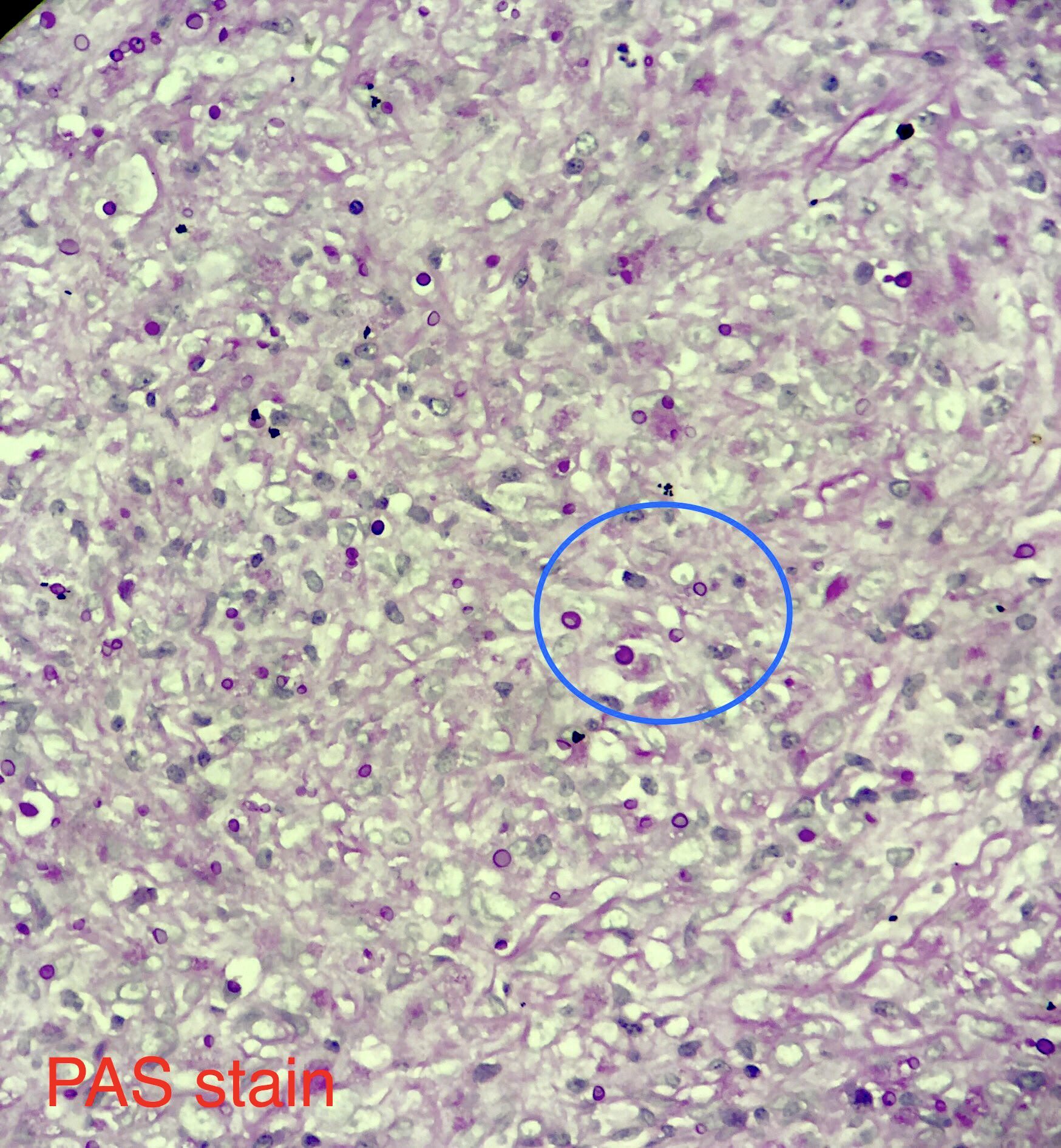
- Periodic acid-Schiff (PAS) and Gomori methenamine silver (GMS) stains highlight organisms
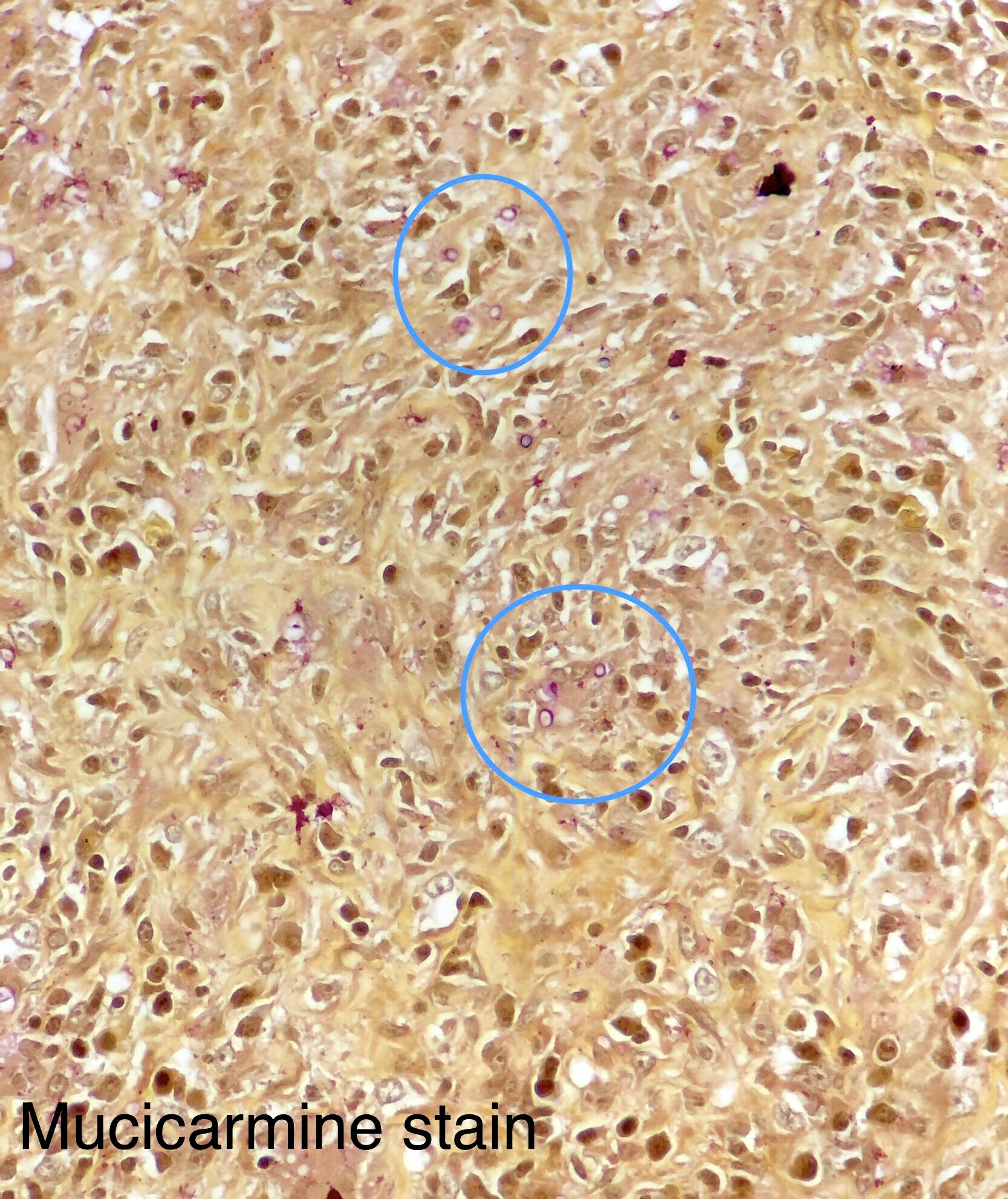
- Mucicarmine may be weakly positive; contrast with a strongly positive Cryptococcus
- Fontana-Masson stain for melanin: negative
- Reference: Semin Respir Crit Care Med 2020;41:31
Microscopic (histologic) images
Positive stains
- Periodic acid-Schiff (PAS) and Gomori methenamine silver (GMS) stains highlight organisms
- Mucicarmine may be weakly positive; contrast with a strong positive Cryptococcus
Molecular / cytogenetics description
- Nucleic acid probe hybridization assays are commercially available (AccuProbe by Hologic, Inc.) for definitive identification on culture
- Additional lab developed PCR tests, sequencing and matrix assisted laser desorption / ionization time of flight (MALDI-TOF) mass spectrometry (MS) may enable identification
Differential diagnosis
- Acute illness:
- Community acquired pneumonia:
- Viral, bacterial, atypical bacterial
- Community acquired pneumonia:
- Chronic illness:
- Tuberculosis, infection with nontuberculous mycobacteria:
- Acid fast organisms
- Histoplasmosis:
- Small, uniform, narrow based budding yeasts; large tuberculate macroconidia
- Coccidioidomycosis:
- Thick walled spherules with endospores in tissue
- Sarcoidosis:
- Diagnosis of exclusion after ruling out all the infectious agents
- Tuberculosis, infection with nontuberculous mycobacteria:
Additional references
Board review style question #1
A 36 year old Caucasian woman from Tennessee develops flu-like symptoms with fever, headache, diffuse joint pain and cough. She currently takes ibuprofen, which does not alleviate symptoms. An Xray shows diffuse bilateral pneumonia. After 1 week at 30 °C, sputum cultures show unique colonies with central yeast-like creamy morphology and peripheral fuzzy mold-like extensions. At 2 weeks, the colonies are almost entirely mold-like and a tape prep shows thin hyaline septate hyphae with lollipop-like structures called aleurioconidia. No large tuberculate macroconidia are noted at 2 weeks but are spotted at 4 weeks. A molecular test is performed to confirm the diagnosis. What is the most likely cause of respiratory illness?
- Blastomyces dermatitidis
- Coccidioides immitis
- Histoplasma capsulatum
- Paracoccidiodes braziliensis
Board review style answer #1
C. Histoplasma capsulatum. Coccidiodes species exhibit alternating barrel shaped arthroconidia. Paracoccidiodes, Blastomyces and Histoplasma species cultured at 30 °C form lollipop-like aleurioconidia but only Histoplasma proceeds to make large tuberculate macroconidia in late cultures.
Comment Here
Reference: Blastomyces
Comment Here
Reference: Blastomyces
Board review style question #2
Which of the following is true for Blastomyces infection?
- Cutaneous lesions demonstrate pseudoepitheliomatous hyperplasia of the epidermis
- Direct exam shows large, thick walled, refractile yeast forms (8 - 10 μm) with narrow based budding
- Extrapulmonary dissemination does not occur in Blastomyces
- Late cultures show large tuberculate macroconidia
Board review style answer #2
A. Cutaneous lesions demonstrate pseudoepitheliomatous hyperplasia of the epidermis
Comment Here
Reference: Blastomyces
Comment Here
Reference: Blastomyces
Blood culture contamination (pending)
[Pending]
Borrelia burgdorferi / Lyme disease
Table of Contents
Definition / general | Epidemiology | Pathophysiology | Etiology | Clinical features | Staging / staging classifications | Diagnosis | Laboratory | Case reports | Treatment | Clinical images | Microscopic (histologic) description | Microscopic (histologic) images | Positive stains | Differential diagnosis | Additional referencesDefinition / general
- Lyme disease is a multisystem disorder caused by spirochete Borrelia burgdorferi
Epidemiology
- In the United States, Lyme disease most commonly occurs in the northeast and upper midwest
- Worldwide, more commonly found in northern Asia and eastern and central Europe
Pathophysiology
- The bite of an infected Ixodes dammini tick causes proliferation of spirochetes in the dermis
- The host dermal inflammatory response causes a rash known as erythema chronicum migrans
- Over days, the spirochetes spread to the nervous system, cardiac tissue and joints via the blood stream
- The spirochetes may induce host cells to produce quinolinic acid, which stimulates NMDA receptors and manifests as malaise in lyme encephalopathy (Neurology 1992;42:43)
- Note: Lyme disease spirochetes are never seen in peripheral blood
Etiology
- Usually transmitted by Ixodes dammini tick
Clinical features
- Causes erythema chronicum migrans (red papule with central clearing that expands slowly), acrodermatitis chronica atrophicans (disease of extremities, usually women, with erythematous, edematous, pruritic phase, followed by sclerosis and atrophy), cutaneous lymphoid hyperplasia of skin
- Also affects heart, joints, nervous system
Staging / staging classifications
- Stage 1: skin lesion (erythema chronicum migrans)
- Stage 2: cardiovascular and nervous system involvement
- Stage 3: arthritis stage characterized by migratory polyarthritis; however, cutaneous lesions and peripheral nervous system involvement are also encountered in this stage
Diagnosis
- Clinical symptoms such as erythema migrans, fever, facial palsy or arthritis
- Biopsy interpretation
Laboratory
- EIA or ELISA for total Lyme titer or IgG and IgM titers
- If EIA / ELISA test results come back positive or equivocal, Western blot IgG and IgM titers are performed
- Lyme titers should be done if the above tests are positive
- PCR in synovial fluid (for spirochetes)
- CSF analysis
- ECG for Lyme carditis
- Darkfield microscopy for spirochetes
Case reports
- Young boy with butterfly rash (Acta Derm Venereol 2010;90:109)
- 17 year old boy with fatal Lyme carditis (Cardiovasc Pathol 2015;24:317)
- 27 year old woman with Lyme disease associated neuroretinitis (Acta Microbiol Immunol Hung 2015;62:403)
- 30 and 50 year old men with "chronic Lyme disease" (JAMA Intern Med 2015;175:132)
- Lyme disease with hearing loss as sole presentation (J Laryngol Otol 2015;129:183)
Treatment
- Early infection: Antibiotics such as doxycycline, amoxicillin, azithromycin
- Late infection: IV ceftriaxone is considered treatment of choice
Clinical images
Microscopic (histologic) description
- Superficial and deep perivascular polymorphic infiltrate of neutrophils, lymphocytes, plasma cells, eosinophils and mast cells
- Vascular proliferation and dermal necrosis may be present
- Identification of spirochetes by silver stain or immunocytochemistry is diagnostic
- Borrelia spirochetes are long spiral bacilli (5 - 20 microns in length) with relatively regular undulations
- Typically Borrelia burgdorferi does NOT reach sufficient levels in blood to be seen on peripheral blood smear
- PCR can also be used for diagnosis
Microscopic (histologic) images
Positive stains
- Warthin-Starry (for spirochetes)
- Immunocytochemistry with monoclonal antibodies against Borrelia species
Differential diagnosis
- Bacteria that are not spirochetes but have curved or wavy rods include Vibrio, Campylobacter, Helicobacter
- Other spirochetes include Treponema and Leptospira, but they are not typically seen in blood
Additional references
- WHO: Lyme Borreliosis [Accessed 28 August 2018], eMedicine: Lyme Disease [Accessed 28 August 2018], Wikipedia: Lyme Disease [Accessed 28 August 2018], Rosai: Rosai and Ackerman's Surgical Pathology, 10th Edition, 2011, Johnston: Weedon's Skin Pathology Essentials, 1st Edition, 2012, Emerg Infect Dis 2007;13:436, Pritt: Creepy Dreadful Wonderful Parasites Blog [Accessed 7 August 2018]
Borrelia recurrentis / relapsing fever (pending)
[Pending]
Candida auris
Table of Contents
Definition / general | Essential features | ICD coding | Epidemiology | Sites | Pathophysiology | Etiology | Clinical features | Diagnosis | Laboratory | Radiology description | Radiology images | Prognostic factors | Case reports | Treatment | Gross description | Microscopic (histologic) description | Microscopic (histologic) images | Cytology description | Cytology images | Positive stains | Negative stains | Electron microscopy description | Electron microscopy images | Molecular / cytogenetics description | Sample pathology report | Differential diagnosis | Additional references | Board review style question #1 | Board review style answer #1 | Board review style question #2 | Board review style answer #2Definition / general
- Candida auris is an emergent, multidrug resistant fungal pathogen that causes infections with a high mortality rate; first described in Japan in 2009 (Arch Pathol Lab Med 2020;144:107, J Clin Microbiol 2011;49:3139)
Essential features
- Candida auris has a similar morphology to other Candida species in infected tissues, except C. glabrata, which does not form pseudohyphae or hyphae (Mycoses 2018;61:377)
- Incidence in the U.S. has been rising with outbreaks in healthcare facilities
ICD coding
Epidemiology
- Outbreaks of C. auris have happened in the U.S. since 2016, after the introduction of multiple strains from different continents; since then, local transmission has taken place (MMWR Morb Mortal Wkly Rep 2016;65:1234, Lancet Infect Dis 2018;18:1377)
- Major outbreaks occurred in Illinois, Chicago, New York and New Jersey (MMWR Morb Mortal Wkly Rep 2020;69:6, Ann Intern Med 2021;174:1554)
- Risks for C. auris infection include (MMWR Morb Mortal Wkly Rep 2017;66:514)
- Immunosuppression (malignancy, chemotherapy, neutropenia, high doses of corticosteroids, AIDS, chronic underlying diseases)
- Prolonged intensive care stays
- Abdominal surgery and anastomotic leak
- Pancreatitis
- Hemodialysis
- Use of broad spectrum antibiotics and azoles (previous fluconazole treatment)
- Total parenteral nutrition
- Injection of illicit drugs
- Preventive measures for healthcare infection transmission, such as contact isolation for colonized or infected patients and laboratory diagnostic surveillance to determine species and antifungal susceptibility / resistance in Candida isolates (MMWR Morb Mortal Wkly Rep 2017;66:514)
Sites
- Candida auris can infect any organ from any body system
- Bloodstream and disseminated infection must be investigated when C. auris is isolated from any sample (Clin Microbiol Rev 2017;31:e00029)
Pathophysiology
- Candida auris infection starts with colonization, followed by tissue invasion and then reaches the bloodstream
- Candida auris forms biofilm on catheter device surfaces (insertion or hub)
- Total parenteral nutrition is rich in lipid emulsions, which enhances the biofilm formation; broad spectrum antibiotics and intestinal / biliary surgery alter normal flora with Candida spp. overgrowth, predisposing to its intestinal translocation, followed by bloodstream dissemination
- Host factors predispose to disseminated C. auris infections (e.g., immune dysfunction [neutropenia, lymphopenia, denutrition, etc.] and mucositis after chemotherapy) (Clin Microbiol Rev 2017;31:e00029)
Etiology
- Retrospective study has shown the first Candida auris isolate is from South Korea
- Sequencing of internal transcribed spacer (ITS) and D1 / D2 regions of ribosomal DNA has shown C. auris is similar to C. haemulonii and C. pseudohaemulonii in C. auris is geographically grouped into 4 clades: East Asia, South Asia, Africa and South America
- Molecular profiles of sequenced strains are more linked with strains from the same country (Clin Microbiol Rev 2017;31:e00029)
Clinical features
- Invasive healthcare associated infections with high mortality
- Pneumonia
- Vascular device associated bloodstream infections
- Skin lesions (papules, ulcers) in disseminated disease
- Pyelonephritis
- Biliary tract infections
- Inhospital sepsis
- Septic shock
- Panophthalmitis in immunocompromised host (AIDS) (Am J Ophthalmol Case Rep 2020;19:100738)
Diagnosis
- Definitive diagnosis of C. auris infection is performed with sequencing of 18S ITS regions or D1 / D2 regions of ribosomal DNA
- Real time polymerase chain reaction (RT PCR) may have high diagnostic accuracy
- Proteomic methods, such as MALDI TOF MS, can be useful
- Significant overlap with other Candida species on cultures, phenotypic and biochemical diagnostic systems (Clin Microbiol Rev 2017;31:e00029)
Laboratory
- Patients with C. auris infection may have neutropenia, anemia, lymphopenia, elevated C reactive protein or signs of multiorgan system failure with altered markers for organ dysfunction (azotemia, hypoxemia, elevated bilirubin, etc.) (Clin Microbiol Rev 2017;31:e00029)
Radiology description
- Nonspecific
- Abscesses, pneumonia, pyelonephritis
- Infective foci suggestive of hematogenous spread
Prognostic factors
- Candida auris infection has a poor prognosis in general
Case reports
- 61 year old immunocompetent man with Candida auris candidemia after posttraumatic brain injury (Cureus 2020;12:e8850)
- 71 year old Japanese man with COVID-19 presented with Candida auris candidemia (J Infect Chemother 2023;29:713)
- First 7 reported cases of Candida auris in the U.S. (MMWR Morb Mortal Wkly Rep 2016;65:1234)
Treatment
- Echinocandins
- C. auris has a higher minimum inhibitory concentration (MIC) than other Candida species
- Lipid formulation amphotericin (Clin Microbiol Rev 2017;31:e00029)
- Often multidrug resistant
Gross description
- Tissue necrosis, abscesses, mucosal ulcers, mucosa covered with whitish or yellowish fibrinous exudate (Emerg Microbes Infect 2020;9:1160, Clin Microbiol Rev 2017;31:e00029)
Microscopic (histologic) description
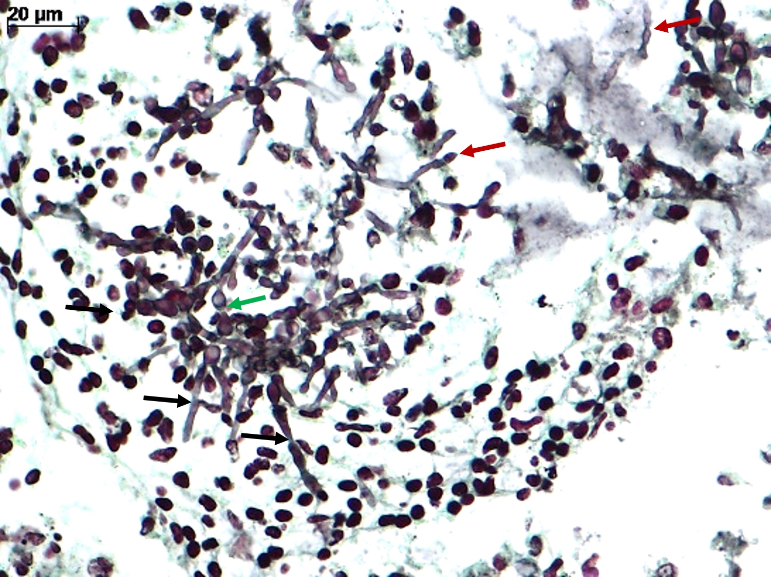
- Spores, pseudohyphae and hyphae (Mycoses 2018;61:377, Clin Microbiol Rev 2011;24:247)
- Yeasts measure 3 - 5 μm in diameter
- Candida auris has a similar morphology to other Candida species in infected tissues, except C. glabrata, which does not form pseudohyphae or hyphae
- Yeasts measure without capsule and with narrow neck budding
- Hyphae with erratic ramification (in general,
- Tissue inflammatory reaction: variable, depending on host immune status; in general, necrosis, cell debris and mixed inflammatory reaction, with neutrophils, microabscesses
- Invasion of mucosa and vessels (angioinvasion); Candida invasive form: hyphae (Emerg Microbes Infect 2020;9:1160)
- Colonization of mucosa (mucosal surface, amid mucous, fibrinous exudate and cell debris), without epithelial invasion (Emerg Microbes Infect 2020;9:1160)
Microscopic (histologic) images
Cytology description
- Round to ovoid, isolated or grouped cells on smear; this method is not species specific and cannot differentiate colonization from invasive infection
Positive stains
- Candida auris has a similar histochemical profile to other Candida species: gram positive, PAS positive and argyrophilic yeasts; amphophilic on hematoxylin and eosin (H&E)
- Silver stains (such as Grocott-Gomori) can give more details about form and budding of the yeast
- Fuchsin (acid fast stains) may stain irregular fungal structures (Emerg Microbes Infect 2020;9:1160, Clin Microbiol Rev 2011;24:247)
Negative stains
- Mucicarmine: Candida spp. do not have cell capsule with mucopolysaccharide
- Fontana-Masson: Candida spp. do not express melanin on the cell wall (Emerg Microbes Infect 2020;9:1160, Clin Microbiol Rev 2011;24:247)
Electron microscopy description
- Ovoid cells with chlamydospore with bilayered cell wall formed by an outer electron transparent primary layer and an inner electron dense secondary layer; single large vacuole, several smaller vacuoles and cytoplasmic organelles (J Gen Microbiol 1981;125:199, J Electron Microsc (Tokyo) 2012;61:343)
Electron microscopy images
Molecular / cytogenetics description
- Multiplex PCR setup using specific primers for glycosylphosphatidylinositol (GPI) protein encoding genes; it is applicable for fluids and fresh tissue samples (Int J Med Microbiol 2018;308:812)
Sample pathology report
- Any tissue, biopsy or autopsy:
- Candida infection (see comment)
- Comment: The fungal structures show yeasts, pseudohyphae and hyphae forms, with single narrow neck budding, with (or without) angioinvasion.
Differential diagnosis
- Cryptococcus spp. (Clin Microbiol Rev 2011;24:247):
- Measure 4 - 10 μm in diameter
- May form germinative tube in highly proliferative infections
- Small yeasts in small tissue samples (e.g., pulmonary biopsies) may not produce a large capsule or mucopolysaccharide, rendering the diagnosis difficult
- Fontana-Masson stain may be helpful (stain melanin on Cryptococcus capsule)
- Histoplasma capsulatum (Clin Microbiol Rev 2011;24:247):
- Small yeasts (measure 2 - 4 μm in diameter) in small samples can be problematic for diagnosis
- Histoplasma spp. may have a central black dot on the Grocott stain
- Thick pseudocapsule on H&E may be helpful
- Malassezia furfur (Clin Microbiol Rev 2011;24:247):
- Fungic structures with spaghetti and meatball appearance on the stratum corneum of the skin
- Hyphomycetes (Clin Microbiol Rev 2011;24:247):
- Wide hyphae with spores and pseudohyphae
- Isolated conidia of Aspergillus in small lung biopsies may be difficult when it is not possible to identify Aspergillus conidial heads; those conidias are amphophilic on H&E, Grocott positive, PAS positive and gram positive, mimicking Candida spp. spores, mainly C. glabrata
Additional references
Board review style question #1
A premature newborn (33 weeks of pregnancy) was hospitalized in the intensive care unit after birth with a low Apgar score and respiratory insufficiency. He was maintained on mechanical ventilation; central venous lines were set and he was fed with a nasogastric tube. On the eighth day, he developed abdominal distension, had bloody stool and was diagnosed with necrotizing colitis, which required large spectrum antibiotics and surgical treatment. He was then started on total parenteral nutrition. After a week, the newborn developed a new sepsis with alveolar - perivascular infiltrates on the lungs. A bronchoalveolar lavage showed mixed inflammatory infiltrate surrounding structures, which are shown in the image above. What is the most likely etiological agent of this sepsis?
- Aspergillus spp.
- Candida spp.
- Gram positive cocci
- Histoplasma capsulatum
Board review style answer #1
B. Candida spp. There are spores, pseudohyphae and hyphae in the figure. Candida spp. are the most common nosocomial fungal infections and their main characteristic is forming spore, pseudohyphae and hyphae in tissues. Answer D is incorrect because H. capsulatum is a yeast in tissue and rarely produces pseudohyphae, even in cases with high fungal burden; moreover, it is not a common fungal nosocomial infection. Answer C is incorrect because gram positive cocci do not have a yeast-like aspect in tissues, as they are much smaller, without budding. Gram positive cocci form colonies in tissues. Answer A is incorrect because Aspergillus spp. are hyaline hyphae fungal agents, with acute dichotomous branching and multiple regular septa. Candida spp. can form hyphae but they are associated with spores and pseudohyphae. The Candida spp. hyphae are amphiphilic, randomly branching and without septa.
Comment Here
Reference: Candida auris
Comment Here
Reference: Candida auris
Board review style question #2
A man with idiopathic pulmonary fibrosis requiring high dose corticosteroids was hospitalized with fever and sepsis. The blood culture identified Candida spp. and in a few days, the strain was identified as C. auris by sequencing the D1 / D2 region of the 28s ribosomal DNA. In parallel, the patient developed bilateral pulmonary infiltrates with respiratory insufficiency and was put under mechanical ventilation. A bronchoalveolar lavage and biopsy were performed, showing round to oval yeasts, with thick walls and narrow based single budding, associated with mononuclear inflammatory reaction. Rare forms showed germinative tubes. Which stain should be requested to confirm the etiology of this fungal infection?
- Gram stain
- Mucicarmine stain
- Von Kossa stain
- Ziehl-Neelsen stain
Board review style answer #2
B. Mucicarmine stain. Cryptococcus spp. has mucicarmine positive capsule. It causes opportunistic infections in patients who receive high doses of corticoids, mainly pneumonia and meningitis. Cryptococcosis can occur with other opportunistic infections in the same patient. When there is high C. neoformans tissue burden, some yeasts can form germ tubes, which mimic Candida spp. pseudohyphae. Answer A is incorrect because Gram stain can label all fungal species (as they are all gram positive) and does not give a specific diagnosis. Answers C and D are incorrect because these stains (von Kossa and Ziehl-Neelsen) do not stain fungal forms.
Comment Here
Reference: Candida auris
Comment Here
Reference: Candida auris
Candida species (pending)
[Pending]
Cimex lectularius (bed bug)
Table of Contents
Definition / general | Case reports | Gross description | Gross images | Differential diagnosis | Additional referencesDefinition / general
- Cimex lectularius is the common human bed bug
- Cimex hemipterus is found primarily in tropical regions
Case reports
- 69 year old woman with bed bugs (Pritt: Creepy Dreadful Wonderful Parasites Blog - Case of the Week 497 [Accessed 9 November 2018])
Gross description
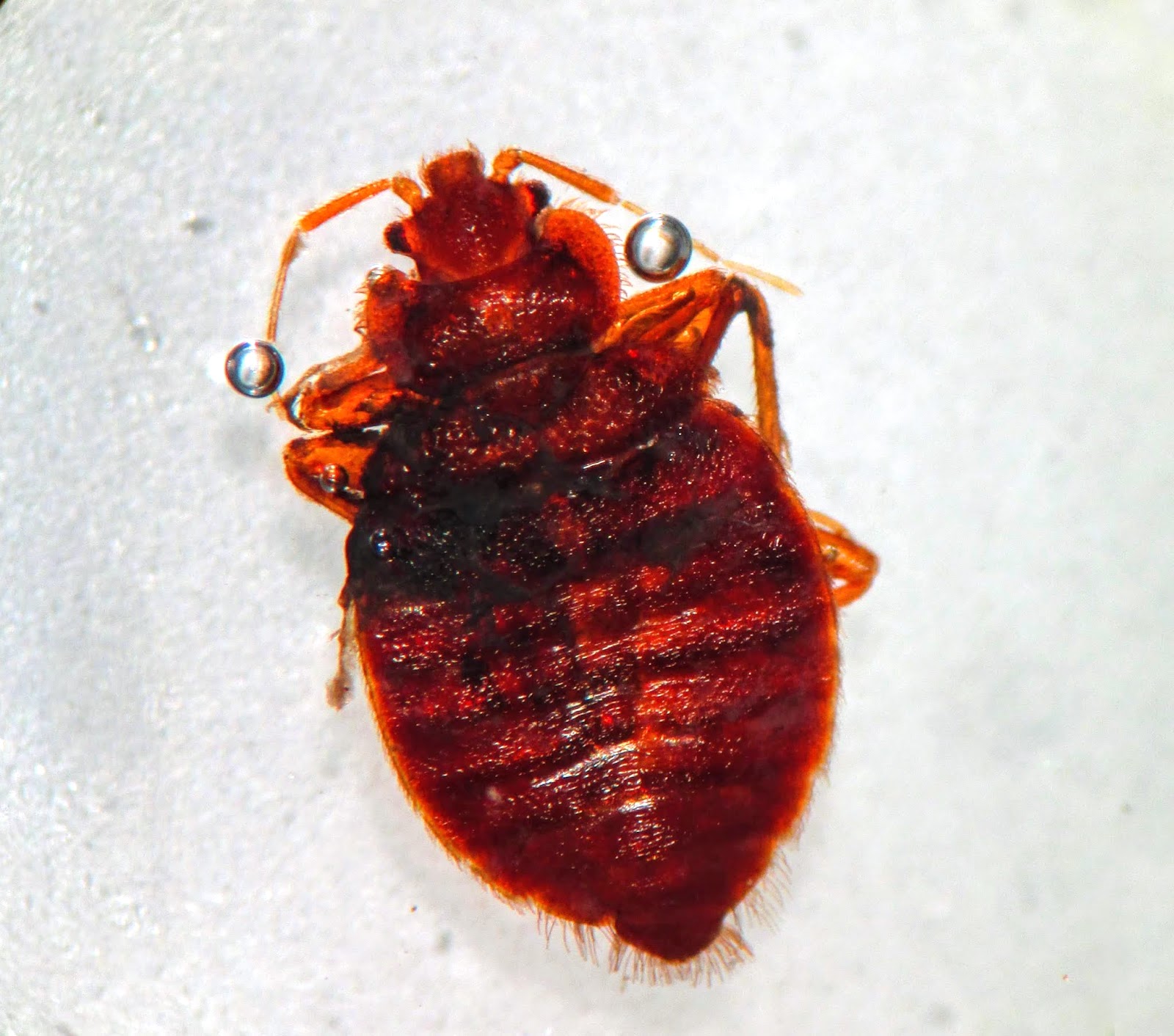
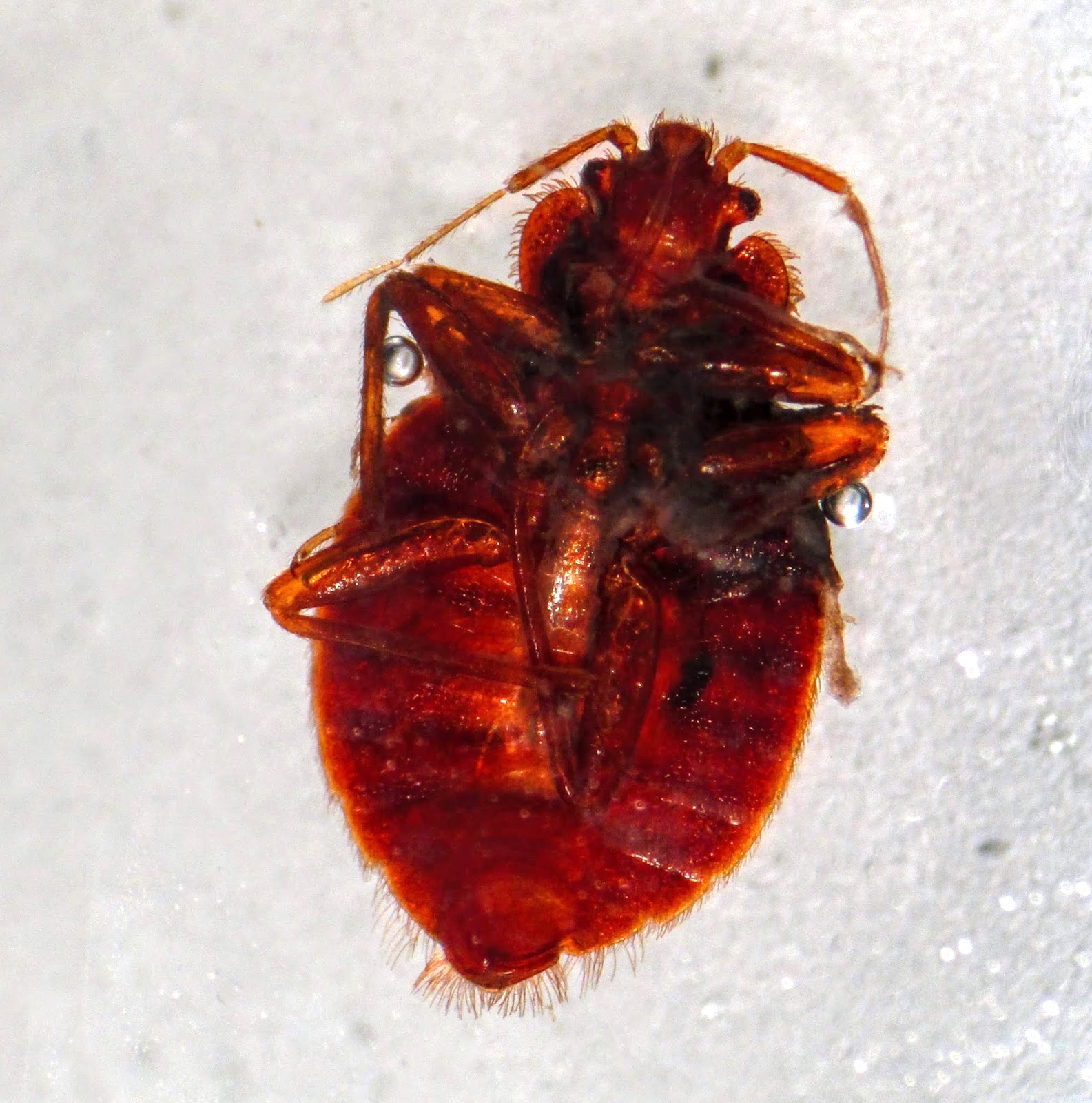
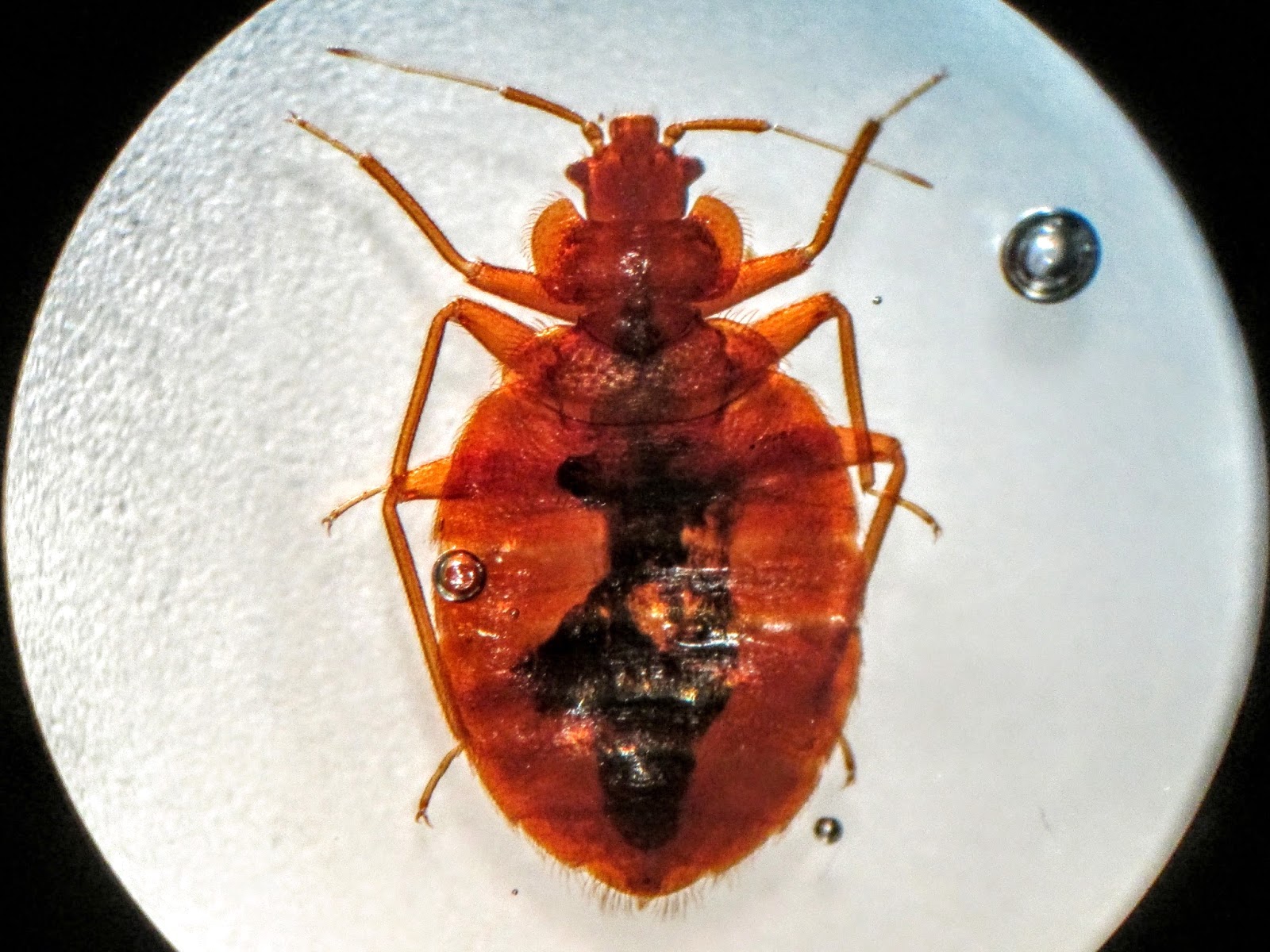
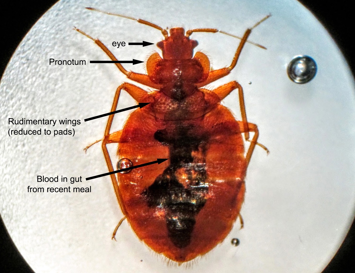
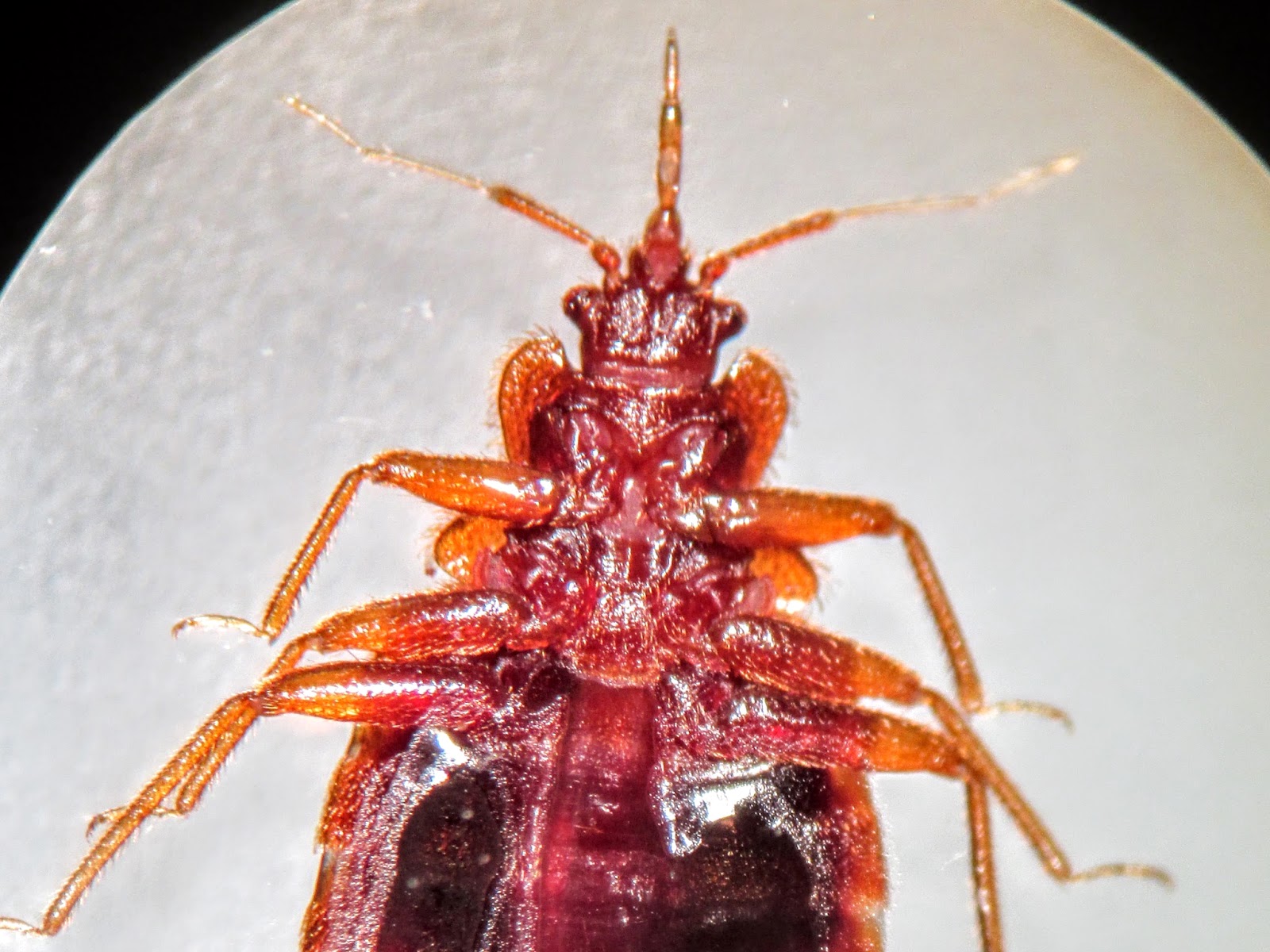
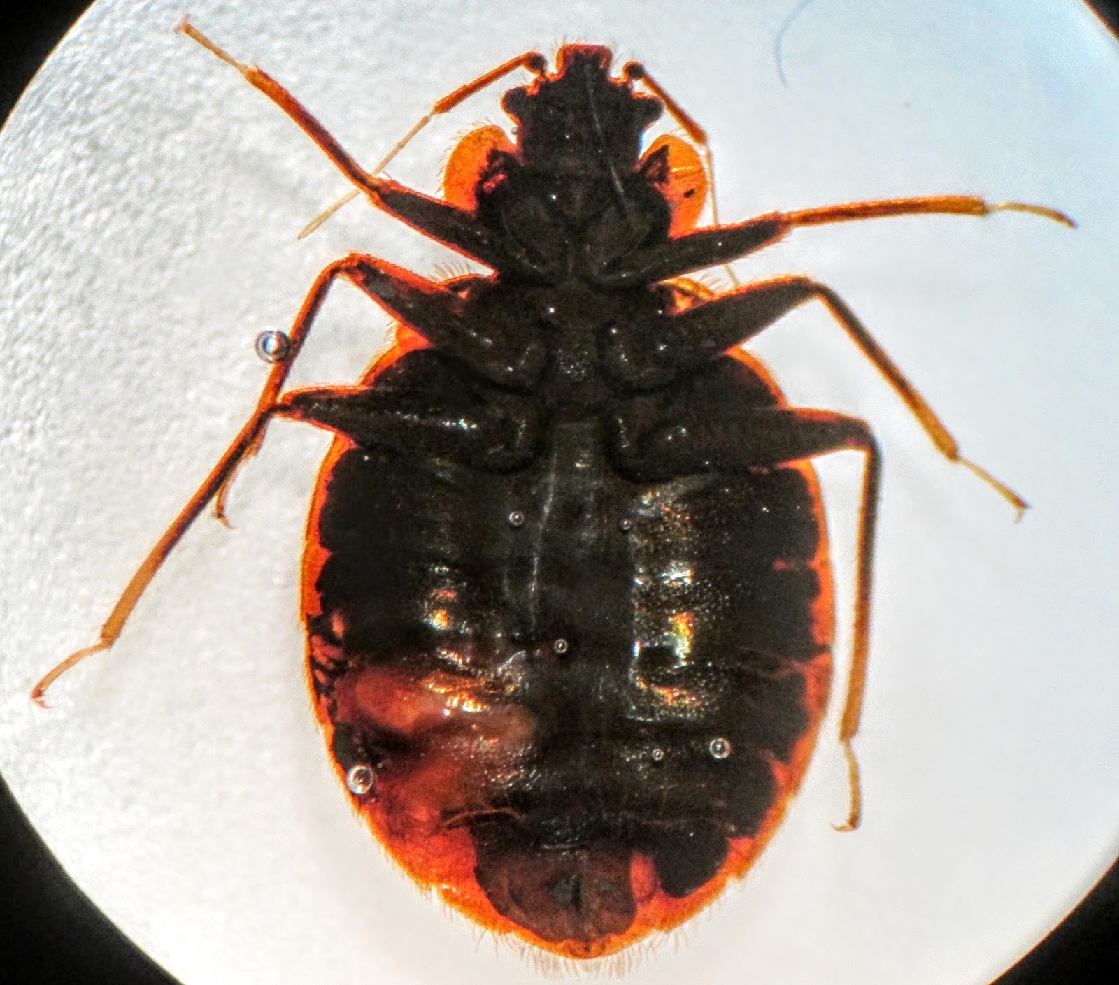
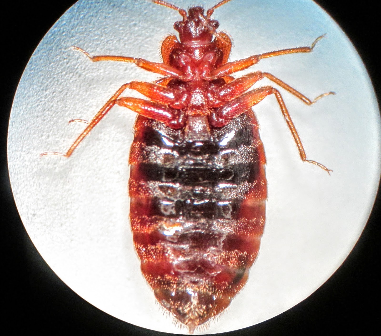
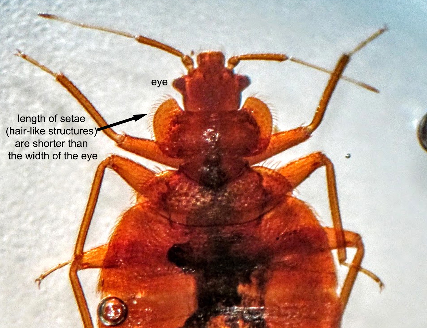
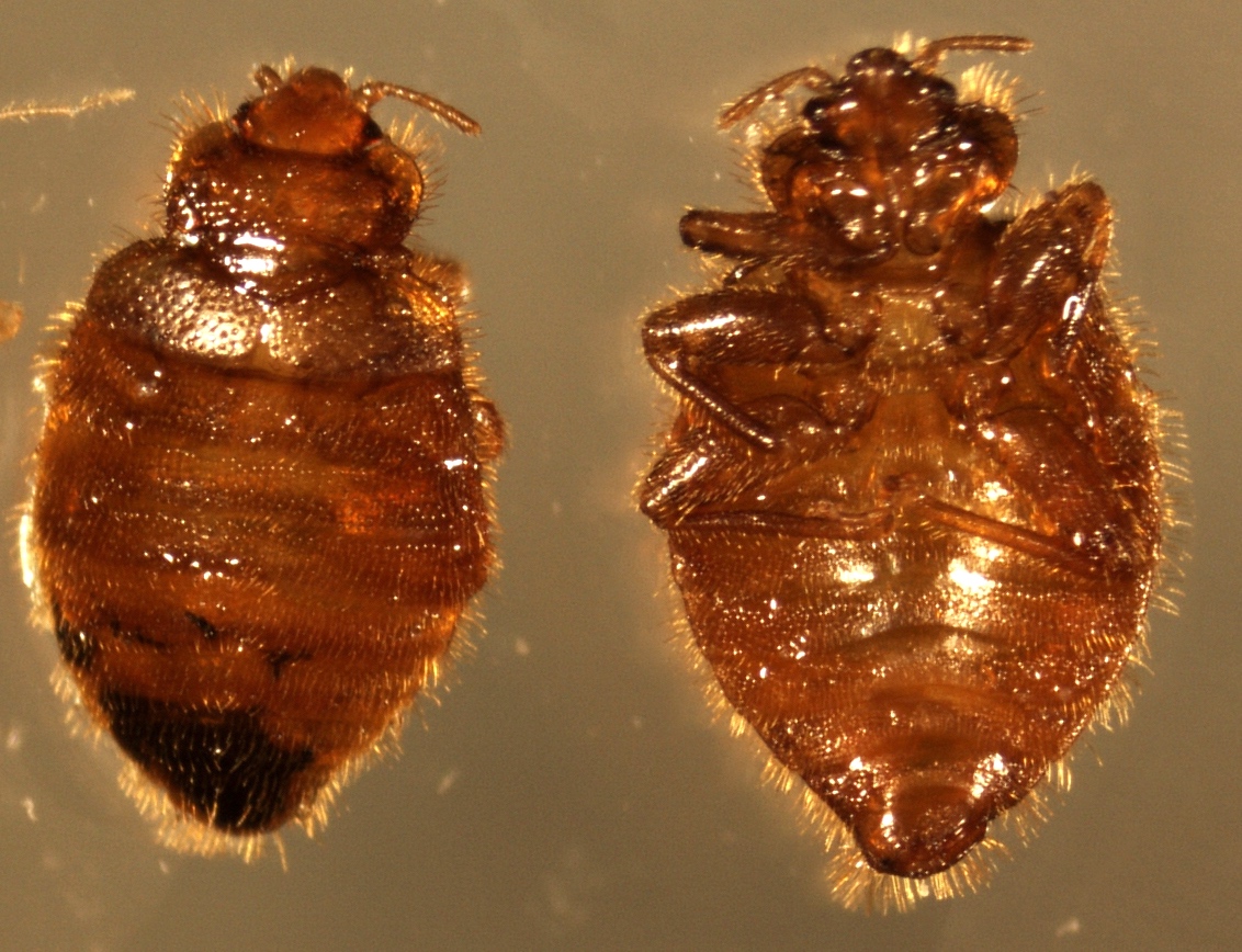
- Broad oval dorsoventrally flattened body, short rudimentary front wings, laterally flared pronotum lined with setae (hair-like structures), which are shorter than the width of the eye (Pritt: Creepy Dreadful Wonderful Parasites Blog - Answer to Case 395 [Accessed 9 November 2018])
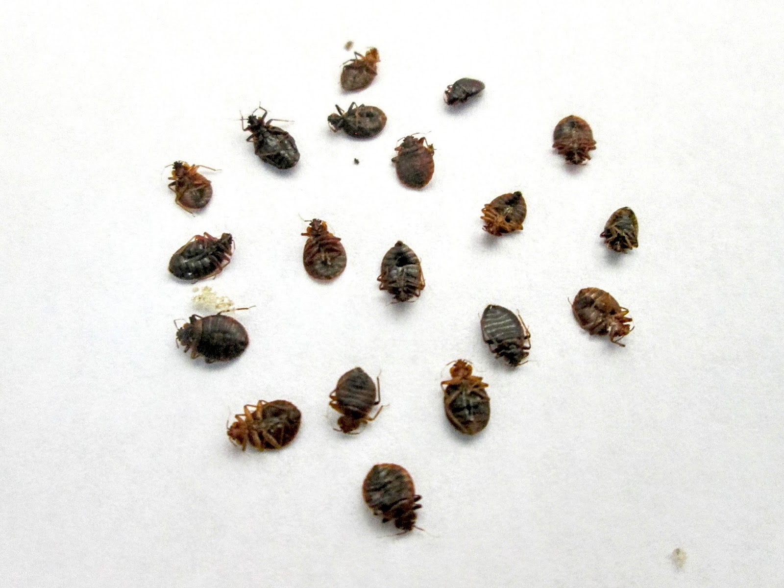
Gross images
Differential diagnosis
- Bat bug: may bite humans if bats are not available; have longer pronotal hairs; important to differentiate since eradication methods are different (Pritt: Creepy Dreadful Wonderful Parasites Blog - Answer to Case 395 [Accessed 9 November 2018])
Additional references
Clostridioides difficile
Table of Contents
Definition / general | Essential features | Terminology | ICD coding | Epidemiology | Sites | Pathophysiology | Laboratory | Case reports | Treatment | Clinical images | Gross description | Gross images | Microscopic (histologic) description | Microscopic (histologic) images | Electron microscopy images | Differential diagnosis | Board review style question #1 | Board review style answer #1 | Board review style question #2 | Board review style answer #2Definition / general
- Gram positive, strict anaerobe bacteria causing pseudomembranous colitis
- Ranges from normal flora of neonates, to asymptomatic carriage in children and adults, to diarrheagenic pathogen
- Transmissible, hand washing necessary (alcohol based sanitizers insufficient to remove spores) and contact precautions are standard
Essential features
- Gram positive bacteria important in antibiotic associated (pseudomembranous) colitis with marked diarrhea
- Anaerobic growth, spore formation
- Morphology (Anaerobe 2016;40:95):
- Microscopic: rods, box car shaped, occasionally in chains
- Colonies: gray-white, low convex, 2 - 5 mm, matte to glossy, pale green fluorescence under UV light (traditional culture rarely employed for ID)
- Toxins required for disease and molecular detection: toxin A, toxin B and regulatory protein TcdC (J Clin Microbiol 2009;47:3729, J Clin Microbiol 2010;48:4519, Nature 2010;467:711)
- Epidemic strains (ribotype 027) have a third binary toxin, C. difficile transferase (CDT) that contributes to virulence (J Infect Dis 2014;209:83)
Terminology
- Previously Clostridium difficile but phylogenetically distant from Clostridium sensu strictu and therefore renamed Clostridioides (Anaerobe 2016;40:95)
- Jargon: C. diff
- CDI – Clostridioides difficile infection; CDAD – Clostridioides difficile associated disease / diarrhea
- Causes pseudomembranous colitis
ICD coding
- A04.7: enterocolitis due to Clostridium difficile
Epidemiology
- Causes illness in ~ 500,000 Americans per year; 15 - 30,000 deaths per year (Clin Infect Dis 2018;66:987)
- Incidence:
- 147/100,000 (Clin Infect Dis 2018;66:987)
- Highest incidence at age 65+
- > 60% of cases are health care associated
- Patients with inflammatory bowel disease, immunocompromise (post solid organ or bone marrow transplant) are at increased risk
- Antibiotic use is a major risk factor due to disruption of gut
microbiota (Clin Infect Dis 2018;66:987)
- Prior antibiotic use may be less of a factor in pediatric populations (Pediatr Infect Dis 1982;1:336)
- Major nosocomial pathogen; patient isolation, hand washing and proper use of gown / gloves important interventions (Clin Infect Dis 2018;66:987)
- Rare cause of illness in children under 2 years; testing not recommended unless noninfectious and other infectious causes excluded (Clin Infect Dis 2018;66:987)
Sites
- Colon
Pathophysiology
- Bacterial proliferation when gut microbial population structure is perturbed
- Toxin production (Gut Microbes 2014;5:579, Nat Rev Microbiol 2016;14:609)
- TcdA and TcdB are secreted, inactivate Rho GTPases and lead to actin depolymerization, cell rounding, apoptosis and loss of epithelial barrier function, as well as inflammasome activation
- CDT is not cytotoxic, is associated with virulence and can also disrupt colonocyte cytoskeleton
- Endospore formation permits persistence (Gut Microbes 2014;5:579, Nat Rev Microbiol 2016;14:609)
- Type IV pilus production promotes epithelial adhesion and may have role in biofilm formation (Nat Rev Microbiol 2016;14:609)
Laboratory
- Molecular diagnostic testing is mainstay of detection; multiple
assays available and perform better than cytotoxin neutralization
assay or enzyme immunoassay
- PCR assays detect either single organism versus multi organism panel
- Example of single organism: Cepheid GeneXpert has primers to multiple targets: tcdB, tcdC, cdtA and cdtB loci with ≥ 97% sensitivity and ≥ 90% specificity (J Clin Microbiol 2010;48:4519, J Clin Microbiol 2009;47:3729)
- Example of multiplex panel: Biofire FilmArray GI Panel detects by qPCR and also has high sensitivity (≥ 94%) and specificity (≥ 97%) for C. difficile (J Clin Microbiol 2015;53:915)
- Molecular assays are not tests of cure as DNA can persist in stool after disease resolution
- Institutional guidelines limiting molecular testing important to avoid overdiagnosis (e.g., detection of C. difficile in patients without diarrhea, indicating carrier state) (Clin Infect Dis 2018;66:987)
- Minimum time for retesting after positive test (i.e., do not use as test of cure due to persistence of DNA)
- Laboratories typically only accept nonformed stool for diagnostic testing
- If no preset institutional nucleic acid amplification testing guidelines, recommendation is to use a stool toxin test as part of multistep algorithm
- Gram stain and culture are rarely used in diagnosis
- Bacteria are gram positive, albeit frequently with variable gram staining
- Requires specific culture conditions; typically see anaerobic growth within 48 hours
- Morphology / colonies: see gross and microscopic descriptions
Case reports
- 28 year old man with chronic refractory ulcerative colitis (J Crohns Colitis 2015;9:367)
- 78 year old woman successfully treated using a combination of fecal microbiota therapy and fidaxomicin (Med Princ Pract 2017;26:182)
- Woman with fulminant pseudomembranous colitis caused by Clostridium difficile PCR ribotype 027 (J Infect Chemother 2014;20:729)
Treatment
- Antibiotic treatment is oral vancomycin or fidaxomicin (not absorbed
and thus no systemic exposure)
- Add IV metronidazole if severe, fulminant infection including ileus or megacolon
- Oral metronidazole if vancomycin or fidaxomicin are unavailable
- Surgical management (subtotal colectomy) may be necessary for severe infections
- Fecal microbiota transplant for patients with ≥ 2 recurrences and for whom medical therapy has not worked (Mayo Clin Proc 2017;92:1617, Am J Transplant 2018 Aug 7 [Epub ahead of print], Anaerobe 2013;19:22)
- Pending FDA approval; interim guidance for use issued by FDA (FDA: Enforcement Policy Regarding Investigational New Drug Requirements [Accessed: August 15, 2018])
Gross description
- Colonies: gray-white, low convex, 2 - 5 mm, matte to glossy, pale green fluorescence under UV light (but traditional culture rarely employed for ID)
Microscopic (histologic) description
- Rods, box car shaped, 0.5 - 1.9 x 3 – 17 microns
- Subterminal spores may be evident as oval, intracellular clearings
- Occasional chains of 2 - 6 cells
Differential diagnosis
- Diarrheagenic E. coli
- Viral diarrheal agents
- Travel history, immunosuppression may point to more exotic pathogens including parasites
Board review style question #1
A 57 year old man with ulcerative colitis presents with 4 days of diarrhea and a
leukocytosis of 14,000 cells/μL two weeks after being treated for bacterial
sinusitis. The best laboratory test to diagnose his condition is:
- Anaerobic and aerobic bacterial culture
- Immunoassay for cytotoxin production
- Nucleic acid amplification testing (NAAT) targeted to toxin B and the CDT toxin of the suspected pathogen
- Stool examination for ova and parasites
Board review style answer #1
C. This patient has a recent history of antibiotic exposure and
ulcerative colitis, both risk factors for Clostridioides difficile infection (CDI).
Diarrhea with a high WBC and this history is strongly suggestive of active
CDI. Nucleic acid amplification testing (NAAT) is more sensitive than cytotoxin immunoassay and faster than
culture (turnaround ~1 hour). Rapid turnaround time is valuable when CDI is
suspected to guide isolation practices. The description does not indicate any clear risk factors for parasitic infection.
Comment Here
Reference: Clostridioides difficile
Comment Here
Reference: Clostridioides difficile
Board review style question #2
A patient with ulcerative colitis presented with diarrhea for 4 days after antibiotic treatment for sinusitis. NAAT and culture were performed. Gram stain showed the following:
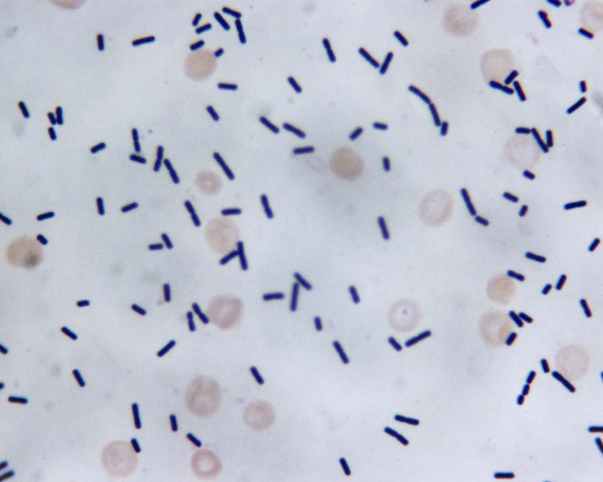

After oral antibiotics, his diarrhea resolved, and his physicians want to repeat the NAAT as a test of cure. What do you recommend?
- Advocate against testing as bacterial DNA may persist after clinical symptoms resolve
- Advocate against testing because it is expensive
- Proceed with testing as a negative NAAT result can shorten the patient's antibiotic exposure
- Proceed with testing because a positive result should prompt fecal microbiota transplant
Board review style answer #2
A. This is Clostridioides difficile infection (CDI). Institutional guidelines are important to prevent repeat testing.
Bacterial DNA may persist longer than symptoms, thus proving a clinical
false positive result on nucleic acid amplification testing (NAAT) (albeit an analytical true positive result!).
Treatment is fairly protocolized with evidence based recommendations for
length of treatment. Fecal microbiota transplant is reserved for patients
with at least 2 recurrences of CDI who have failed medical therapy and
would not (yet) be indicated for this patient. In addition, a positive test after
only a few days of treatment would NOT constitute evidence of a
recurrence but most likely represents persistent C. difficile DNA in the
stool.
Comment Here
Reference: Clostridioides difficile
Comment Here
Reference: Clostridioides difficile
Clostridium perfringens / C. septicum
Table of Contents
Definition / general | Essential features | Terminology | Epidemiology | Sites | Pathophysiology | Clinical features | Diagnosis | Laboratory | Case reports | Treatment | Clinical images | Microscopic (histologic) description | Microscopic (histologic) images | Videos | Differential diagnosis | Board review style question #1 | Board review style answer #1 | Board review style question #2 | Board review style answer #2Definition / general
- Common anaerobic, gram positive pathogen (Proc Natl Acad Sci U S A 2002;99:996)
Essential features
- Gram positive, spore forming, rod shaped, anaerobic
- Ubiquitous in environment, soil and human intestinal tracts
- Cause: significant trauma related gas gangrene, food poisoning and sepsis
Terminology
- Clostridia, Eubacteriales, Clostridiaceae, Clostridium, Clostridium perfringens / septicum
Epidemiology
- Clostridium perfringens: diabetic, immunocompromised patients and history of trauma (J Bone Joint Surg Am 2003;85:1454)
- Clostridium septicum: strongly associated with malignancy, most commonly of colonic or hematological origin (Int Semin Surg Oncol 2006;3:12)
Sites
- Sites of trauma (subcutaneous tissue, skeletal muscle)
- Large intestine (enteritis)
- Blood stream infections
- Reference: J Bone Joint Surg Am 2003;85:1454
Pathophysiology
- Production of alpha toxin (a phospholipase C) important virulence factor for myonecrosis
- Collagenases, hyaluronidase, fibrinolysin, hemagglutinin and hemolysins mediates increase vascular permeability systemic spread of the infection
- Food poisoning mediated by enterotoxin production (Future Microbiol 2014;9:361)
Clinical features
- Traumatic gas gangrene, blood stream infections, food poisoning and preterm necrotizing enterocolitis (Clostridium perfringes)
- Nontraumatic gas gangrene and blood stream infections (Clostridium septicum)
- References: J Bone Joint Surg Am 2003;85:1454, Clin Infect Dis 2016;62:863
Diagnosis
- Diagnosis is based on clinical observations
- Necrotizing tissue Gram stain and anaerobic bacterial culture
- Blood cultures (aerobic and anaerobic)
- Reference: J Bone Joint Surg Am 2003;85:1454
Laboratory
- Anaerobic growth in routine culture conditions, 37 °C
- Growth on blood agar
- Catalase negative
- Small to medium sized gray colonies
- Clostridium perfringens double zone hemolysis, differentiates from other Clostridium spp.
- Matrix assisted laser desorption ionization time of flight (MALDI-TOF) mass spectrometry
- Rapid alternative to 16S rRNA sequencing for identifying anaerobes isolated from solid culture media
- Reference: J Bone Joint Surg Am 2003;85:1454
Case reports
- Newborn boy with Clostridium perfringens meningitis following caudal anesthesia (A A Pract 2020;14:e01188)
- 17 year old boy with sustained intraocular penetrating injury (BMC Ophthalmol 2018;18:88)
- 60 year old woman with food poisoning (Toxins (Basel) 2019;11:138)
- Elderly woman who presented in shock with a progressive abdominal pain (Br J Haematol 2020;190:641)
Treatment
- Surgical debridement of traumatic injuries
- Wound cleansing, removal of dead tissue
- Broad spectrum beta lactams
- Hyperbaric oxygen may be useful
- Food poisoning, self limiting
- Reference: J Bone Joint Surg Am 2003;85:1454
Microscopic (histologic) description
- 1 x 6 micron large, rectangular rod shaped, gram positive bacteria
Videos
Clostridium perfringens - an osmosis preview
Differential diagnosis
- Group A Streptococcus infection:
- Appears as gram positive cocci on Gram stain
- Vibrio vulnificus myositis:
- Associated with clinical history of traumatic water injury
- Pyomyositis due to Staphylococcus aureus:
- Appears as gram positive cocci in clusters on Gram stain
- Rhabdomyolysis:
- Associated with clinical history of trauma or strenuous exercising
- Culture negative
- Bacillus cereus food poisoning:
- Toxin producing facultatively anaerobic gram positive bacterium
- Association with reheated food exposure
Board review style question #1
A 44 year old male construction worker presented to the emergency room with acute onset severe right thigh pain and altered mental status. Physical examination showed large bulla at the location of his pain and crepitance on palpation. Gram stain performed on the bulla aspirate showed large gram positive rods. What is the most likely organism?
- Clostridium difficile
- Clostridium perfringens
- Pseudomonas aeruginosa
- Streptococcus pyogenes
Board review style answer #1
B. Clostridium perfringens. Gas gangrene is a necrotic infection of soft tissue with high mortality rate, often necessitating amputation and commonly associated with Clostridium perfringens. While Streptococcus pyogenes could cause gangrene / necrotizing fasciitis, it is gram positive cocci. Pseudomonas aeruginosa is gram negative rod. While Clostridium difficile is gram positive rod, it is the causative agent of antibiotic associated diarrhea and not linked to necrotizing fasciitis.
Comment Here
Reference: Clostridium perfringens / Clostridium septicum
Comment Here
Reference: Clostridium perfringens / Clostridium septicum
Board review style question #2
For Clostridium septicum bacteremia, association with which of the following clinical conditions should be investigated?
- Bronchiectasis
- Colon cancer
- Diverticulitis
- Inflammatory bowel disease
Board review style answer #2
B. Colon cancer. Clostridium septicum is known to be associated with malignancy, most commonly of colonic or hematological origin and therefore in patients where this organism is isolated, efforts must be made to exclude an occult underlying condition. Association with any of the other mentioned choices has not been described.
Comment Here
Reference: Clostridium perfringens / Clostridium septicum
Comment Here
Reference: Clostridium perfringens / Clostridium septicum
Cordylobia rodhaini (Lund fly)
Table of Contents
Epidemiology | Pathophysiology / etiology | Case reports | Gross description | Gross images | Differential diagnosisEpidemiology
- Found in the African subtropics (Pritt: Creepy Dreadful Wonderful Parasites Blog - Answer to Case 547 [Accessed 12 July 2019])
Pathophysiology / etiology
- Adult flies deposit their eggs on soil or clothing (Pritt: Creepy Dreadful Wonderful Parasites Blog - Answer to Case 547 [Accessed 12 July 2019])
- Contact with warm blooded host then causes the larvae to hatch and penetrate the skin
- Therefore, it is common practice in endemic areas to iron clothes
Case reports
- 1 cm long structure was extracted from a Belgian patient returning from Ghana (Pritt: Creepy Dreadful Wonderful Parasites Blog - Case of the Week 547 [Accessed 12 July 2019])
Gross description
- Weakly pigmented cuticular spines and sinuous spiracles (Pritt: Creepy Dreadful Wonderful Parasites Blog - Answer to Case 547 [Accessed 12 July 2019])
Gross images
Differential diagnosis
- Cordylobia anthropophaga or botfly / tumbu fly (Pritt: Creepy Dreadful Wonderful Parasites Blog - Answer to Case 547 [Accessed 12 July 2019]):
- More commonly seen on humans
- Also found in African subtropics
- C. rodhaini has more sinuous spiracles than C. anthropophaga
Corynebacterium diphtheriae (pending)
[Pending]
COVID-19 (SARS-CoV-2) testing
Table of Contents
Definition / general | Clinical features | Essential features of laboratory testing | Testing priorities | Evolution of diagnostic testing | Technology platforms | FDA's list of serological assays that should not be distributed | ICD, CPT and Medicare rate | Diagrams / tables | Board review style question #1 | Board review style answer #1 | Board review style question #2 | Board review style answer #2Definition / general
- Coronavirus disease 2019 (COVID-19) is caused by a single stranded RNA virus belonging to the genus Betacoronavirus
- Causative agent is named SARS-CoV-2 (severe acute respiratory syndrome coronavirus 2) due to similarities to the virus that caused the SARS epidemic in 2003 (J Microbiol Immunol Infect 2020 Mar 31 [Epub ahead of print])
- Public health emergency declared by the U.S. Secretary of Health and Human Services on January 31, 2020 (CDC: Coronavirus Disease 2019 - Diagnostic Testing [Accessed 24 April 2020])
- Pandemic declared by WHO on March 11, 2020
- Genome likely of bat origin (Proc Natl Acad Sci U S A 2020 Apr 8 [Epub ahead of print])
Clinical features
- Symptoms mostly mild to moderate but can be severe or fatal, especially among the elderly and those with underlying illnesses
- Typical signs and symptoms: fever, cough and dyspnea
- Other symptoms include chills, muscle pain, sore throat and loss of taste or smell
- Incubation period: 2 - 14 days (Cureus 2020;12:e7560)
- Children of all ages are at risk for COVID-19 infection but complications generally appear to be less common than in adults (CDC: Coronavirus Disease 2019 - Information for Pediatric Healthcare Providers [Accessed 19 May 2020])
- Symptoms may be similar to those of common viral respiratory infections, requiring appropriate suspicion for COVID-19 as well as consideration for other infectious etiologies
- A serious multisystem inflammatory syndrome in children (MIS-C) has been reported (up to 21 years of age)
- May present with fever, elevated serum inflammatory markers and multiorgan involvement, such as rash, abdominal pain and myocarditis (NYS DOH: Pediatric Multi-System Inflammatory Syndrome Potentially Associated with COVID-19 [Accessed 19 May 2020])
- Bears similarities with toxic shock syndrome and Kawasaki disease
- Febrile illness characterized by vasculitis
- Manifesting as rash, skin peeling, cervical lymphadenopathy, swelling of the hands & feet and gastrointestinal symptoms
- Elevated interleukin 6 (median 135 pg/mL) and d-dimer (median 5284 ng/mL) in one cohort of 35 MIS-C patients ( Circulation 2020 May 17 [Epub ahead of print])
- MIS-C may begin weeks after infection with SARS-CoV-2
Essential features of laboratory testing
- Emergency use authorization (EUA), put into place by U.S. Congress, allows for expedited Food and Drug Administration (FDA) review (within 24 hours in many cases) based on less stringent validation standards than in nonurgent situations (FDA: Coronavirus (COVID-19) Update - FDA Expedites Review of Diagnostic Tests to Combat COVID-19 [Accessed 24 April 2020])
- Emergency use authorization adoption has led to rapid expansion of testing capacity in the U.S. since mid March, 2020
- Increasing demand continues to outpace assay reagents and supplies
- FDA has provided further flexibility by allowing commercial launch of a diagnostic assay as early as 15 business days prior to emergency use authorization submission as of March 1, 2020 (FDA: Coronavirus (COVID-19) Update - FDA Provides More Regulatory Relief During Outbreak, Continues to Help Expedite Availability of Diagnostics [Accessed 24 April 2020])
- Samples in a laboratory should be handled in a class II or higher biological safety cabinet (FDA: CDC 2019 - Novel Coronavirus (2019-nCoV) Real-Time RT-PCR Diagnostic Panel [Accessed 24 April 2020])
Testing priorities
- Guidelines for patient testing established by the U.S. Centers for Disease Control (CDC)
- Previously considered “nonpriority”, some asymptomatic individuals are now considered “priority” for testing due to the possibility of asymptomatic infection and viral shedding that can lead to disease transmission
- Guidance updated on May 3, 2020 to test certain asymptomatic individuals as a part of public health surveillance or a mitigation strategy in long term care facilities and other settings (CDC: Evaluating and Testing Persons for Coronavirus Disease 2019 (COVID-19) [Accessed 18 June 2021])
- Broader testing guidelines reflect the need for more aggressive infection control measures as well as the increased availability of commercial assays, reagents and supplies
- High priority:
- Hospitalized patients with symptoms
- Healthcare facility workers, workers in congregate living settings and first responders with symptoms
- Residents in long term care facilities or other congregate living settings, including prisons and shelters, with symptoms
- Priority:
- Persons with symptoms of potential COVID-19 infection including fever, cough, shortness of breath, chills, muscle pain, new loss of taste or smell, vomiting or diarrhea or sore throat
- Persons without symptoms who are prioritized by health departments or clinicians, for any reason, including but not limited to public health monitoring, sentinel surveillance or screening of other asymptomatic individuals according to state and local plans
- Special considerations for healthcare personnel:
- Testing may be considered if there has been exposure to a person with suspected (not yet confirmed) COVID-19
- Even mild signs and symptoms (e.g., sore throat) of COVID-19 should be evaluated in healthcare personnel who may be potentially exposed, in order to protect vulnerable patients they may come in contact with
- Special considerations for children and young adults with MIS-C
- May represent a post-viral syndrome or cytokine storm caused by body’s immune response
- Incubation period may be up to 4 weeks (longer than that seen in adults with respiratory symptoms)
- Serology testing more likely positive than RT-PCR for SARS-CoV-2 in one small cohort of 10 Italian children (Lancet 2020 May 13 [Epub ahead of print])
- Special considerations for neonates (CDC: Evaluation and Management Considerations for Neonates At Risk for COVID-19 [Accessed 28 May 2020])
- Testing recommended for all neonates born to mothers with confirmed or suspected COVID-19, whether or not there are any signs and symptoms in the neonate or the mother
- RNA testing by RT-PCR on nasopharyngeal, oropharyngeal or nasal swab samples
Evolution of diagnostic testing
- CDC was the first in the U.S. to develop a SARS-CoV-2 diagnostic assay
- Emergency use authorization received on February 4, 2020
- Real time reverse transcriptase polymerase chain reaction (rRT-PCR)
- Gene target: SARS-CoV-2 nucleocapsid (N) gene (FDA: CDC 2019 - Novel Coronavirus (2019-nCoV) Real Time RT-PCR Diagnostic Panel [Accessed 24 April 2020])
- Platform: Applied Biosystems 7500 Fast DX Real Time PCR System (Coronavirus Disease 2019 - Diagnostic Testing [Accessed 24 April 2020])
- Encountered manufacturing issues with a failed reagent released to public health laboratories, which limited availability of testing outside the CDC (FDA:Coronavirus (COVID-19) Update - FDA Expedites Review of Diagnostic Tests to Combat COVID-19 [Accessed 24 April 2020])
- New York state department of public health developed its own molecular assay to satisfy unmet demands; granted second emergency use authorization on February 29, 2020 (FDA: Emergency Use Authorizations - In Vitro Diagnostics EUAs [Accessed 24 April 2020])
- Virus samples became available to private assay developers for validation in late February, 2020
- Rapidly growing list of commercial assays since mid March, 2020 (exceeding 25 as of April 10, 2020) (FDA: Emergency Use Authorizations - In Vitro Diagnostics EUAs [Accessed 24 April 2020])
Technology platforms
- Current commercial in vitro diagnostic assays typically feature:
- Qualitative rRT-PCR for amplification of viral RNA in respiratory samples
- Dual or triple amplification targets may include genetic sequences from nucleocapsid (N) gene, envelope (E) gene, spike (S) protein or ORF1ab
- First commercial molecular assay to receive emergency use authorization: Roche cobas (March 12, 2020)
- Rapid point of care assay by Abbott Diagnostics granted emergency use authorization on March 27, 2020 based on isothermal nucleic acid amplification
- Serology assay, typically either a rapid diagnostic test (lateral flow assay) or enzyme linked immunoassay (ELISA) to detect IgM and IgG antibody immune response in the blood
- May be helpful for retesting symptomatic / suspected patients who have tested negative by molecular method
- Can help to determine who has immunity and who may donate convalescent plasma in the proper clinical context (FDA: Coronavirus (COVID-19) Update - Serological Tests [Accessed 24 April 2020])
- May help to identify asymptomatic infection as a part of contact tracing or mitigation strategy
- Seroconversion of 50% at 13 days (median) and 100% at 19 days after symptom onset in 1 cohort of 285 patients (Nat Med 2020 Apr 29 [Epub ahead of print])
- Not for sole diagnosis of acute illness
- FDA review waived for serology assays as of March 16, 2020, upon assay validation, notification of the FDA and inclusion of a disclaimer (FDA: Coronavirus (COVID-19) Update - Serological Tests [Accessed 24 April 2020])
- First serology assay received emergency use authorization on April 1, 2020, by Cellex for testing serum, plasma or whole blood from venipuncture in the laboratory (FDA: Cellex - qSARS-CoV-2 IgG/IgM Rapid Test [Accessed 24 April 2020])
- Many are being developed or released in the U.S., some of which have been in use in other countries
- First next generation sequencing (NGS) assay under COVID-19 EUA (6/9/2020): Illumina COVIDSeq Test
- High throughput sequencing of amplified viral RNA
- Detects 98 targets on SARS-CoV2 genome
- 12 hours of processing time (up to 3072 samples on NovaSeq or 384 samples on NextSeq instruments)
- Nasopharyngeal, oropharyngeal and nasal swabs
FDA's list of serological assays that should not be distributed
- U.S. FDA recommends health care providers to "be aware that not all marketed serological tests have been evaluated by the FDA"
- 53 EUA serological assays have been removed from the FDA website notification list and should no longer be used for COVID-19 testing (FDA: What Tests Should No Longer Be Distributed for COVID-19? [Accessed 29 June 2020])
- An independent study conducted by a federally funded national laboratory has found that 15 of 20 commercial SARS-CoV-2 antibody assays should not be marketed (FDA: Independent Evaluations of COVID-19 Serological Tests [Accessed 29 June 2020])
- Study assays were each compared against 110 frozen serum samples with known IgM and IgG status (30 positive and 80 negative)
- Performance characteristics were evaluated, including sensitivity, specificity, positive predictive value and negative predictive value (assumed prevalence of 5%)
- EUA authorization was granted for 5 serological assays based on this study:
- Biohit Healthcare (Hefei) Co., Ltd. SARS-CoV-2 IgM/IgG Antibody Test Kit
- Euroimmun SARS-COV-2 ELISA (IgG)
- Hangzhou Biotest Biotech, Co., Ltd. Covid-19 IgG/IgM Rapid Test Cassette
- Hangzhou Laihe Biotech Co., Ltd. Novel Coronavirus (2019-nCoV) IgM/IgG Antibody Combo Test Kit (Colloidal Gold)
- Healgen COVID-19 IgG/IgM Rapid Test Cassette
- 15 serological test kit manufacturers received “should not be distributed” status or market withdrawal notice based on unacceptable assay performance:
- Abacus Pharma International; Accudiagnostics; Atlas-Link (Beijing); Aurora Biomed Inc.; Biomedomics; ChemBio; Chemtron Biotech, Inc.; GP Getein Biotech, Inc.; Phamatech; SD BIOSENSOR, Inc.; Shanghai Fosun Long March Medical Science Co., Ltd.; TESTSEALABS; Tianjin Beroni Biotechnology Co., Ltd.; W.H.P.M, Inc.; Zhongshan Bio-Tech Co LTD
ICD, CPT and Medicare rate
- The first ICD-10-CM diagnostic code listed below can be used for positive COVID-19 laboratory test results, while the last three below for negative or unknown test results:
- The following CPT billing codes and their corresponding Medicare reimbursement rates can be used for SARS-CoV-2 laboratory testing (CMS: Medicare Administrative Contractor (MAC) COVID-19 Test Pricing [Accessed 7 August 2020], ACOG: Coding for COVID-19 Testing [Accessed 7 August 2020]):
CPT code Medicare rate Description 86328 $45.23 Immunoassay for SARS-CoV-2 (COVID-19) antibody(ies), qualitative or semiquantitative, single step method (e.g. reagent strip used in the point-of-care setting) 86769 $42.13 Immunoassay for SARS-CoV-2 (COVID-19) antibody(ies); qualitative or semiquantitative; multiple step method 87635 $51.31 Infectious agent detection by nucleic acid (DNA or RNA); SARS-CoV-2 (COVID-19), amplified probe technique 87426 $45.23 Antigen detection by immunoassay, (e.g., enzyme immunoassay [EIA], enzyme linked immunosorbent assay [ELISA], immunochemiluminometric assay [IMCA]) qualitative or semiquantitative, multiple step method; SARS-CoV or SARS-CoV-2 U0001 $35.91 2019 novel coronavirus real time RT-PCR diagnostic test panel at a CDC lab U0002 $51.31 2019-nCoV coronavirus, SARS-CoV-2/2019-nCoV (COVID-19) using any technique, multiple types of subtypes at a non-CDC lab U0003 $100 Infectious agent detection by nucleic acid (DNA or RNA); SARS-CoV-2 (COVID-19), amplified probe technique, making use of high throughput technologies as described by CMS-2020-01-R U0004 $100 2019-nCoV Coronavirus, SARS-CoV-2/2019-nCoV (COVID-19), any technique, multiple types or subtypes (includes all targets), non-CDC, making use of high throughput technologies as described by CMS-2020-01-R 0223U $416.78 QIAstat-Dx Respiratory SARS-CoV-2 (proprietary laboratory analyses), infectious disease (bacterial or viral respiratory tract infection), pathogen specific nucleic acid (DNA or RNA), 22 targets including SARS-CoV-2, qualitative RT-PCR, nasopharyngeal swab, each pathogen reported as detected or not detected 0202U $416.78 BioFire® respiratory panel 2.1 (RP2.1) (proprietary laboratory analyses; contractor priced until a national fee schedule can be established), infectious disease (bacterial or viral respiratory tract infection), pathogen specific nucleic acid (DNA or RNA), 22 targets including severe acute respiratory syndrome coronavirus 2 (SARS-CoV-2), qualitative RT-PCR, nasopharyngeal swab, each pathogen reported as detected or not detected
Diagrams / tables
- Below is a partial list of commercially available SARS-CoV-2 assays in the U.S. and a comparison of the key features based on manufacturers’ claims gleaned from package inserts and news releases (FDA: Emergency Use Authorizations - In Vitro Diagnostics EUAs [Accessed 24 April 2020])
- Independent studies are needed to verify the comparative performances of these assays; due to the rapid rollout of these emergency use authorization assays, peer reviewed publications will take time to catch up
- The analytical sensitivities (limits of detection) vary widely among assays
Table 1. Performance Characteristics of Some EUA SARS-CoV-2 Assays
| Manufacturer | In Vitro Diagnostic EUAs | Technology | Claimed LoD | Sensitivity | Specificity |
| Abbott | Abbott RealTime SARS-CoV-2 Assay | rRT-PCR | 100 copies/mL | 100% at 1-2x LoD | 100% |
| Abbott | ID NOW COVID-19 Test | Isothermal nucleic acid amplification | 125 copies/mL | 100% at 2-5x LoD | 100% |
| Becton Dickonson | BD SARS-CoV-2 Reagents | rRT-PCR | 40 copies/mL | 95% at 1-2x LoD, 100% at 3-5x LoD | 100% |
| Becton Dickonson | BioGX SARS-CoV-2 Reagents | rRT-PCR | 40 copies/mL | 95% at 1-2x LoD, 100% at 3-5x LoD | 100% |
| BGI Genomics | Real-Time Fluorescent RT-PCR Kit for Detecting SARS-2019-nCoV | rRT-PCR | Throat: 150 copies/mL; BALF: 100 copies/mL | Throat: 95% at 1x LoD; BALF: 100% at 1x LoD | 100% |
| *Bio-Rad | Platelia SARS-CoV-2 Total Ab assay | ELISA (IgM, IgA, IgG) | 100% serum (n=27), 83% plasma (n=24) | 99.6% | |
| BioFire | BioFire COVID-19 Test | rRT-PCR | 330 copies/mL | 100% at 1x LoD | 100% |
| *Cellex | qSARS-CoV-2 IgG/IgM Rapid Test | Lateral flow immunoassay (IgM, IgG) | 94% (n=128, no symptoms to severe) | 95.6% | |
| Cepheid | Xpert Xpress SARS-CoV-2 | rRT-PCR | 250 copies/mL | 100% at 2x LoD | 100% |
| Co-Diagnostics | Logix Smart Coronavirus Disease 2019 (COVID-19) Kit | rRT-PCR | 4290 copies/mL of sputum | 100% at 1x LoD | 100% |
| DiaCarta | QuantiVirus SARS-CoV-2 Test Kit | rRT-PCR | ABI QuantStudio 5: 200 copies/mL; ABI 7500 Fast Dx: 100 copies/mL | ABI QuantStudio: 95% at 1x LoD; ABI 7500 Fast Dx: 100% at 1x LoD | 100% |
| DiaSorin | Simplexa COVID-19 Direct Assay | rRT-PCR | 500 copies/mL | 100% at 1x LoD | 100% |
| GenMark | ePlex SARS-CoV-2 Test | RT-PCR, electrochemical detection | 1 x 10⁵ copies/mL | 94.4% at 1x LoD | 100% |
| Gnomegen | Gnomegen COVID-19 RT-Digital PCR Detection Kit | rRT-PCR, digital, nanofluidic chip | 60 copies/mL | 100% at 1-2x LoD | 100% |
| Hologic | Panther Fusion SARS-CoV-2 Assay | rRT-PCR | 0.01 TCID50/mL | 100% at 1-5x LoD | 100% |
| InBios | Smart Detect SARS-CoV-2 rRT-PCR Kit | rRT-PCR | 7500 Fast Dx: 1100 copies/mL; CFX96 Touch: 860 copies/mL | 100% at 1x LoD | 100% |
| Ipsum | COV-19 IDx Assay | rRT-PCR | 8.5 x 10³ copies/mL | 100% at 1x LoD | 100% |
| Luminex | NxTAG CoV Extended Panel Assay | rRT-PCR | 5.0 x 10³ copies/mL | 95% at 2x LoD | 100% |
| Luminex | ARIES SARS-CoV-2 Assay | rRT-PCR | 7.5 x 10⁴ copies/mL | 100% at 1x LoD | 100% |
| Mesa Biotech | Accula SARS-CoV-2 | rRT-PCR, lateral flow | 200 copies/60uL reaction (pooled nasal & throat) | 100% at 2-50x LoD | 100% |
| NeuMoDx | NeuMoDx SARS-CoV-2 Assay | rRT-PCR | 150 copies/mL | 100% at 1.5x LoD | 100% |
| *Ortho | Vitros Anti-SARS-CoV-2 Total Reagent Pack and Total Calibrator | Immunometric luminescent reaction (IgG) | 92.3% at 1-5 days, 88.9% at 6-15 days, 75% at 16-22 days post-PCR | 100% | |
| PerkinElmer | PerkinElmer New Coronavirus Nucleic Acid Detection Kit | rRT-PCR | ORF1ab: 8.3 copies/mL; N: 27.2 copies/mL | ORF1ab: 100% at 1.5x LoD; N: 100% at 1.5x LoD | 100% |
| Primerdesign | Primerdesign Ltd COVID-19 genesig Real-Time PCR assay | rRT-PCR | 330 copies/mL | 94.7% at 1-2x LoD, 100% at 3-5x LoD | 100% |
| QIAGEN | QIAstat-Dx Respiratory SARS-CoV-2 Panel (22 bacterial & viruses) | rRT-PCR | 500 copies/mL | 100% at 1-2x LoD | 100% |
| Quidel | Lyra SARS-CoV-2 Assay | rRT-PCR | 800 copies/mL | 100% at 1x LoD | 100% |
| Roche | cobas SARS-CoV-2 Assay | rRT-PCR | 0.007 TCID50/mL | 100% at 1.5x LoD | 100% |
| *Roche | Elecsys Anti-SARS-CoV-2 | Sandwich immunoassay (antibodies) | 65.5% at ≤ 6 days, 88.1% at 7-13 days, 100% ≥ 14 days post-PCR | 99.8% | |
| Thermo Fisher | TaqPath COVID-19 Combo Kit | rRT-PCR | 10 copies/140uL | 100% at 1x LoD | 100% |
Abbreviations: LoD = limit of detection; rRT-PCR = real time reverse transcriptase polymerase chain reaction
Table 2. Categories of SARS-CoV-2 EUA Assays and Examples
| Category | In Vitro Diagnostic EUAs | Instruments | Throughput | Cycle Time |
| Point of Care Setting | ||||
| Abbott ID NOW COVID-19 Test | ID NOW | Nonbatch | 5 min (+), 13 min (-) | |
| Cepheid Xpert Xpress SARS-CoV-2 | GeneXpert Xpress System | Nonbatch modular | 45 min per cartridge | |
| Mesa Biotech Accula SARS-CoV-2 | Accula/Silaris Dock | Nonbatch cassette | 30 min | |
| Respiratory Pathogen Panel | ||||
| Qiagen QIAstat-Dx Respiratory SARS-CoV-2 Panel (22 bacterial & viruses) | QIAstat Dx Analyzer System | Nonbatch modular | About 1 hour per cartridge | |
| Serology | ||||
| Bio-Rad Platelia SARS-CoV-2 Total Ab | None | 96 wells per microplate (manual) | 1st incubation: 60 min 2nd incubation: 30 min | |
| Cellex qSARS-CoV-2 IgG/IgM Rapid Test | None | Nonbatch cassette | 15-20 min per cassette | |
| Ortho VITROS Immunodiagnostic Anti-SARS-CoV-2 Total Reagent and Calibrator | VITROS Eci/EDiQ/3600 or VITROS 5600/XT 7600 | Automated | 37 min incubation plus 48 min testing | |
| Roche Elecsys Anti-SARS-CoV-2 | cobas e411, e601, e602, e801 | Automated | 18 min | |
| Modular Cartridge / Cassette / Pouch | ||||
| BioFire COVID-19 Test | FilmArray 2.0 or Torch | Nonbatch modular | 50 min per pouch | |
| Cepheid Xpert Xpress SARS-CoV-2 | GeneXpert Dx / Infinity | Nonbatch modular | 45 min per cartridge | |
| GenMark ePlex SARS-CoV-2 Test | ePlex | Nonbatch modular | cartridge | |
| Luminex ARIES SARS-CoV-2 Assay | ARIES System | Nonbatch cassette | 2 hours per cassette | |
| Qiagen QIAstat-Dx Respiratory SARS-CoV-2 Panel (22 bacterial & viruses) | QIAstat Dx Analyzer System | Nonbatch modular | About 1 hour per cartridge | |
| High Throughput Capability | ||||
| Abbott RealTime SARS-CoV-2 Assay | m2000 RealTime | 96 wells per batch | ||
| BGI Real-Time Fluorescent RT-PCR Kit for Detecting SARS-2019-nCoV | ABI 7500 Real Time PCR | 96 wells per batch | ||
| DiaCarta QuantiVirus SARS-CoV-2 Test Kit | ABI QuantStudio 5 / 7500 Fast Dx Real-Time PCR | Batch | ||
| DiaSorin Simplexa COVID-19 Direct Assay | LIAISON MDX | Batch | ||
| Hologic Panther Fusion SARS-CoV-2 Assay | Panther Fusion | 1150 samples per 24 hours | < 3 hours | |
| InBios Smart Detect SARS-CoV-2 rRT-PCR Kit | ABI 7500 Fast Dx Real-Time PCR / CFX96 Touch Real-Time PCR | 96 wells per batch | ||
| Luminex NxTAG CoV Extended Panel Assay | Luminex MAGPIX & bioMerieux easyMAG / EMAG | 96 wells per batch | < 2.5 hours thermal cycler run time | |
| NeuMoDx SARS-CoV-2 Assay | NeuMoDx 96 and 288 Molecular Systems | Random access, modular | ||
| Ortho VITROS Immunodiagnostic Anti-SARS-CoV-2 Total Reagent and Calibrator | VITROS Eci/EDiQ/3600 or VITROS 5600/XT 7600 | 100 wells per reagent pack | 37 min incubation plus 48 min testing | |
| PerkinElmer New Coronavirus Nucleic Acid Detection Kit | ABI 7500 Real-Time PCR | 96 wells per batch | ||
| Primerdesign Ltd COVID-19 genesig Real-Time PCR Assay | ABI 7500 / Bio-Rad CFX Connect / Roche LightCycler 4800 II | Batch | ||
| Quidel Lyra SARS-CoV-2 Assay | ABI 7500 Fast Dx/Standard, Roche LightCycler 480, Qiagen Rotor-Gene Q, BioRad CFX96 Touch, Thermofisher Quantstudio 7 Pro | Batch | ||
| Roche cobas SARS-CoV-2 Assay | cobas 6800/8800 | 384/960 per 8 hours | 3.5 hours | |
| Thermal Fisher TaqPath COVID-19 Combo Kit | Applied Biosystems 7500 Fast Dx | 96 wells per batch | 4 hours (incl. sample prep) | |
Board review style question #1
Which of the following is true regarding serology (nonmolecular) testing for SARS-CoV-2?
- Dual or triple genetic sequences are targeted to increase the performance of the assay
- It is typically based on reverse transcription followed by nucleic acid amplification
- Elevated IgA antibody to COVID-19 indicates suitability for convalescent plasma donation
- Suitable respiratory specimens include the nasopharyngeal swab and bronchial lavage
- The lack of an antibody response does not rule out acute infection
Board review style answer #1
E. The lack of an antibody response does not rule out acute infection
Comment Here
Reference: Diagnostic testing of SARS-CoV-2
Comment Here
Reference: Diagnostic testing of SARS-CoV-2
Board review style question #2
Which of the following groups of individuals is considered a top priority for COVID-19 testing according to the CDC?
- Children with earache
- Pre-employment screening for the retail industry
- Symptomatic healthcare workers and hospitalized patients
- Asymptomatic transportation personnel with contact exposure to a person with COVID-19
Board review style answer #2
C. Symptomatic healthcare workers and hospitalized patients are a top priority for COVID-19 testing
Comment Here
Reference: Diagnostic testing of SARS-CoV-2
Comment Here
Reference: Diagnostic testing of SARS-CoV-2
Cryptococcus neoformans & gattii
Table of Contents
Definition / general | Essential features | Epidemiology | Sites | Pathophysiology | Clinical features | Diagnosis | Laboratory | Case reports | Treatment | Clinical images | Microscopic (histologic) description | Microscopic (histologic) images | Positive stains | Molecular / cytogenetics description | Differential diagnosis | Additional references | Board review style question #1 | Board review style answer #1 | Board review style question #2 | Board review style answer #2Definition / general
- Taxonomy: phylum Basidiomycetes (similar to mushrooms); order Tremellales; family Tremellaceae
- 7 species total:
- C. neoformans (formerly C. neoformans var grubiii)
- C. deoneoformans (formerly C. neoformans var neoformans)
- C. gattii species complex (formerly C. neoformans var gattii)
- C. gattii sensu stricto (ss; formerly molecular type VGI)
- C. deuterogattii (formerly molecular type VGII)
- C. bacillisporus (formerly molecular type VGIII)
- C. tetragattii (formerly molecular type VGIV)
- C. decagattii (formerly recognized as VGIII / VGIV)
- References: Mycoses 2019;62:1029, Emerg Infect Dis 2018;24:2095
Essential features
- Variably sized yeasts with thin walls and narrow based budding
- Encapsulated by thick sugar capsule composed of glucuronoxylomannan (GXM)
- Histopathology: methenamine silver+, mucicarmine+, Fontana-Masson+, India ink+
- Diseases range from cutaneous to severe pulmonary and central nervous system disease
Epidemiology
- C. neoformans: immunocompromised; bird droppings, soil, widespread distribution (UpToDate: Microbiology and epidemiology of Cryptococcus neoformans infection [Accessed 05 August 2020])
- C. gattii: immunocompetent; tropical / subtropical distribution; eucalyptus trees; Vancouver Island, BC; endemic in the Southeastern U.S.; South America, parts of Africa and Southeast Asia (UpToDate: Cryptococcus gattii infection - Microbiology, epidemiology, and pathogenesis [Accessed 05 August 2020])
Sites
- Immunocompromised host
- Lung / pneumonia
- CNS infection / meningitis / meningoencephalitis
- Cutaneous manifestation: pustules, papules, superficial granulomas, cellulitis (UpToDate: Cryptococcus neoformans infection outside the central nervous system [Accessed 05 August 2020])
- Can resemble molluscum contagiosum
- Disseminated infection
- Immunocompetent host
Pathophysiology
- Antiphagocytic and poorly immunogenic sugar capsule composed mainly of glucuronoxylomannan (GXM), which is a major virulence factor
- Phenol oxidase production of the antioxidant melanin
- Reference: Emerg Infect Dis 1998;4:71
Clinical features
- Opportunistic infection
- C. neoformans infection in immunocompetent individuals, however, C. gattii infects immunocompetent individuals
- Major illness in patients with HIV / AIDS with an estimated 220,000 annual cases of cryptococcal meningitis worldwide (Lancet Infect Dis 2017;17:873)
- Pulmonary disease can be subacute and indolent
- Cough and dyspnea with or without constitutional symptoms
- Pulmonary nodules; old cryptococcomas
- CNS disease presents with increased intracranial pressure, seizures and focal neurologic deficits
- Spectrum of diffuse meningeal disease and focal lesions including soap bubble lesions
- Not a known zoonotic disease
Diagnosis
- CrAg Lateral Flow Assay: Dipstick sandwich immunochromatographic assay: qualitative or semiquantitative detection of the capsular polysaccharide antigens of Cryptococcus species complex
- If CrAg is present in the specimen, then it binds to the gold conjugated, antiCrAg antibodies; gold labeled antibody antigen complex forms a sandwich at the test line causing a visible line to form
- Positive test results create 2 lines (test and control); negative test results form only 1 line (control); if a control line fails to develop, then the test is not valid
- Testing on BAL / pleural fluid is limited to large reference labs that have validated these sample types
- Unchanged or increased titer of antigen in CSF is correlated with clinical and microbiological failure to respond to treatment
- Titer >1:8
- Rise in CSF antigen titer during suppressive therapy is associated with relapse of cryptococcal meningitis (Clin Infect Dis . 1994;18:789)
Laboratory
- Gram stain shows variably sized yeasts with narrow based budding
- India ink highlights organisms (rarely used in clinical practice)
- Grows well on sheep blood, chocolate agar and fungal media, including sabouraud dextrose agar
- Rapid growth within 24 hours at 37 °C
- Creamy, butyrous, mucoid colonies (due to sugar capsule)
- Rapid urease positive
- Brown colonies on birdseed agar due to melanin production
- Matrix assisted laser desorption / ionization time of flight mass spectrometry (MALDI-TOF MS) can distinguish C. neoformans from C. gattii
- Can distinguish C. gattii by deep blue colonies on canavanine glycine bromothymol blue (CGB) media
Case reports
- 28 year old woman from rural India admitted for an abrupt deterioration of mental status, behavioral changes and hallucinations (Cases J 2009;2:9084)
- 56 year old man presents for evaluation of a progressively worsening headache (PLoS One 2011;6:e28625)
- 68 year old man presents for outpatient evaluation of dyspnea and new onset atrial fibrillation 9 months after undergoing bilateral lung transplantation (Transpl Infect Dis 2019;21:e13137)
- 76 year old asymptomatic woman with incidental pulmonary nodule (Med Mycol Case Rep 2018;21:23)
Treatment
- Intrinsic resistance to echinocandins
- Induction therapy with liposomal amphotericin B and flucytosine for 14 days
- Consolidation with high dose fluconazole for 8 weeks
- Maintenance with lower dose fluconazole for 6 - 12 months
- C. gattii treatment regimens are similar but more intense
- Reference: J Fungi (Basel) 2018;4:79
Clinical images
Microscopic (histologic) description
- Variably sized (3.5 - 8 μm in diameter)
- Round to oval encapsulated yeasts with thin cell walls
- Narrow based budding
- Chronic lesions with unencapsulated forms mimic Blastomyces
- Corpora amylacea with retraction artifact (neural tissues) mimics Cryptococcus species
- Predominantly chronic inflammation; pyogranulomatous not infrequent
Microscopic (histologic) images
Positive stains
- Methenamine silver, mucicarmine and Fontana-Masson
Molecular / cytogenetics description
- Detected on some meningitis molecular panels; relatively high false positive rate
- 28S rDNA sequencing
Differential diagnosis
- Unencapsulated strains mimic Blastomyces and Candida species:
- Fontana-Masson positivity is helpful
- Corpora amylacea in neural tissue:
- Concentric lamellations are helpful
Additional references
Board review style question #1
A 45 year old previously healthy man is brought to the emergency department with fever, malaise and altered mental status. MRI reveals dilated perivascular spaces with soap bubble-like lesions in the basal ganglia. CSF analysis shows variably sized encapsulated yeasts with narrow based budding. Which culture media is useful in distinguishing C. neoformans from C. gattii?
- Sabouraud dextrose agar
- Birdseed agar
- Canavanine glycine bromothymol blue agar
- Sheep blood agar
Board review style answer #1
C. Canavanine glycine bromothymol blue agar. Both species produce the same features on all listed media except canavanine glycine bromothymol blue agar. Only C. gattii produces deep blue colonies on CGB agar.
Comment Here
Reference: Cryptococcus neoformans and gattii
Comment Here
Reference: Cryptococcus neoformans and gattii
Board review style question #2
A 67 year old woman presented with a solitary lung nodule. Biopsy showed variably sized yeast with no discernible capsule. The organism stained positive for mucicarmine and Fontana-Masson but serum cryptococcal antigen was negative. Two 10 μm formalin fixed, paraffin embedded (FFPE) scrolls were sent for sequencing and high confidence BLASTs resulted Cryptococcus neoformans. What is the most likely explanation for the negative antigen test?
- Cryptococcal antigen test is less sensitive than nucleic acid amplification tests (NAAT)
- Too much antigen was present resulting in hook effect
- Unencapsulated Cryptococcus isolates produce significantly less capsule resulting in less antigen shedding
- Cryptococcus antigen test was negative because the organism is actually Blastomyces dermatitidis
Board review style answer #2
C. Unencapsulated Cryptococcus isolates produce significantly less capsule resulting in less antigen shedding. The Cryptococcus antigen test is more sensitive than nucleic acid amplification tests; however, unencapsulated Cryptococcus isolates produce minimal amounts of GXM antigen and are not readily detected by antigen assays and lack discernible capsule on H&E. Unencapsulated Cryptococcus still retains some mucicarmine and Fontana-Masson staining. Hook effect occurs with too much antigen. Blastomyces has a thick refractile cell wall that is characteristic on H&E and should be mucicarmine and Fontana-Masson negative (occasionally faint positive). 28S rRNA sequencing is the definitive gold standard.
Comment Here
Reference: Cryptococcus neoformans and gattii
Comment Here
Reference: Cryptococcus neoformans and gattii
Cutibacterium acnes
Table of Contents
Definition / general | Essential features | Epidemiology | Sites | Pathophysiology | Clinical features | Diagnosis | Laboratory | Case reports | Treatment | Clinical images | Microscopic (histologic) description | Microscopic (histologic) images | Positive stains | Differential diagnosis | Board review style question #1 | Board review style answer #1Definition / general
- Gram positive anaerobic bacilli that are a normal component of skin flora
- Rare cause of infection, predominantly prosthetic joint infections (PJI)
Essential features
- Anaerobic and lipophilic Gram positive bacterium that colonizes sebaceous glands in the skin
- Common cause of bacterial culture contamination
- May cause infection in association with abiotic surfaces such as prosthetic joints and shunts
- PJI with Cutibacterium acnes are typically indolent and may not exhibit common signs of infection
Epidemiology
- Normal component of human skin flora (Am J Clin Dermatol 2020;21:18, J Shoulder Elbow Surg 2009;18:897)
- Colonizes sebaceous glands
- Maintains skin health by producing propionic acid; acidic skin pH protects from pathogens
- Most prevalent on skin in high sebum areas including nose, shoulders and back
- Men have greater prevalence of detectable skin colonization than women
- An opportunistic pathogen, infrequently causing infection in the absence of surgical implants
- ~10% of PJI are attributed to C. acnes (Orthopedics 2020;43:52)
- Men are more likely than women to experience PJI due to C. acnes
- ~1% of arthroplasties result in infection (Clin Microbiol Rev 2014;27:302)
Sites
- Significant cause of PJI (Orthopedics 2020;43:52)
- Shoulders are most common site of infection, likely due to high abundance of C. acnes colonization in skin of that area
- Second most common microorganism causing shoulder PJI after coagulase negative Staphlococcus
- Rare cause of hip, knee and ankle PJI
- Also causes spinal infections in association with hardware
- Other common sources of infection include ventriculoperitoneal shunts, implanted cardiac devices and breast implants
- Contributes to the development of acne vulgaris (Am J Clin Dermatol 2020;21:18)
- Presence of C. acnes alone does not cause acne
- Some causes of acne linked to dysbiosis of normal flora, loss of microbial diversity and overabundance of some C. acnes strains
Pathophysiology
- PJI likely develops from introduction of C. acnes during surgical arthroplasty (Orthopedics 2020;43:52)
- Likely due to contamination of implant or surgical tools from subdermal layers of the patient's skin
- Standard skin preparation may not eradicate C. acnes prior to surgery
- Longer procedure time is associated with higher risk of infection
- C. acnes readily forms biofilms on abiotic surfaces (Biofilm 2021;4:100063)
- Increased production of virulence factors, such as lipases, in biofilm
- Resists immune clearance and antibiotic treatment
- In vivo biofilm formation linked to treatment failure in PJI
Clinical features
- PJI due to C. acnes are typically indolent infections that develop slowly and present weeks to months after surgery (Orthopedics 2020;43:52)
- Most common complaints are persistent pain and stiffness
- May mimic aseptic loosening in presentation
- May lack typical signs of infection such as erythema, purulence and drainage or formation of sinus tracts
Diagnosis
- Culture and Gram stain of affected joint for PJI
- Multiple periprosthetic tissue specimens are recommended along with hardware as available, for improved sensitivity and specificity
- Explanted joints or hardware may also be cultured using sonication or a vortex mixer to disrupt biofilms while preserving microbial viability
- Sensitivity of culture for PJI may be reduced due to previous antibiotic therapy, the presence of biofilms, insufficient incubation time or lack of anaerobic culture conditions
- Recovery in culture may indicate contamination, which necessitates clinical correlation
- May indicate contamination during collection or processing or may represent colonization of an abiotic surface without infection
- 2 or more deep specimens or higher colony counts for semiquantitative cultures may correlate with true infection
- Typical inflammatory markers such as C reactive protein, erythrocyte sedimentation rate and white blood cell count may be within normal limits
- Molecular approaches such as broad range 16s sequencing or targeted C. acnes PCR may aid diagnosis but should be interpreted with caution due to risk of detecting contamination
- No FDA cleared assays for molecular detection of C. acnes
- References: Clin Microbiol Rev 2014;27:302, Orthopedics 2020;43:52, J Shoulder Elbow Surg 2020;29:1920
Laboratory
- Culture (Carroll: Manual of Clinical Microbiology, 12th Edition, 2019)
- Optimal recovery via anaerobic culture conditions, though may grow poorly under aerobic conditions
- Can be recovered on rich media (e.g., sheep's blood agar), anaerobic media supplemented with hemin (e.g., Brucella agar) and anaerobic media selective for Gram positives (e.g., phenylethyl alcohol blood agar [PEA])
- Recovery from periprosthetic tissue specimens may be improved by using thioglycolate broth or blood culture media
- Disruption of biofilms from explanted prosthetic joints or hardware, using vortex mixer or sonication, improves culture sensitivity (J Clin Microbiol 2009;47:1878, J Clin Microbiol 2013;51:591)
- Should be performed in a sterile, sealed, hard walled container (not bag) with sterile saline or Ringer's solution
- Culture from sonicate fluid can be more sensitive than periprosthetic tissue
- Recommended incubation time of 14 days for surgical specimens (Clin Microbiol Rev 2014;27:302)
- Primary isolation of C. acnes from specimens may require a longer time period than growth from subculture due to slow growth from metabolically dormant biofilms
- Time to recovery may also depend on media type
- Identification (Carroll: Manual of Clinical Microbiology, 12th Edition, 2019)
- Gram stain: pleomorphic, thin, nonbranching, non-spore forming Gram positive rods
- May be coryneform
- Bacilli clump to form arrangements that resemble spiders
- Colony morphology: white, shiny, slightly domed colonies
- May be beta, slightly alpha or nonhemolytic (Shoulder Elbow 2020;12:390)
- Catalase test positive
- Can be identified using MALDI-TOF mass spectrometry
- Gram stain: pleomorphic, thin, nonbranching, non-spore forming Gram positive rods
Case reports
- 27 and 43 year old immunocompetent women with C. acnes breast implant infection (J Surg Case Rep 2023;2023:rjad042)
- 29 year old man with recurrent C. acnes prosthetic valve endocarditis (BMJ Case Rep 2021;14:e243878)
- 71 year old man with spinal hardware associated infection (J Bone Jt Infect 2019;4:163)
- 82 year old man with shoulder PJI (BMJ Case Rep 2021;14:e239020)
Treatment
- Recommended first line treatment for C. acnes PJI is penicillin or third generation cephalosporins (e.g., ceftriaxone) (Clin Infect Dis 2013;56:e1)
- Alternative options include vancomycin and clindamycin (Clin Infect Dis 2013;56:e1)
- Surgical intervention such as revision or explant of the prosthetic joint may be indicated
- Combination therapy with rifampin has demonstrated increased efficacy against biofilms in vitro but clinical efficacy has not been shown (Antibiotics (Basel) 2022;11:1801)
- Sensitivity to clindamycin is variable with increasing resistance (Orthop Traumatol Surg Res 2018;104:S19)
- C. acnes is intrinsically resistant to metronidazole (Orthop Traumatol Surg Res 2018;104:S19)
Clinical images
Microscopic (histologic) description
- Thin, pleomorphic, non-spore forming Gram positive rod
- Bacilli clump in arrangements described as squashed spiders
Microscopic (histologic) images
Positive stains
- See Gram stain description under Laboratory - identification
Differential diagnosis
- Common agents of PJI (Clin Microbiol Rev 2014;27:302):
- Staphylococcus epidermidis:
- Aerobic Gram positive cocci in clusters
- Coagulase negative
- Staphylococcus aureus:
- Aerobic Gram positive cocci in clusters
- Coagulase positive
- Streptococcus and Enterococcus species:
- Aerobic Gram positive cocci in pairs or chains
- Staphylococcus epidermidis:
- Aseptic joint failure or aseptic loosening (Clin Infect Dis 2013;56:e1):
- Septic joint loosening often associated with elevated inflammatory markers, including C reactive protein and erythrocyte sedimentation rate
- These markers may be negative in C. acnes PSI
- Synovial fluid analysis (including cell count and differential leukocyte count) indicating a predominance of neutrophils
- Identification of one organism from multiple deep cultures supports infection
- Purulence or presence of a sinus tract likely indicates infection
- Septic joint loosening often associated with elevated inflammatory markers, including C reactive protein and erythrocyte sedimentation rate
Board review style question #1
A 57 year old patient presents with 9 weeks of shoulder pain and stiffness following total shoulder arthroplasty. Periprosthetic tissue specimens are collected during surgery and are sent for bacterial culture. After 6 days, the anaerobic cultures are growing a pleiomorphic, non-spore forming Gram positive rod from the thioglycolate broth but the aerobic cultures are not. Which of the following is most likely to be the organism?
- Bacillus subtilis
- Clostridium perfringens
- Cutibacterium acnes
- Staphylococcus epidermidis
Board review style answer #1
C. Cutibacterium acnes. The question describes an anaerobic, non-spore forming Gram positive rod, which is true of C. acnes. The Gram stain shown has clumped arrangements of Gram positive rods that are characteristic of C. acnes. C. acnes is a leading cause of prosthetic joint infections, commonly in the shoulder. Answer A is incorrect because Bacillus subtilis is spore forming and will grow in aerobic conditions and it is also a rare cause of invasive infections. Answer B is incorrect because Clostridium perfringens spore forming does not display the morphology seen on the Gram stain and is a rare cause of joint infection. Answer D is incorrect because S. epidermidis, though a common cause of joint infections, is a Gram positive cocci and will grow in aerobic cultures.
Comment Here
Reference: Cutibacterium acnes
Comment Here
Reference: Cutibacterium acnes
Cyclospora cayetanensis
Table of Contents
Definition / general | Diagrams / tables | Diagnosis | Case reports | Microscopic (histologic) description | Microscopic (histologic) imagesDefinition / general
- Coccidian parasite associated with municipal water systems, first reported in late 1970s, causes protracted diarrhea (Clin Microbiol Rev 2010;23:218)
Diagnosis
- Oocyst in stool with modified acid fast stain
- Diagnosis can be made by the shape and size of oocysts (~8 micrometers in diameter) (Pritt: Creepy Dreadful Wonderful Parasites Blog - Answer to Case 551 [Accessed 8 August 2019])
Case reports
- Objects were seen on a stool specimen stained by Wright-Giemsa to look for fecal leukocytes (Pritt: Creepy Dreadful Wonderful Parasites Blog - Case of the Week 551 [Accessed 8 August 2019])
Microscopic (histologic) description
- Resembles Isospora
Microscopic (histologic) images
Dematiaceous molds
Table of Contents
Definition / general | Essential features | Epidemiology | Sites | Pathophysiology | Clinical features | Diagnosis | Laboratory | Case reports | Treatment | Clinical images | Microscopic (histologic) description | Microscopic (histologic) images | Molecular / cytogenetics description | Differential diagnosis | Additional references | Board review style question #1 | Board review style answer #1 | Board review style question #2 | Board review style answer #2Definition / general
- Heterogeneous molds characterized by dark pigmentation of hyphae (Clin Microbiol Rev 2010;23:884)
- Hundreds of species known to cause disease in humans
- Phaeohyphomycosis: invasion of tissue by pigmented hyphae
- Chromoblastomycosis: chronic subcutaneous infection characterized by pigmented round structures termed copper pennies
- Mycetoma: subcutaneous collection of pigmented hyphae that expands within the tissue (tumor-like growth)
Essential features
- Phaeohyphomycosis
- Alternaria sp.
- Bipolaris sp.
- Curvularia sp.
- Exserohilium sp. (outbreak associated with steroid injections)
- Lomentospora prolificans (resistant to all antifungal agents)
- Cladophialophora bantiana (neurotropic; immunocompetent)
- Exophiala dermatitidis (neurotropic; black yeast)
- Rhinocladiella mackenzeii (neurotropic)
- Verruconis gallopava (neurotropic)
- Chromoblastomycosis
- Fonsecaea pedrosoi and other species
- Phialophora sp.
- Cladophialophora sp.
- Mycetomas
- Madurella sp.
- Rhinocladiella
- Exophiala jeanselmei (black yeast)
- Allergic disease
- Many ubiquitous black molds
- Reference: Clin Microbiol Rev 2010;23:884
Epidemiology
- Organisms present in soil with worldwide distribution
- Mycetoma and chromoblastomycosis with highest incidence in tropical and subtropical zones
- Direct subcutaneous inoculation or spore inhalation
- CARD9 genetic deficiency associated with severe disease (Front Immunol 2018;9:1836)
- A type of primary immunodeficiency affecting the caspase recruitment domain family member 9 regulating apoptosis and inflammation through modulation of signals from pro and anti-inflammatory cytokines
- Individuals have increased susceptibility to fungal infections, especially phaeohyphomycetes, dermatophytes and candida
- The condition follows an autosomal recessive mode of inheritance
Sites
- Subcutaneous
- Pulmonary
- CNS / brain abscess
- Systemic or disseminated, especially in immunocompromised hosts (Kauffman: Essentials of Clinical Mycology, 2nd Edition, 2011)
Pathophysiology
- Melanin production in hyphae scavenges free radicals and inhibits phagocytosis (Kauffman: Essentials of Clinical Mycology, 2nd Edition, 2011)
- Acute inflammatory cells fail to clear infection, leading to pyogranulomatous inflammation
- Characterized by neutrophils infiltrating a chronically inflamed area containing mononuclear cells
Clinical features
- Phaeohyphomycotic cyst: well circumscribed subcutaneous nodule with pigmented hyphae
- Phaeohyphomycosis: pigmented hyphae invading within tissue (typically skin, lung, brain)
- Chromoblastomycosis: psoriasis-like chronic skin inflammation and hyperkeratinization with pigmented round sclerotic bodies (copper pennies) in tissue; no hyphae present (Bennett: Mandell, Douglas, and Bennett's Principles and Practice of Infectious Diseases, 8th Edition, 2014)
- Mycetoma: a focal subcutaneous collection of pigmented hyphae (fungus ball)
- All lesions are characterized by pyogranulomatous inflammation
Diagnosis
- Culture and microscopic evaluation of tape preps is the standard identification method; antifungal susceptibility testing is rarely performed but available in specialized reference laboratories
Laboratory
- Black pigmentation on the backside of the culture plate is due to melanin production in hyphae and is pathognomonic for dematiaceous mold (Kauffman: Essentials of Clinical Mycology, 2nd Edition, 2011)
- Rapidly growing molds (~2 - 4 days) include:
- Alternaria sp.
- Bipolaris sp.
- Curvularia sp.
- Exserohilum sp.
- Lomentospora prolificans
- Most other pathogenic dematiaceous molds are slow growing
- Black yeast colonies initially appear yeast-like, develop a fuzzy edge and convert to molds
Case reports
- 7 week old boy admitted to NICU for management of prematurity and failure to thrive (J Clin Microbiol 2002;40:2207)
- 21 year old soldier with no significant medical history presents for new onset generalized seizures (Clin Neurol Neurosurg 2014;124:179)
- 55 year old diabetic man with slurred speech and left sided paresia (JMID 2012;2:171)
- 60 year old man admitted for progressive verrucous skin lesion (Am J Trop Med Hyg 2018;99:124)
- 67 year old Iranian woman with history of diabetes mellitus and Behçet disease admitted for weakness, lethargy, left sided paresia and radicular pain (Mycoses 2018;61:261)
- Outbreak of Exserohilum rostratum caused by intrathecal injection of a contaminated steroid solution (Med Mycol 2014;52:376)
Treatment
- No standardized treatment; however, itraconazole, voriconazole and posaconazole have been used successfully
- Infection with chromoblastomycosis can be treated with combination itraconazole and terbinafine but may require surgery, chemotherapy or thermotherapy (Kauffman: Essentials of Clinical Mycology, 2nd Edition, 2011)
- Treatment often occurs over an extended period of time; months to years
Microscopic (histologic) description
- Phaeohyphomycosis: pigmented septate hyphae with occasional globose swellings
- Black yeasts like Exophiala dermatitidis form pigmented pseudohyphae in tissue (Clin Microbiol Rev 2011;24:247)
- Chromoblastomycosis: dark sclerotic bodies with thick walled septations
- Mycetoma: black mycotic granules or grains surrounded by dense extracellular matrix
- May exhibit Splendore-Hoeppli phenomena due to antigen / antibody protein complex deposition
- Also known as asteroid bodies, these formations of eosinophilic material around microorganisms (fungi, parasites, bacteria) and foreign material are observed in multiple mucocutaneous and noninfectious conditions (J Cutan Pathol 2008;35:979)
- Pigmentation may be faint; Fontana-Masson stain for melanin may prove helpful
Microscopic (histologic) images
Molecular / cytogenetics description
- 28S rDNA sequencing
Differential diagnosis
- Hyaline septate molds (e.g. Aspergillus):
- Note pigmentation and Fontana-Masson positivity (Clin Microbiol Rev 2011;24:247)
- Actinomycosis:
- Especially in some mycetoma infections
- Note width larger than filamentous bacteria
- Sporotrichosis:
- Note pigmented fungal elements
- Squamous cell carcinoma:
Additional references
Board review style question #1
A 64 year old man with medical history significant for hypertension, atrial fibrillation and deep vein thrombosis presents to the hospital with generalized right sided weakness and right sided facial droop for 12 days. Neurological deficits are noted on physical exam but lab results are within normal limits. He is afebrile and denies nausea, vomiting or headache. Imaging reveals a 3 cm x 2 cm ring enhancing mass in the left frontal lobe. Biopsy reveals abscess formation with necrotizing granulomatous inflammation and irregular brown hyphal elements on H&E.
What is the most likely etiology?
What is the most likely etiology?
- Actinomyces meyeri
- Cladophialophora bantiana
- Rhizopus oryae
- Toxoplasma gondii
Board review style answer #1
B. Cladophialophora bantiana. Anaerobic filamentous bacteria like Actinomyces meyeri form large aggregates (sulfur granules) in tissue and are not pigmented. Rhizopus hyphae are broad and pauciseptate but not pigmented. T. gondii is a parasite and does not make hyphae. The neurotropic mold C. bantiana, like other dematiaceous molds causing phaeohyphomycosis, have hyphae with melanin that appear light to dark brown / black in tissue.
Comment Here
Reference: Dematiaceous molds
Comment Here
Reference: Dematiaceous molds
Board review style question #2
A 42 year old woman from Brazil presented with a red verrucous lesion on her wrist which has been progressively increasing in size over the last 4 months. Further evaluation reveals erythematous plaques with silvery scales on the extensor surfaces of her upper extremities. Biopsy reveals pyogranulomatous inflammation and dark brown round structures but no hyphae. 4 weeks later, 5 mold colonies appear on the plate all with dark brown pigmentation on the reverse of the plate.
What is the most likely etiology?
What is the most likely etiology?
- Malignancy: squamous cell carcinoma
- Fungal infection: phaeohyphomycosis
- Autoimmune: plaque psoriasis
- Fungal infection: chromoblastomycosis
Board review style answer #2
D. Fungal infection: chromoblastomycosis. Chronic skin lesions caused by chromoblastomycosis can grossly mimic squamous cell carcinoma but most closely resemble the scaly plaques of psoriasis. A diagnosis of phaeohyphomycosis requires the detection of invasive pigmented hyphae. Chromoblastomycosis is caused by slow growing dematiaceous molds such as Fonsecaea pedrosoi that elicit acute and chronic inflammation (pyogranulomatous). No hyphae are seen in the tissue and instead the mold forms darkly pigmented muriform structures termed copper pennies.
Comment Here
Reference: Dematiaceous molds
Comment Here
Reference: Dematiaceous molds
Dengue fever
Table of Contents
Definition / general | Clinical presentation and diagnosis | Case reports | Treatment | Clinical images | Microscopic (histologic) description | Microscopic (histologic) images | Electron microscopy images | Additional referencesDefinition / general
- Transmitted by Stegomyia aegypti (formerly Aedes aegypti) mosquito (Wikipedia: Aedes aegypti Biting Human [Image])
- Causes bone marrow suppression, DIC and thrombopathy (Baillieres Best Pract Res Clin Haematol 2000;13:261)
- Also known as "breakbone" fever
Clinical presentation and diagnosis
- Characteristic skin rash similar to measles
- Fever, headache, muscle and joint pain
- Rarely presents with life threatening hemorrhagic fever
- Leukopenia, lymphocytosis with atypical lymphocytes in peripheral blood
- Confirmation by serology or PCR
Case reports
- Patient with hemoglobin H disease and acute myeloid leukemia precipitated by dengue virus infection (Haematologica 2001;86:E17)
Treatment
- Usually supportive (oral rehydration, intravenous fluids, etc.)
Microscopic (histologic) description
- Bone marrow hypoplasia or hyperplasia
- Megakaryocytic hypoplasia with thrombocytopenia as the most common hematologic manifestation
- Occasional extreme plasmacytosis (Arch Pathol Lab Med 2003;127:1026)
- Hemophagocytosis and dysplastic changes (Kaohsiung J Med Sci 2005;21:34)
Microscopic (histologic) images
Additional references
Dermatophagoides
Table of Contents
Terminology | Case reports | Microscopic (histologic) images | Videos | Differential diagnosisTerminology
- Dust mite
Case reports
- Arthropod was found in the urine specimen of a man with HIV and a history of dysuria (Pritt: Creepy Dreadful Wonderful Parasites Blog - Case of the Week 548 [Accessed 19 July 2019])
Microscopic (histologic) images
Videos
Creepy Dreadful Wonderful Parasites Case
Differential diagnosis
- Sarcoptes scabiei (Pritt: Creepy Dreadful Wonderful Parasites Blog - Answer to Case 548 [Accessed 19 July 2019]):
- Distinctive features include short triangular legs and round body shape
Dermatophytes (pending)
Table of Contents
Trichophyton species | Microsporum species | Epidermophyton floccosum | Nondermatophyte moldsTrichophyton species
[Pending]
Microsporum species
[Pending]
Epidermophyton floccosum
[Pending]
Nondermatophyte molds
[Pending]
Diphyllobothrium latum
Table of Contents
Definition / general | Epidemiology | Pathophysiology | Diagrams / tables | Clinical features | Diagnosis | Case reports | Treatment | Gross images | Microscopic (histologic) images | Differential diagnosisDefinition / general
- Humans may be infected by one of several species of the fish tapeworm Diphyllobothrium, which normally infect piscivorous mammals and possibly birds
- Although Diphyllobothrium latum is the most common species to infect humans, differentiation cannot be made based on morphology - molecular methods are required (Emerg Infect Dis 2014;20:1955)
Epidemiology
- Occurs in Northern Hemisphere (Europe, North America, Asia) and South America (Uruguay and Chile)
- Humans acquire larvae by ingesting raw or incompletely cooked fish that have spent at least part of their life in fresh water
Pathophysiology
- Cestode Diphyllobothrium latum (the fish or broad tapeworm) is largest human tapeworm
- Inhabits small intestine, grows to 10 meters or longer, can persist for years
Clinical features
- Adult worms mature and initiate egg production in ~1 month
- Infection may be asymptomatic, with passage of a length of strobila being the initial complaint (Korean J Parasitol 2007;45:219)
- In others, a variable degree of abdominal discomfort and diarrhea may be present
- Rarely, intestinal obstruction occurs
- In endemic areas in Northern Europe, a small percentage of patients develop vitamin B12 deficiency and associated megaloblastic anemia
Diagnosis
- Diagnosis made by finding the typical brown, oval, operculate eggs in feces using standard recovery techniques
- Eggs: measure 58 - 76 μm by 40 - 51 μm and in addition to the operculum, have a small round knob-like projection on the abopercular end
- Scolex: elongated; displays a pair of longitudinal grooves known as bothria, which replace the usual suckers
- Gravid proglottids: wider than long, have genital pores located midventrally, adjacent to centrally located, rosette shaped uterus
Case reports
- 20 and 50 year old men with Diphyllobothrium nihonkaiense infection (Korean J Parasitol 2014;52:197)
- Molecular detection of Diphyllobothrium nihonkaiense in humans (Emerg Infect Dis 2014;20:315)
Treatment
- Praziquantel: adults and children, one dose, 5 - 10 mg/kg orally
- Niclosamide: adults 2 gm orally once; children 50 mg/kg (maximum 2 gm) orally once
Microscopic (histologic) images
Differential diagnosis
- Other intestinal cestodes
Dipylidium caninum
Table of Contents
Definition / general | Diagrams / tables | Life cycle | Clinical features | Diagnosis | Case reports | Treatment | Gross description | Gross images | Microscopic (histologic) images | Differential diagnosis | Additional referencesDefinition / general
- Common tapeworm of dogs and cats in most parts of the world which may infect humans, especially children
- Human infections have been reported in Europe, the Philippines, China, Japan, Argentina and United States
Life cycle
- Usually tapeworm eggs are ingested by flea larvae, which infest areas frequented by dogs or cats
- Cysticercoid larvae persist as the flea undergoes metamorphosis to the adult stage
- Dogs, cats and humans ingest the adult flea containing the infectious cysticercoid
- Children are at highest risk for infection because of their close contact with pets
- Worms mature in the human small intestine and grow up to 70 cm in length
- Infection produces few symptoms
Clinical features
- Most infections are asymptomatic but mild gastrointestinal disturbances may occur
- Most striking feature in animals and children consists of the passage of proglottids, found in the perianal region, in feces, on diapers and occasionally on floor coverings and furniture
- Proglottids are motile when freshly passed and may be mistaken for maggots or fly larvae
- Pets may exhibit behavior to relieve anal pruritis, such as scraping anal region across grass or carpeting
Diagnosis
- Based on finding characteristic eggs, egg packets or proglottids in feces
- Spherical eggs contain a six hooked embryo, measure from 24 - 40 μm in diameter and occur singly or in packets
- Scolex (head) is somewhat elongated with four suckers and a small retractable rostellum
- Proglottids are barrel shaped and possess two genital pores, one on each lateral margin, which give rise to the common name double pored tapeworm
Case reports
- 2 and 4 year old boys with Dipylidium caninum infection (Rev Chilena Infectol 2008;25:465, Indian J Med Microbiol 2013;31:82)
Treatment
- Infection is self limiting in humans and typically clears spontaneously in 6 weeks
- Praziquantel:
- Adults, 5 - 10 mg/kg orally in a single dose
- Not approved for children less than 4 years old but has been used successfully in children as young as 6 months
- Niclosamide is effective but unavailable in United States
- No purge or follow up stool examination is indicated but appearance of proglottids after therapy is indication for retreatment
Gross description
- Dipylidium caninum adults measure 10 - 70 cm long
- As proglottids mature, they break off from the parent stroblia
Microscopic (histologic) images
Differential diagnosis
- Other intestinal tapeworms
Additional references
Dirofilaria immitis (pending)
Table of Contents
Definition / generalDefinition / general
(pending)
Dirofilaria repens
Table of Contents
Definition / general | Epidemiology | Diagrams / tables | Clinical features | Diagnosis | Case reports | Gross description | Gross images | Microscopic (histologic) description | Microscopic (histologic) images | Peripheral smear description | Peripheral smear images | Differential diagnosisDefinition / general
- Dirofilariasis is an emerging roundworm infection caused by Dirofilaria sp. (Pritt: Creepy Dreadful Wonderful Parasites Blog - Answer to Case 509 [Accessed 19 October 2018])
- Should be considered whenever a patient presents with a macroscopic worm moving across the cornea
Epidemiology
- Widely distributed throughout parts of Africa, Asia and Europe, including temperate climates (Pritt: Creepy Dreadful Wonderful Parasites Blog - Answer to Case 509 [Accessed 19 October 2018])
Clinical features
Diagnosis
- Clinical presentation, geographic exposure and morphology of the microfilariae in blood; Note: Dirofilaria only rarely produce microfilaria in humans (Pritt: Creepy Dreadful Wonderful Parasites Blog - Answer to Case 509 [Accessed 19 October 2018])
- Biopsy or worm extraction (CDC: Dirofilaria repens Nematode Infection with Microfilaremia in Traveler Returning to Belgium from Senegal [Accessed 19 October 2018])
- Length of the adult worm (males: 5 - 7 cm; females: 10 - 17 cm) and its cuticle (longitudinal ridges) (CDC: Biology - Life Cycle of D. repens [Accessed 19 October 2018], Pritt: Creepy Dreadful Wonderful Parasites Blog - Answer to Case 509 [Accessed 19 October 2018])
Case reports
- 76 year old European man with recent travel to Senegal presented with right conjunctivitis (Pritt: Creepy Dreadful Wonderful Parasites Blog - Case of the Week 509 [Accessed 19 October 2018], CDC: Dirofilaria repens Nematode Infection with Microfilaremia in Traveler Returning to Belgium from Senegal [Accessed 19 October 2018])
Gross description
- Adult females are usually 10 - 17 cm long by 460 - 650 μm wide; males are usually 5 - 7 cm long by 370 - 450 μm wide (CDC: Biology - Life Cycle of D. repens [Accessed 19 October 2018])
Microscopic (histologic) description
- Cuticle: Dirofilaria has longitudinal ridges, resembling tree bark (Pritt: Creepy Dreadful Wonderful Parasites Blog - Answer to Case 509 [Accessed 19 October 2018])
Microscopic (histologic) images
Peripheral smear description
- Microfilariae are rarely seen in blood in human Dirofilaria infections (Pritt: Creepy Dreadful Wonderful Parasites Blog - Answer to Case 509 [Accessed 19 October 2018]):
- D. repens microfilariae do not have a sheath and the nuclei do not go all the way to the tip of the tail
Peripheral smear images
Differential diagnosis
- Loa loa (Pritt: Creepy Dreadful Wonderful Parasites Blog - Answer to Case 509 [Accessed 19 October 2018]):
- Dirofilaria can be easily differentiated from L. loa by examining its cuticle:
- Dirofilaria has longitudinal ridges, while L. loa does not
- L. loa has irregularly spaced cuticular bosses or bumps that are located along along the long axis of the worm
- Microfilariae are only rarely seen in human Dirofilaria infections; unlike L. loa, the microfilariae of D. repens do not have a sheath and the nuclei do not go all the way to the tip of the tail
- L. loa can reach a maximum length of ~7 cm, while D. repens can grow to ~17 cm long (CDC: Dirofilaria repens Nematode Infection with Microfilaremia in Traveler Returning to Belgium from Senegal [Accessed 19 October 2018], CDC: Biology - Life Cycle of D. repens [Accessed 19 October 2018])
- D. repens is widely distributed throughout parts of Africa, Asia and Europe, while L. loa has a limited geographic distribution (West and Central Africa) (Trans R Soc Trop Med Hyg 2013;107:273)
- Dirofilaria can be easily differentiated from L. loa by examining its cuticle:
DNA Viruses
Table of Contents
Definition / general | Terminology | Etiology | Laboratory | Treatment | Molecular / cytogenetics descriptionDefinition / general
- Common human pathogens
- Includes Herpes viruses, Hepadnaviruses, Adenoviruses, Papillomaviruses, Polyomaviruses, Parvoviruses, Pox viruses (HHAPPPP mnemonic)
- May be categorized by:
- Organization of genome (linear / circular, single - stranded / double - stranded)
- Replication strategy
- Structure (enveloped / naked)
Terminology
- Naming regulated by the International Committee on Taxonomy of Viruses (ICTV), see MicrobeWiki Viral Nomenclature
Etiology
- Herpesviridae
- HSV - 1 / HSV - 2 (NIH-Herpes):
- Ocular
- Oral / mucosa
- Genital
- Cutaneous infections
- Encephalitis
- Meningitis
- Varicella - zoster:
- Chicken pox
- Shingles
- Epstein - Barr:
- Infectious mononucleosis
- Burkitt lymphoma
- Hodgkin lymphoma
- Others
- Cytomegalovirus:
- Congenital chorioretinitis / CNS abnormalities
- Severe infection in immunosuppressed adults
- Human Herpes Virus - 8:
- Kaposi sarcoma
- Multifocal Castleman's disease
- Primary effusion lymphoma
- HSV - 1 / HSV - 2 (NIH-Herpes):
- Hepadnaviridae:
- Hepatitis B virus
- Denoviridae:
- Upper / lower respiratory tract infections
- Papillomaviridae (NIH - HPV):
- Warts
- Cervical cancer
- Penile cancer
- Condyloma
- Head and neck cancers
- Polyomaviridae:
- Hemorrhagic cystitis in transplant recipients
- Progressive multifocal leukoencephalopathy
- Others
- Parvoviridae:
- Parvovirus B19
- Fifth disease / slapped - cheek disease
- Aplastic anemia
- Poxviridae:
- Molluscum contagiosum
- Small pox
- Cow pox
- Monkey Pox
- Orf
- Many DNA viruses have cancer associations (Arch Virol 2013;158:1433)
Laboratory
- Culture conditions:
- Most viruses must be collected in viral transport medium (VTM) for culture
- Ability to culture, and optimal conditions vary by virus
- Culture techniques include growth in standard eukaryotic cell lines such as Hep - 2 or human diploid fibroblasts
- Infected cell cultures may be examined for characteristic viral cytopathic effects (inclusions, multinucleation, etc.)
- Newer molecular techniques increasingly preferred (PCR, multiplex PCR) over viral culture (Clin Microbiol Rev 2008;21:716)
- Serology (change in titers, IgM to IgG switch) may assist in determining exposure and/or time course of disease for some DNA viruses
- Direct fluorescent antibody (DFA) kits
- Enzyme immunoassay (EIA)
- Tzanck smear is Giemsa or Wright stain used for morphologic identification of suspected herpes infection (light microscopic evaluation for multinucleated cells from a clinically suspected herpetic lesion)
Treatment
- DNA synthesis inhibitors (ganciclovir, galaciclovir, acyclovir) often used for treatment of herpes viruses, cytomegalovirus infections (Cochrane Database Syst Rev 2013;28:2)
Molecular / cytogenetics description
- Molecular identification techniques (nucleic acid amplification testing) replacing traditional diagnostics in some settings (Curr Issues Mol Biol 2007;9:87)
E. coli
Table of Contents
Definition / general | Terminology | Etiology | Laboratory | Treatment | Gross description | Molecular / cytogenetics description | Differential diagnosis | Additional referencesDefinition / general
- Gram stain: negative
- Morphology: bacilli
- Facultative anaerobe
- Normal component of bowel microflora
- Glucose fermenting, oxidase negative, nitrate reducer
- Motile
Terminology
- Named after Theodor Escherich (Clin Infect Dis 2007;45:1025)
- Serotyping based on O antigen (somatic) and H antigen (flagellar)
Etiology
- Urinary tract infections
- Bacteremia
- Meningitis
- O157 : H7 strain associated with Hemolytic Uremic Syndrome (TTP / HUS) from contaminated beef
- Diarrhea-producing strains:
- Shiga Toxin producing E. coli (STEC)
- Enterohemorrhagic E. coli (EHEC)
- Enterotoxin producing E. coli (EPEC)
- Enteroinvasive E. coli (EIEC)
- Enteropathogenic E. coli (EPEC)
- Enteroaggregative E. coli (EAEC)
Laboratory
- Culture conditions:
- 37, MacConkey agar (selective for gram negative bacteria)
- Gram negative
- IMViC (Indole, Methyl Red, Voges-Proskauer, Citrate) pattern: (+), (+), (-), (-)
- Non - O157 strains ferment lactose on MacConkey agar, O157 serotypes lose ability to ferment sorbitol
- No selective culture medium exists for non - O157 strains; test for Shiga toxin using EIA (enzyme immunoassay) or use chromogenic agar
- Acid/Acid, gas - producer pattern on Triple Sugar Iron (TSI) slant test
Treatment
- Urinary tract infections traditionally treated with:
- Fluoroquinolones
- Cephalosporins
- Trimethoprimsulfamethoxazole
- Resistance to fluoroquinolones and cephalosporins emerging in extraintestinal E. coli (Expert Rev Anti Infect Ther 2012;10:1165)
- Bacteremia often treated with empirical combination antibiotic regimen directed against gram negative bacteria (i.e., B - lactam plus aminoglycoside or fluoroquinolone) (Antimicrob Agents Chemother 2010;54:1742)
- Plasmid - mediated AmpC B - lactamases (e.g., CMY), extended - spectrum B - lactamases (e.g., CTX - M), and carbapenemases seen increasingly (Front Microbiol 2012;3:9)
Gross description
- White
Molecular / cytogenetics description
- Multiplex PCR for detection of pathogenic E. coli becoming available (Front Cell Infect Microbiol 2012;2:8)
Differential diagnosis
- Other members of Enterobacteriaceae (Salmonella, Shigella, etc.)
Additional references
Ebola (pending)
[Pending]
Echinococcal cyst
Ehrlichia
Table of Contents
Definition / general | Clinical presentation and diagnosis | Case reports | Treatment | Microscopic (histologic) description | Microscopic (histologic) images | Peripheral smear images | Electron microscopy images | Additional referencesDefinition / general
- Ehrlichia: obligate intracellular gram negative bacteria belonging to recently reorganized family Anaplasmataceae
- Species producing disease in humans (3):
- E. chaffeensis: human monocytic ehrlichiosis (HME)
- Anaplasma phagocytophilia: human granulocytotropic anaplasmosis (HGA, formally termed human granulocytic ehrlichiosis)
- E. ewingii: rare granulocytic disease (increased frequency in transplant patients)
- Vector borne disease transmitted through bite of Ixodes ticks
- Bacteria is obligate intracellular pathogen that binds to P selectin glycoprotein ligand 1 (PSGL1 / CD162)
- Susceptibility also associated with expression of CD15s (J Clin Invest 1999;103:407)
- First described in USA in 1994
- Geographic distribution of E. chaffeensis (HME) reflects regions of US where their hard tick vectors reside: south central, southeastern and mid Atlantic states
Clinical presentation and diagnosis
- HME is more often serious with hospitalization in 50% of patients; septic shock-like syndrome possible, especially if immunosuppressed
- Presents with fever, leukopenia, thrombocytopenia (70 - 90%) and elevated liver enzymes
- Mortality rate is 2 - 3% for HME
- Particularly severe infections occur in elderly / immunocompromised
- Characteristic intracytoplasmic morulae (morula is Latin for mulberry): cytoplasmic membrane bound vacuoles with irregular edges containing hundreds to thousands of clustered gram negative bacteria
- Infected cells typically contain only 1 or 2 morulae although as many as 15 may be seen in immunosuppressed individuals
- Morulae are present in less than 0.2% of circulating WBCs in HME infection; examination of buffy coats facilitates detection
- Greatly variable percentage of peripheral blood films with detectable morulae in the literature (3 - 80%) with a higher number seen with immunosuppressed individuals
Case reports
- 3 pancreas transplant recipients with HGA / human granulocytic ehrlichiosis (Transpl Infect Dis 2001;3:34)
Treatment
- Most patients are seronegative during first few weeks of acute infection (60 - 97%), so therapeutic decisions must be based on clinical suspicion, peripheral blood findings and PCR (sensitivity is 60 - 85%, high degree of false positive results)
- Became a nationally reportable disease to US Centers for Disease Control in 1999
- Organisms are susceptible to tetracyclines and their derivatives, particularly doxycycline
Microscopic (histologic) description
- Peripheral blood: buffy coat examination may reveal intracytoplasmic inclusions (morulae - spherical structures with irregular edges) within neutrophils or monocytes
- Bone marrow: epithelioid granulomas; usually normo or hypercellular with intact trilineage maturation; rare hypoplasia; possible increased megakaryocytes
- Histopathologic bone marrow findings: inconsistent and likely to change during the course of the disease
Microscopic (histologic) images
Electron microscopy images
Additional references
Enterococcus (pending)
[Pending]
Enteromonas hominis
Table of Contents
Case reports | Microscopic (histologic) description | Microscopic (histologic) images | Differential diagnosisCase reports
- Small trophozoites were seen on a trichrome stained stool specimen (Pritt: Creepy Dreadful Wonderful Parasites Blog - Case of the Week 552 [Accessed 9 August 2019])
Microscopic (histologic) description
- Small trophozoites, measuring only 5 - 7 micrometers in greatest dimension (Pritt: Creepy Dreadful Wonderful Parasites Blog - Answer to Case 552 [Accessed 9 August 2019])
Microscopic (histologic) images
Differential diagnosis
- Chilomastix mesnili:
- E. hominis trophozoites have smaller size, less elongated shape without a well defined terminal point, and larger karyosome (Pritt: Creepy Dreadful Wonderful Parasites Blog - Answer to Case 552 [Accessed 9 August 2019])
Fasciola
Table of Contents
Definition / general | Epidemiology | Etiology | Diagrams / tables | Clinical features | Diagnosis | Case reports | Treatment | Gross description | Gross images | Microscopic (histologic) description | Microscopic (histologic) images | Differential diagnosis | Additional referencesDefinition / general
- Disease of the intestine caused by infection by the trematode (liver fluke) Fasciola hepatica
- Infection by F. gigantica is often included in this category
Epidemiology
- Disease is more common in animals; however, worldwide an estimated 2 million people are infected
- Disease is uncommon in the continental United States and Canada
- Disease is more common in Europe, Asia, Africa, the Caribbean, and South America
- The highest rates of infection are in Bolivia and Peru in the Andean highlands
- Disease has been reported in Hawaii
- Infection may be endemic or imported
- Disease related to infection of the colon is very rare
Etiology
- Life cycle:
- Adult hermaphroditic worms in mammalian bile ducts, usually sheep or cattle, pass immature eggs in feces in fresh water
- The eggs hatch releasing miracidium that infect Lymnaeidae snail hosts
- In 5 to 7 weeks the snails release cercariae that lose their tails and become metacercariae with a hard outer cyst wall that attach to plants and can survive for long periods
- Cysts may also float on water
- Human disease is relatively commonly caused by ingestion of raw watercress
- The plants are eaten by cattle, sheep, or people
- The metacercariae hatch in the duodenum, penetrate the intestinal wall, and migrate across the peritoneum to the liver
- They burrow into the liver for 2-3 months, mature into adults, and enter bile ducts completing the cycle
- Worms may live 10 years in bile ducts
- Uncommonly, infection has occurred from ingestion of undercooked goat or sheep liver
Clinical features
- Acute or invasive fascioloidiasis generally causes right upper quadrant pain and discomfort, fever, hepatomegaly, and eosinophilia
- Less commonly immune mediated disease of the heart, lungs, or nervous system may occur
- Chronic disease is associated with chronic biliary obstruction, ascending cholangitis, and jaundice
- In contrast to acute disease, eosinophilia may be mild or absent
- Cirrhosis or malignancy may ensue. Disease caused by ectopic flukes is uncommon, it has been reported in subcutaneous tissue, lymph nodes, epididymis, duodenum, appendix, stomach and colon
- Only rare case reports of colonic disease exist - these patients have presented with abdominal pain and right sided colon masses
Diagnosis
- In general, diagnosis is from identification of eggs in stool although serologic testing exists
- In colonic cases the diagnosis has been made from identification of worms and eggs during tissue examination
Case reports
- 19 year old woman with ectopic fascioliasis (Kisaengchunghak Chapchi 1982;20:191)
- 27 year old woman with human ectopic fascioliasis in the cecum (Am J Surg Pathol 1984;8:73)
- 55 year old man with ectopic fascioliasis mimicking a colon tumor (World J Gastroenterol 2007;13:2633)
Treatment
- Triclabendazole, available through the CDC under a special investigational protocol
- Resistance has been reported (Emerg Infect Dis 2012;18:1028)
Gross description
- In the colon, masses mimicking malignancy are described
- Mechanical bowel obstruction has been reported
- In bile ducts, the adult worm is leaf shaped and up to 2 cm in greatest dimension
Microscopic (histologic) description
- In the colon sinus tracts, eosinophilia, Charcot Leyden crystals, necrosis, fibrosis, granulomatous inflammation, lymphocytic and plasma cell infiltrates
Differential diagnosis
- In the liver depending on the circumstance the differential diagnosis includes ascending cholangitis, cysts caused by bile duct dilation caused by other infections (other fasciola, clonorchiasis), non-paracytic cysts, primary biliary cirrhosis, and sarcoidosis
- During parasitic exam F. hepatica must be distinguished from F. buski
Additional references
- CDC - Fascioliasis, Travel Med Infect Dis 2014;12:636, Clin Infect Dis 2001;33:1, Emerg Infect Dis 2005;11:1507, Clin Microbiol Rev 2009;22:466, Kradin: Diagnostic Pathology of Infectious Disease, 1st Edition, Bennett: Mandell, Douglas, and Bennett's Principles and Practice of Infectious Diseases, 2nd Edition
Filariasis
Table of Contents
Definition / general | Essential features | Terminology | ICD coding | Epidemiology | Sites | Pathophysiology | Etiology | Diagrams / tables | Clinical features | Diagnosis | Laboratory | Radiology description | Radiology images | Prognostic factors | Case reports | Treatment | Clinical images | Gross description | Microscopic (histologic) description | Microscopic (histologic) images | Cytology description | Positive stains | Videos | Sample pathology report | Differential diagnosis | Additional references | Board review style question #1 | Board review style answer #1Definition / general
- Lymphatic filariasis is caused by a chronic mosquito borne parasitic infection that involves the lymphatic vessels and lymph nodes of a human host (Trends Parasitol 2017;33:83)
Essential features
- Lymphatic filariasis is transmitted by mosquito bite
- Infection is caused by the migration of filaria larvae into lymph nodes and lymphatic obstruction
Terminology
- Elephantiasis
- Bancroftian filariasis
ICD coding
- ICD-10:
- ICD-11:
Epidemiology
- Lymphatic filariasis is a human parasitic infection (animal reservoirs are of minor epidemiologic importance or absent)
- Lymphatic filariasis affects over 120 million people in 73 countries
- Endemic in Central Africa, Southeast Asia, the Western Pacific, South America and the Caribbean (Nurs Clin North Am 2019;54:181)
- In the USA, Charleston, South Carolina was the last known place with lymphatic filariasis; the infection was eradicated in the USA in the early 20th century (CDC: Lymphatic Filariasis [Accessed 18 October 2021])
- M:F = 10:1 (Asian Pac J Trop Med 2011;4:581)
Sites
- Inguinal (femoral) lymph nodes and lower extremities are most commonly involved but can also occur in the arms, breasts and genitalia (Clin Microbiol Rev 1998;11:366)
- Microfilariae have been reported in multiple aspiration sites, cervicovaginal smears, urine, bronchial washings and effusions (Acta Cytol 1983;27:432, Diagn Cytopathol 2011;39:8)
Pathophysiology
- Bite from an infected mosquito introduces third stage infective filarial larvae onto the human skin
- They penetrate into the bite wound to reach the lymphatics where they reside and develop into adults
- Adult females give birth to numerous microfilariae (Asian Pac J Trop Med 2011;4:581)
- Intact adult worms produce minimal tissue reaction but can cause lymphatic obstruction leading to lymphedema (peau d'orange skin - mimics malignancy)
- Disrupted lymphatic drainage increases the susceptibility to repeated infections, most commonly streptococcal and fungal infections
- Acute and chronic inflammation lead to fibrosis, hyperpigmentation and hyperkeratosis (Microcirculation 2013;20:349)
- Degenerating / dying worms provoke an inflammatory reaction in the tissue, forming a mass with eosinophilic and granulomatous inflammation
- Life cycle continues as microfilariae actively move through lymph and blood channels and are ingested by mosquitoes during a blood meal
- After ingestion, the microfilariae develop into an infective larvae inside the mosquito through a 3 stage process
- Third stage infective larvae migrate through the mosquito's proboscis and can infect another human when the mosquito takes a blood meal
Etiology
- 3 species of nematode (roundworm) parasites cause lymphatic filariasis in humans (Trends Parasitol 2017;33:83):
- Wuchereria bancrofti is the most common cause of infections worldwide (accounting for > 90% of cases)
- Brugia malayi and Brugia timori can be the causative organisms in Asia
- Infection is transmitted by mosquito vectors (Culex, Anopheles, Aedes and Mansonia species)
- Humans are the definitive host
Clinical features
- May be asymptomatic
- Incubation period is variable and can be 8 - 16 months
- Breast (local) (Asian Pac J Trop Med 2011;4:581)
- Unilateral, nontender swelling, commonly in the upper outer quadrant
- May be mobile from quadrant to quadrant and mimic fibroadenoma
- May be filarial abscess
- May be associated with enlarged lymph nodes and simulate breast carcinoma
- Overlying skin has induration, hyperpigmentation, dilated veins
- Systemic (filariatic fever) (Korean J Parasitol 2008;46:119)
- Headache, fever, chills and general malaise
- Vaginal bleeding is common; lymphedema is common at late stages
Diagnosis
- Standard method to diagnose active infection is microscopic blood smear examination; sensitivity can be increased by concentration techniques
- Peripheral blood smear (preferably taken after 8 p.m. and stained with either Giemsa or H&E stain) (Asian Pac J Trop Med 2011;4:581)
- PCR (detection parasite antigen)
- Serologic enzyme immunoassay test (detection of antifilarial IgG1 and IgG4)
- Imaging with CT, MRI and ultrasonography (Filaria J 2003;2:3)
- Histologic evaluation of surgically removed tissue
Laboratory
- Antifilarial IgG1 and IgG4 antibody in peripheral blood by enzyme linked immunoassay (ELISA)
- DNA sequencing and nucleic acid amplification by PCR
- In chronic cases with lymphedema, lab tests are usually negative
- Reference: CDC: Lymphatic Filariasis [Accessed 24 February 2021]
Radiology description
- Real time ultrasound may show the movement of echogenic particles ("filarial dance") (Filaria J 2003;2:3)
Radiology images
Images hosted on other servers:
Prognostic factors
- Chronic lymphedema is irreversible (Korean J Parasitol 2008;46:119)
- Good prognosis if recognized and treated early
Case reports
- 21 year old woman with swelling and pain in both breasts (Med J Armed Forces India 2015;71:S240)
- 55 year old woman with a nontender palpable mass in the right axilla (Breast Care (Basel) 2012;7:487)
- 59 year old woman with a nodule in the right breast (Indian J Pathol Microbiol 2008;51:85)
- 69 year old woman presenting for a baseline mammography (Radiology 2002;222:515)
- Woman with breast mass (Arch Pathol Lab Med 1987;111:757)
- Microfilariae of Wuchereria bancrofti identified in a cervicovaginal smear (Acta Cytol 1998;42:840)
- Microfilaria in cervicovaginal smear (Cytopathology 2005;16:156, J Obstet Gynaecol 2004;24:843)
- Vaginal parasitosis (Acta Cytol 1987;31:866)
Treatment
- Medical treatments
- Diethylcarbamazine (DEC) is recommended as potential monotherapy (Trends Parasitol 2017;33:83)
- Single combined dose of ivermectin, albendazole and DEC resulted in clearance of microfilaria in 96% of affected patients for up to 3 years (currently recommended by the WHO as triple therapy) (Clin Infect Dis 2020;71:e68)
- Surgical treatment may be an option (J Cutan Med Surg 2018;22:611)
- Chronic management
- Skin hygiene, regular washing with soap and water, using compressive bandages, cold / heat therapy, antibiotic and antifungal creams to prevent flares of lymphangitis (PLoS Negl Trop Dis 2015;9:e0004171)
Clinical images
Gross description
- Adult worm
- Long, cylindrical, slender and smooth with rounded ends
- White in color and almost transparent
- Females: 8 - 10 cm; males: 2 - 3 cm
- Microfilaria (miniature adult)
- Retains the egg membrane as a sheath
- Often considered as an advanced embryo (Magill: Hunter's Tropical Medicine and Emerging Infectious Diseases, 9th Edition, 2013)
Microscopic (histologic) description
- Microfilariae are the diagnostic form
- Intact adult filariae in lymph nodes are pathognomonic; may see dead filariae with surrounding granulomatous inflammation and calcifications (Clin Microbiol Rev 1998;11:366)
- Eggs may be seen
- Background shows increased eosinophils, neutrophils, plasma cells, macrophages and even granulomas with lymphatic vessel dilatation and fibrosis
- Microfilarial morphology is better appreciated with a Giemsa stain
- Presence or absence of a sheath and the pattern of nuclei in their tail are the main features used to distinguish the various species
- Microfilariae of Wuchereria bancrofti are sheathed with no nuclei in the tip of the tail
Microscopic (histologic) images
Cytology description
- Microfilaria may appear as coiled structures with visible nuclei (Diagn Cytopathol 2011;39:8)
- Fragments of adult worms may be present
Positive stains
- Filaria are visible on Giemsa stain
Videos
"Filarial dance"
BMJ Case Rep 2019;12:e229956
Sample pathology report
- Left breast, excisional biopsy:
- Foreign body reaction to larval organisms showing a granulomatous inflammation with longitudinal calcified corpuscles, consistent with filariasis
Differential diagnosis
- Granulomatous mastitis:
- Shows well formed granulomas within lobules or adjacent to ducts
- May show giant cells and chronic inflammation
- Microabscesses may be present with lipid vacuoles
- Bacterial or fungal lymphadenitis:
- Presence of bacteria on Gram or silver stain
- Presence of fungal forms on silver stain
- Cysticercosis:
- Caused by larval cysts of the tapeworm Taenia solium
- Larvae with similar stroma but contain a scolex, hooks and surrounding fluid filled "bladder"
- Clinically presents with multiple lesions
- Breast carcinoma:
- Poorly defined palpable mass or area of thickening
- Neoplastic epithelial proliferation with variable architecture, including glandular, cribriform, cords, trabeculae, papillae or solid growth
- Fibroadenoma:
- Painless, slowly growing, mobile, well defined, palpable mass
- Calcifications may be present and appear as a cluster
- Admixed benign glandular and stromal elements
- Wuchereria bancrofti (1):
- Sheath, no nuclei in the tip of the tail
- Brugia malayi (2):
- Sheath, 2 distinct nuclei in the tip of the tail
- Loa loa (3):
- Sheath, nuclei extending to the tip of the tail
- Onchocerca volvulus (4):
- No sheath, no nuclei in the tip of the tail
- Mansonella perstans (5):
- No sheath, nuclei extending to the tip of the tail
- Mansonella ozzardi (6):
- No sheath, no nuclei in the tip of the tail
- Mansonella streptocerca (7):
- No sheath, nuclei extending to the tip of the tail
Additional references
Board review style question #1
A 37 year old woman presents with a nodule in her left breast associated with enlarged axillary lymph nodes. The nodule is surgically removed and shows the image above on review. What histological finding is most specific for lymphatic filariasis of the breast?
- Adult worm with eggs in lymph node
- Chronic lymphangitis with fibrosis
- Eosinophilic lymphadenitis
- Granulomatous lymphadenitis with calcifications
- Lymphatic vessel dilatation
Board review style answer #1
A. Adult worm with eggs in lymph node
Lymphatic filariasis of the breast due to Wuchereria and Brugia can show chronic lymphangitis with lymphatic vessel dilatation, eosinophilic lymphadenitis and granulomatous inflammation; however, these are not specific for filariasis and can be often seen due to other conditions. A key histologic feature supportive of filariasis is the presence of adult worms in lymph nodes.
Comment Here
Reference: Filariasis
Fusobacterium necrophorum
Table of Contents
Definition / general | Essential features | Terminology | Epidemiology | Sites | Pathophysiology | Diagrams / tables | Clinical features | Diagnosis | Laboratory | Case reports | Treatment | Clinical images | Microscopic (histologic) description | Microscopic (histologic) images | Board review style question #1 | Board review style answer #1 | Board review style question #2 | Board review style answer #2Definition / general
- Gram negative anaerobe; common oropharyngeal pathogen
- Taxonomy: genus Fusobacterium, species necrophorum, subspecies necrophorum / fundiliforme (GMS Infect Dis 2018;6:Doc03)
Essential features
- Gram negative, rod shaped, obligate anaerobe; nonspore forming, nonmotile
- Most common causal agent of Lemierre syndrome; oropharyngeal infection leads to septic thrombophlebitis of the internal jugular vein
- Debated if commensal or exogenous invader of orophopharynx (Anaerobe 2006;12:165, Anaerobe 2015;34:120)
Terminology
- Lemierre syndrome: invasive F. necrophorum disease, postanginal sepsis, necrobacillosis
- Historical: nekrosebazillus (Anaerobe 2006;12:165)
- Postanginal: following a purulent pharyngitis or peritonsillar abscess
Epidemiology
- Lemierre syndrome is rare; F. necrophorum tonsillitis / pharyngitis rate of progression to Lemierre syndrome unknown
- Demographic: previously healthy adolescents and young adults, ages 14 - 24
- M:F = 2:1 (Anaerobe 2006;12:165)
- Trends: Lemierre syndrome dramatic increase in last several decades
- Decreased antibiotic use for sore throat
- Decreased indication for tonsillectomy (Clin Otolaryngol Allied Sci 2004;29:161)
- Mortality: 5 - 18% (Case Rep Crit Care 2016;2016:1264283)
Sites
- Oropharyngeal: acute tonsillitis, peritonsillar abscess
- Tonsillitis, historically called angina tonsillaris, hence postanginal sepsis
- Head and neck: acute otitis
- Spread to meninges / meningitis
- Female urogenital tract
- Metastasis from septic thrombophlebitis of the internal jugular vein
- Lungs: pneumonia from septic pulmonary emboli
- Joints: septic arthritis
- Heart: endocarditis (mortality: 75%) (Anaerobe 2015;34:120)
Pathophysiology
- Lemierre syndrome:
- F. necrophorum invades into adjacent internal jugular vein and causes nidus of septic thrombophlebitis (leukotoxin induces thrombosis by aggregating platelets) (Infect Immun 2002;70:4609)
- Thromboemboli travel hematogenously, causing pneumonia / pleural empyema / distant metastatic lesions (J Microbiol Immunol Infect 2020;53:513)
- Virulence factors:
- Endotoxins: lipopolysaccharide (LPS) in the cell wall, coagulase enzyme
- Exotoxins: leukotoxin, hemolysin, lipase (Anaerobe 2006;12:165)
Diagrams / tables
Clinical features
- Lemierre syndrome: multistep disease progression
- Pharyngitis / tonsillitis: sore throat, cervical lymphadenopathy
- F. necrophorum local invasion of tissue / internal jugular vein (IJV): tender neck, high fever, rigors
- IJV thromboemboli seed other organs: cavitating pulmonary infarcts, septic arthritis, soft tissue infection / abscesses, endocarditis, meningitis
- Thrombocytopenia, sepsis / septic shock (J Clin Microbiol 2017;55:1147, Clin Microbiol Infect 2020;26:1089.e7)
Diagnosis
- Invasive infections:
- Positive blood culture
- 16S rDNA sequencing
- Culture from sterile sites (Clin Microbiol Infect 2020;26:1089.e7)
- Criteria for Lemierre syndrome:
- Tonsillitis / pharyngitis in the preceding 4 weeks
- Metastatic lesions in lungs or another site
- Internal jugular vein thrombophlebitis or isolation of F. necrophorum or Fusobacterium sp. from a normally sterile site
- Imaging: CT / MRI / US internal jugular vein thrombophlebitis
Laboratory
- Anaerobic growth in routine culture conditions, 37 °C
- Fastidious anaerobe agar (FAA) or blood agar / vitamin K / hemin / menadione
- Pale yellow, round, smooth colonies, fluoresce green-yellow under long wave UV
- Produces butyric acid from glucose: smells like overcooked cabbage or rancid butter (Anaerobe 2006;12:165)
- Beta hemolytic
- Positive: indole, lipase
- Negative: catalase, hydrogen sulfide, esculin
- MALDI TOF mass spectrometry, 16S rDNA sequencing, PCR targeting rpoB gene (Anaerobe 2016;42:89)
Case reports
- 15 year old boy with prior tonsillitis presents with neck pain (Malays Fam Physician 2018;13:37)
- 19 year old man with life threatening Lemierre syndrome following sore throat (Case Rep Crit Care 2016;2016:1264283)
- 21 year old woman presents in septic shock after feeling burst in her neck (Cureus 2021;13:e14713)
- 22 year old woman, who presents with flu-like syndrome, has fatal gynecological Lemierre syndrome (Med Mal Infect 2019;49:72)
- 37 year old woman presents with suspected COVID-19 acute respiratory syndrome (Cureus 2021;13:e15984)
- 4 cases of Lemierre syndrome (Cureus 2021;13:e18436)
Treatment
- Antibiotics: beta lactam agents (penicillins), metronidazole
- Surgery: debridement and drainage of abscesses (Cureus 2021;13:e18436)
Microscopic (histologic) description
- Slender fusiform rods with tapered ends, with or without pleomorphic filaments / coccoid (Medicine (Baltimore) 2002;81:458)
Board review style question #1
A 22 year old woman presented to the emergency department with a sore throat. She now has pain with swallowing and was found to have a left tonsillar abscess. PCR for group A strep (Streptococcus pyogenes) was negative. The abscess was drained and sent for aerobic and anaerobic bacterial cultures. Culture grew Fusobacterium necrophorum. What is true about this organism?
- Associated with Lemierre syndrome
- Gram positive cocci on Gram stain
- Indole negative
- The organism is obligate aerobe
Board review style answer #1
A. Associated with Lemierre syndrome. Fusobacterium necrophorum is a strict anaerobic, pleomorphic, gram negative bacillus. The organism is catalase negative and indole positive.
Comment Here
Reference: Fusobacterium necrophorum
Comment Here
Reference: Fusobacterium necrophorum
Board review style question #2
The clinical microbiology laboratory has isolated a gram negative rod from the blood culture of a young patient admitted with suspected septic thrombophlebitis. The isolate forms a pale yellow colony with slight level of hemolysis on complete blood count agar and produces a blue spot when placed on filter paper and treated with indole. Which of the following is the organism?
- Bacteroides fragilis
- Fusobacterium necrophorum
- Klebsiella pneumoniae
- Streptococcus pyogenes
Board review style answer #2
B. Fusobacterium necrophorum. The patient presentation is consistent with that of Lemierre syndrome. Fusobacterium necrophorum is strict anaerobic and indole positive. While Bacteroides fragilis is an anaerobic gram negative bacillus, it is indole negative. Streptococcus pyogenes is a gram positive coccus and Klebsiella pneumoniae is a facultative anaerobe.
Comment Here
Reference: Fusobacterium necrophorum
Comment Here
Reference: Fusobacterium necrophorum
Haemophilus influenzae
Table of Contents
Definition / general | Essential features | Epidemiology | Sites | Pathophysiology | Clinical features | Diagnosis | Laboratory | Case reports | Treatment | Clinical images | Microscopic (histologic) description | Microscopic (histologic) images | Peripheral smear description | Peripheral smear images | Differential diagnosis | Additional references | Board review style question #1 | Board review style answer #1 | Board review style question #2 | Board review style answer #2 | Board review style question #3 | Board review style answer #3Definition / general
- Haemophilus influenzae is a fastidious, opportunistic pathogen of the respiratory tract that can cause invasive infections in susceptible populations
- Primary diagnostic techniques include culture and species differentiation based on growth requirements of hemin (factor X) and NAD+ (factor V)
- Gram negative, coccobacilli shaped bacteria
- Taxonomy (Database (Oxford) 2020;2020:baaa062)
- Kingdom: bacteria
- Phylum: Pseudomonadota
- Class: Gammaproteobacteria
- Order: Pasteurellales
- Family: Pasteurellaceae
- Genus: Haemophilus
- 13 species (Database (Oxford) 2020;2020:baaa062)
- H. influenzae (primary clinical pathogen) belongs to the genus Haemophilus
- Other species include H. aegyptius, H. ducreyi, H. felis, H. haemoglobinophilus, H. haemolyticus, H. parainfluenzae, H. paracuniculus, H. parahaemolyticus, H. paraphrohaemolyticus, H. pittamaniae, H. piscium, H. sputorum (commensal flora and rare clinical pathogens)
Essential features
- Gram negative coccobacilli, oxidase positive, facultative anaerobe
- Fastidious
- Requires both X and V factors for growth (Baron: Medical Microbiology, 4th Edition, 1996)
- Opportunistic pathogen and normal oropharyngeal colonizer
- Causes infection in neonates, immunocompromised persons and patients with chronic respiratory issues (Int J Infect Dis 2024;146:107120)
- 6 different serogroups (StatPearls: Haemophilus influenzae Infection [Accessed 20 August 2024])
- Named A - F
- Serogroup B (Hib) is common in invasive disease
- Nontypeable (NTHi) strains lack capsule
- Named A - F
- Community outbreaks and sporadic infections of invasive disease
Epidemiology
- Children < 5 years old (Pediatr Infect Dis J 2021;40:1028)
- Serious Hib infection most common in this age group
- Carriage > 50%
- Elderly patients with invasive disease have a higher fatality rate than children
- Immunocompromised persons (including stem cell transplant and malignancy) (Pediatr Infect Dis J 2021;40:1028)
- Persons with cystic fibrosis
- Sickle cell disease
- Asplenia
- Complement deficiency
- Nontypeable Haemophilus influenzae (NTHi) most common
Sites
- Respiratory system (community acquired pneumonia and tracheobronchitis)
- Bloodstream, brain and spinal cord (meningitis)
- Epiglottis
- Skin (cellulitis)
- Joint (infectious arthritis)
- Otitis media
- Sinusitis
- References: Sci Rep 2021;11:11, Pneumonia (Nathan) 2015;6:26, J Orthop 2022;31:6, Case Rep Infect Dis 2024;2024:5571104
Pathophysiology
- Normal oropharyngeal flora (BMC Microbiol 2015;15:6)
- Transmitted by respiratory droplets via coughing or sneezing
- Virulence factors (J Med Microbiol 2022;71, Schaechter: Encyclopedia of Microbiology, 3rd Edition, 2009)
- Protein H, Haemophilus surface fibrils (Hsf) and pili aid in adhesion (Infect Immun 2000;68:3362)
- Polyribosyl ribitol phosphate (PRP) capsule is antiphagocytic and helps to evade the host's neutrophil response
- IgA proteases help with evasion of host immune response
- Lipooligosaccharide outer membrane proteins P1, P2, P5 (endotoxin activity)
- Outer membrane protein engages toll-like receptor 2 (TLR2)
- Sialyltransferases and galactosyltransferases (helps evade host immune response)
- Genetic phase variation (helps evade host immune system) (Folia Med 2012;54:19)
- Capsule containing strains can penetrate the epithelium and capillary endothelium
- Spread from mucosa via lymphatic drainage or damaged mucosa
- Invasive infections
- Pneumonia
- Meningitis, which may result in brain damage or hearing loss
- Joint infections
- Sepsis
- Noninvasive infections
- Mucosal infections
- Exacerbation of underlying respiratory disease
Clinical features
- Prophylaxis with rifampin is recommended by the U.S. Centers for Disease Control and Prevention (CDC) for unvaccinated close contacts of someone with invasive Haemophilus influenzae type B disease (CDC: Haemophilus influenzae Disease [Accessed 13 August 2024])
- Risk factor: American Indian and Alaskan native children have a higher incidence (Clin Infect Dis 2021;73:1617)
Diagnosis
- Culture based methods (Baron: Medical Microbiology, 4th Edition, 1996)
- Gold standard
- For serotyping and monitoring antimicrobial susceptibility
- But fastidious growth requirements may reduce sensitivity
- Polymerase chain reaction (PCR) methods on direct specimens (Clin Microbiol Infect 2020;26:281, J Infect Dis 2024;229:214, J Clin Microbiol 2023;61:e0042623)
- Respiratory or cerebrospinal fluid (CSF)
- Enables rapid diagnosis
- For example, FilmArray® multiplex panels by BioFire Diagnostics
- Signs and symptoms for community acquired noninvasive infections
- Otitis media, conjunctivitis, etc.
Laboratory
- Aerobic growth on media containing erythrocyte factors: X (hemin) and V (nicotinamide adenine dinucleotide) containing media at 37 °C (Baron: Medical Microbiology, 4th Edition, 1996)
- Chocolate media
- X and V disks
- Haemophilus ID quad plates
- Small, round or spread, wet tan or yellow colonies
- Oxidase positive
- Indole variable used to differentiate biotypes
- Nitrate reduction positive
- Can satellite on blood agar around colonies that are beta hemolytic
- Wet mouse smell
- Matrix assisted laser desorption / ionization time of flight (MALDI TOF) (J Clin Microbiol 2017;55:2679)
- Serotyping by PCR or whole genome sequencing (WGS) (Genome Med 2022;14:13)
- Molecular syndromic panels for pneumonia and meningitis (Clin Microbiol Infect 2020;26:281, J Infect Dis 2024;229:214, J Clin Microbiol 2023;61:e0042623)
Case reports
- 25 year old man with epiglottitis secondary to SARS-CoV-2 infection (Clin Pract Cases Emerg Med 2023;7:158)
- 32 year old woman with chorioamnionitis (IDCases 2023;32:e01751)
- 50 year old woman with meningitis and bacteremia following conjunctivitis and 75 year old woman with otomastoiditis (Case Rep Infect Dis 2024;2024:5571104)
- 56 year old man with necrotizing fasciitis in the setting of SARS-CoV-2 pneumonia (Int J Surg Case Rep 2023;106:108264)
- 58 year old woman with epiglottitis that led to purpura fulminants (Cureus 2024;16:e55016)
Treatment
- Prevention (HHS: Hib (Haemophilus Influenzae Type B) [Accessed 13 August 2024], CDC: Hib Vaccination [Accessed 13 August 2024])
- Hib vaccine
- Protein conjugate vaccine
- Given to children < 5 years old
- Bone marrow transplant patients
- No vaccine for other serogroups or NTHi
- Hib vaccine
- Treatment
- Beta lactams (ampicillin, second or third generation cephalosporins) (Clin Infect Dis 2011;53:617)
- Beta lactam / beta lactamase inhibitors (amoxicillin clavulanate)
- Fluoroquinolones and tetracycline resistance is low (Can J Infect Dis Med Microbiol 2019;2019:6542919)
- Macrolide and ampicillin resistance is common (J Antimicrob Chemother 2024;79:2194)
- Ampicillin resistance (caused by beta lactamases or alterations in penicillin binding proteins)
- Dexamethasone is often included in treatment regimens for patients with meningitis
- Beta lactams (ampicillin, second or third generation cephalosporins) (Clin Infect Dis 2011;53:617)
Clinical images
Microscopic (histologic) description
- Tiny Gram negative coccobacilli
- Tiny Gram negative rods
Microscopic (histologic) images
Peripheral smear description
- Tiny Gram negative coccobacilli
- Tiny Gram negative rods
Peripheral smear images
Differential diagnosis
- All organisms are able to be differentiated from H. influenzae based on Gram stain results or growth patterns in culture
- Meningitis (Pediatr Rev 2024;45:305):
- Community acquired pneumonia (N Engl J Med 2015;373:415):
- Legionella species
- Mycoplasma pneumoniae
- Streptococcus pneumoniae
- Moraxella catarrhalis
- Chlamydia pneumoniae
- Respiratory viruses
- Joint infections (Pediatr Int 2022;64:e14993):
Additional references
Board review style question #1
A 1 year old child presents to the emergency department with fever, lethargy and positive Kernig sign. A Gram stain of the cerebrospinal fluid (CSF) revealed Gram stain morphology as shown in the figure that only grew on chocolate agar. Which of the following is the most likely causative agent?
- Escherichia coli
- Haemophilus influenzae
- Neisseria gonorrhoeae
- Neisseria meningitidis
Board review style answer #1
B. Haemophilus influenzae. H. influenzae is the only Gram negative coccobacillus organism listed. Answer A is incorrect because Escherichia coli has Gram negative rods. Answers C and D are incorrect because Neisseria meningitidis and N. gonorrhoeae are both Gram negative diplococci. In addition, E. coli, N. meningitidis and N. gonorrhoeae can all grow on blood agar.
Comment Here
Reference: Haemophilus influenzae
Comment Here
Reference: Haemophilus influenzae
Board review style question #2
A 65 year old smoker presents to the emergency department with a persistent cough, purulent sputum and fever for > 1 week. Chest Xray reveals a right lower lobe consolidation. The microbiology laboratory reports abundant H. influenzae from the sputum culture. What does H. influenzae require for growth?
- Hemin and NAD+
- Only V factor
- Only X factor
- Red cells and NAD+
Board review style answer #2
A. Hemin and NAD+. H. influenzae requires both the presence of hemin (X factor) and NAD+ (V factor) for growth. Answers B and C are incorrect because only 1 of the 2 necessary factors are listed. Answer D is incorrect because while red cells contain both hemin and NAD+, these nutrients are only available to the bacteria if the red cells are lysed.
Comment Here
Reference: Haemophilus influenzae
Comment Here
Reference: Haemophilus influenzae
Board review style question #3
An unvaccinated 8 month old presents to urgent care with fever, otitis media, congestion and purulent nasal discharge. The Gram stain from the sinus wash shows 4+ Gram negative coccobacilli. The patient is discharged on amoxicillin clavulanate. 2 weeks later, the patient is admitted for symptoms of meningitis in which the cerebrospinal fluid grew H. influenzae. Which virulence factor of H. influenzae is primarily responsible for the invasive nature of this child’s disease?
- Capsular polysaccharide
- IgA protease
- Lipopolysaccharide (LPS)
- Pili
Board review style answer #3
A. Capsular polysaccharide. The capsular polysaccharide prevents phagocytosis and allows the organism to cross the epithelial and endothelial barriers and become invasive. Cerebrospinal fluid infection is an invasive infection. Answer B is incorrect because IgA protease leaves IgA to allow for successful colonization. Answer C is incorrect because lipopolysaccharide triggers the inflammatory process. Answer D is incorrect because pili aids in attachment to mucosal epithelia.
Comment Here
Reference: Haemophilus influenzae
Comment Here
Reference: Haemophilus influenzae
Histoplasma capsulatum
Table of Contents
Definition / general | Essential features | Epidemiology | Sites | Pathophysiology | Clinical features | Laboratory | Case reports | Treatment | Microscopic (histologic) description | Microscopic (histologic) images | Molecular / cytogenetics description | Differential diagnosis | Board review style question #1 | Board review style answer #1 | Board review style question #2 | Board review style answer #2Definition / general
- Taxonomy:
- Class: Ascomycetes
- Order: Onygenales
- Family: Ajellomycetaceae
- Common species
- Histoplasma capsulatum var capsulatum: most common cause of histoplasmosis
- Histoplasma capsulatum var duboisii: cause of African histoplasmosis
Essential features
- Dimorphic mold
- Environmental: mold forming tuberculate macroconidia (ship's wheel)
- Body temperature: intracellular uniform small oval yeasts with rare narrow based budding
- Immunocompetent: tuberculosis-like illness with necrotizing granulomas
- Immunocompromised: disseminated disease with poor prognosis
Epidemiology
- Causal agent of histoplasmosis, a global disease, endemic to 6 continents
- Endemic in the valleys of the Ohio, Missouri and Mississippi rivers in the United States
- Internationally, within river valleys in North and Central America, Eastern and Southern Europe and parts of Africa, Eastern Asia and Australia
- Environmental niche includes nitrogen rich soils and attics with bird or bat droppings
- Disseminated infections in immunocompromised individuals
- Reference: Clin Chest Med 2017;38:403
Sites
- Lungs: solitary nodules can mimic neoplasms
- Mediastinal lymph nodes: can mimic lymphoma
- Skin: resembles Molluscum contagiosum
- Tongue: ulcerative lesion with heaped edges
- Bone marrow: anemia of unknown cause
- Disseminated disease: immunocompromised
Pathophysiology
- Inhaled fungal spores germinate into yeasts
- Alveolar macrophages and neutrophils are unable to clear infection
- Yeasts persist within macrophages and spread to lymphatics
- If immunocompetent, T cell activation promotes effective granuloma formation
- If compromised, organism continues to proliferate within reticuloenothelial system and disseminates
- Reference: Infect Dis Clin North Am 2016;30:207
Clinical features
- Pulmonary histoplasmosis:
- Asymptomatic infection or self limited respiratory illness
- Acute disease is characterized by fever, chills, dyspnea and cough
- Subacute disease presents as persistent community acquired pneumonia, despite standard antibiotic treatment
- Chronic disease presents as productive cough, low grade fever, weight loss and night sweats (Infect Dis Clin North Am 2016;30:207)
- Mediastinal histoplasmosis:
- Mediastinal adenitis: most commonly asymptomatic and self limited, with fever and adenopathy
- Mediastinal granuloma: cluster of necrotic lymph nodes often paratracheal or subcarinal that are coalesced into a semiliquid conglomerate mass (caseation necrosis)
- Fibrosing mediastinitis: dense fibrotic mass with obstruction of pulmonary vessels or airways (Infect Dis Clin North Am 2016;30:207)
- Progressive disseminated histoplasmosis:
- Immunocompromised patients are at increased risk
- Fever, fatigue, night sweats, dyspnea and weight loss
- Dissemination to the liver, spleen, gastrointestinal tract and bone marrow (Infect Dis Clin North Am 2016;30:207)
Laboratory
- Direct exam shows small uniform, narrow based budding yeasts in clusters within host cells
- Slow growing yeasts that transition to a fluffy white mold colony within 1 - 6 weeks
- Early tape prep will show yeast cells (2 - 4 μm), some of which are transitioning into hyphal forms
- Early hyphae may show aleurioconidia (lollipop-like structures; 2 - 4 μm) similar to Blastomyces and Paracoccidioides
- Later cultures will show characteristic large tuberculate macrocondia (8 - 16 μm)
- Molecular confirmation using the Gen-Probe or other technique is essential to rule out nonpathogenic environmental molds that morphologically mimic Histoplasma, such as Sepedonium spp
- Histoplasma galactomannan antigen tests on urine and serum enable noninvasive diagnosis of active disease
- Enzyme immunoassay; quantitative result
- Best for acute or disseminated disease, including HIV patients without detectable antibodies against the fungus
- Very low sensitivity for old granulomas
- Known cross reactivity with Blastomyces, Paracoccidioides and antigens from several other fungi (Clin Chest Med 2017;38:403)
- Serology tests are available but have mostly been replaced by screening for antigen in urine
Case reports
- 22 year old HIV negative African woman with disseminated histoplasmosis caused by Histoplasma capsulatum var duboisii (J Mycol Med 2015;25:159)
- 41 year old immunocompetent man with acute pulmonary histoplasmosis infection (Rev Mal Respir 2020;37:422)
- 42 year old man with disseminated histoplasmosis infection presenting as adrenal insufficiency (Diagn Cytopathol 2011;39:446)
- 48 year old woman with psoriasis, developing cholestasis and disseminated histoplasmosis (BMC Gastroenterol 2020;20:141)
- 62 year old man with an untreated myeloproliferative neoplasm, presenting with an altered mental status (Cureus 2019;11:e4238)
Treatment
- Asymptomatic mild disease and incidental old granulomas (histococcomas) may not require treatment
- Treatment may last from 6 weeks to 12 months
- Immunosuppressed individuals may require lifelong treatment
- Itraconazole is first line therapy
- Liposomal amphotericin B is added for severe disease
- Reference: Semin Respir Crit Care Med 2015;36:729
Microscopic (histologic) description
- Small uniform oval, narrow based budding yeasts with eccentric acorn-like nuclei clustered within histiocytes (intracellular)
- Granulomatous immune response, ranging from nonexistent in immunocompromised individuals to large necrotizing forms
- Organisms may remain clustered beyond the death of their host cell
- Difficult to see organisms on H&E; retraction artifact characteristically present
- GMS, PAS stains are helpful to highlight organisms
- Tissue mimics include Candida glabrata, Talaromyces marneffei and amastigotes of Leishmania and Trypanosoma spp
- Yeasts that do not form pseudohyphae elicit neutrophilic inflammation; T. marneffei yeasts undergo fission characterized by septations separating dividing cells; Leishmania and Trypanosoma amastigotes exhibit a second dot (kinetoplast) within each cell
- Reference: Clin Chest Med 2017;38:403
Microscopic (histologic) images
Contributed by Sixto M. Leal, Jr., M.D., Ph.D.
Molecular / cytogenetics description
- Hologic Gen-Probe hybridization assay (AccuProbe) is routinely used to confirm diagnosis from culture
- Nucleic acid hybridization technique: single-stranded DNA probe with a chemiluminescent label binds to fungal rRNA to form a stable DNA:RNA hybrid
- Internal transcribed spacer (ITS) / 28S rRNA sequencing may identify organisms to the species level
- Reference: J Fungi (Basel) 2019;6:1
Differential diagnosis
- Acute illness:
- Other causes of community acquired pneumonia
- Chronic illness:
- Tuberculosis, infection with nontuberculous mycobacteria:
- Acid fast organisms
- Blastomycosis:
- Broad based budding yeasts with refractile double layered cell walls
- Coccidioidomycosis:
- Thick walled spherules with endospores in tissue
- Sarcoidosis:
- Diagnosis of exclusion after ruling out all the infectious agents
- Tuberculosis, infection with nontuberculous mycobacteria:
Board review style question #1
An outdoor enthusiast in the Midwest United States develops flu-like symptoms with fever, headache, muscle pain and a cough. Azithromycin (Z pack) treatment for community acquired pneumonia does not alleviate symptoms. An Xray shows diffuse bilateral pneumonia and CBC shows anemia. After 2 weeks, sputum cultures show small tan-white yeast-like colonies with peripheral mold-like features. A tape prep shows a mixture of small uniform oval yeast and hyphae with rare aleurioconidia. What is the most likely cause of respiratory illness?
- Aspergillus fumigatus
- Candida glabrata
- Coccidioides immitis
- Histoplasma capsulatum
Board review style answer #1
D. Histoplasma capsulatum. Coccidioides and Aspergillus species do not form characteristic yeast-like colonies. Candida glabrata only forms yeasts without hyphae or pseudohyphae.
Comment Here
Reference: Histoplasma capsulatum
Comment Here
Reference: Histoplasma capsulatum
Board review style question #2
A patient with a prior history of histoplasmosis presents with a recurrent inflammatory bowel disease flare requiring corticosteroids and immunosuppression with biologic agents. While on treatment, she develops dyspnea, ulcerative lesions on the tongue and Molluscum contagiosum-like lesions on the skin. You are concerned about disseminated histoplasmosis and would like to consider noninvasive diagnostic options. What test may meet this niche?
- Beta (D) glucan test
- Cryptococcal GXM antigen test
- Fungal culture performed on bronchoalveolar lavage fluid (BAL)
- Urine galactomannan antigen test
Board review style answer #2
D. Urine galactomannan antigen test. BAL specimens are invasive, beta glucan is a panfungal marker lacking specificity for Histoplasma and the cryptococcal antigen test has high specificity for Cryptococcus spp. Urine Histoplasma galactomannan antigen test can be helpful in this situation to determine the presence or absence of the organism and quantitatively track treatment progression.
Comment Here
Reference: Histoplasma capsulatum
Comment Here
Reference: Histoplasma capsulatum
HIV testing (pending)
[Pending]
Hookworm
Table of Contents
Definition / general | Essential features | Epidemiology | Sites | Pathophysiology | Diagrams / tables | Clinical features | Diagnosis | Laboratory | Case reports | Treatment, prevention and control | Clinical images | Gross description | Gross images | Microscopic (histologic) description | Microscopic (histologic) images | Cytology description | Peripheral smear description | Positive stains | Differential diagnosis | Additional references | Board review style question #1 | Board review style answer #1 | Board review style question #2 | Board review style answer #2Definition / general
- Intestinal hookworm infection (necatoriasis and ancylostomiasis) is caused by the nematodes Necator americanus, Ancylostoma duodenale and Ancylostoma ceylanicum
- A. caninum (dog hookworm) may cause eosinophilic enteritis in humans but does not reach reproductive maturity
- Ancylostomiasis is historically known as uncinariasis
- Estimated 500 million people infected with hookworms worldwide (Lancet 2015;386:743)
- Extraintestinal hookworm infection (cutaneous larva migrans), caused by A. caninum, A. brazilense and Uncinaria stenocephala, is discussed in the Skin nontumor chapter
Essential features
- Intestinal hookworm infection attributed to the hookworms Necator americanus, Ancylostoma duodenale and Ancylostoma ceylanicum
- Predominates in tropical / subtropical regions with poor sanitation / hygiene
- Chronic iron deficiency anemia and malnutrition are major clinical features of infection, though hookworm dermatitis (ground itch at the site of initial penetration) and pneumonitis (Löeffler syndrome) are also important manifestations
Epidemiology
- Risk factors: extreme poverty, poor sanitation and hygiene, open defecation, discharge of raw sewage to the ground outside of a treatment system (straight piping), rural habitation, walking barefoot on contaminated soil, travel to / immigration from endemic areas (PLoS Med 2014;11:e1001620, Am J Trop Med Hyg 2005;73:386)
- Use of human fecal sludge fertilizer (night soil) (Trop Med Int Health 2018;23:692)
- Age: hookworm infection intensity increases with age, plateaus in adulthood (like strongyloidiasis; unlike other soil transmitted helminth infections) (N Engl J Med 2004;351:799)
- Geographic location
- Tropics and subtropical coastal regions with poor sanitation (N Engl J Med 2004;351:799)
- Necator americanus and Ancylostoma duodenale: Asia, Latin America, Caribbean, Africa, Australia, possibly the rural United States (Am J Trop Med Hyg 2017;97:1623, Am J Trop Med Hyg 2011;85:680, Trop Med Infect Dis 2023;8:212)
- Only N. americanus is found in southern India; N. americanus also predominates in the Americas
- In the Middle East, North Africa and northern India, only A. duodenale is found
- Recent detection of N. americanus in the rural United States was based on PCR only and not confirmed by microscopy
- Ancylostoma ceylanicum: Southeast Asia (Laos, Malaysia, Thailand), Australia, Japan, South Africa, Madagascar, Guyana, Suriname, Pacific Islands, United Arab Emirates (Int J Parasitol 2013;43:1009, Clin Microbiol Infect 2015;21:426, Emerg Infect Dis 2017;23:252)
Sites
- Larval stage: skin (ground itch) → vascular system → lungs (Löeffler syndrome) → bronchi → trachea → esophagus (N Engl J Med 2004;351:799)
- Adult worms: small intestines
- Eggs: intestine → fecal material → environment
Pathophysiology
- Eggs passed in feces to environment hatch into rhabditiform larvae (L1) within 48 hours, molt twice to L3 larvae (infectious stage) and quest for host (can survive for 3 - 4 weeks)
- L3 larvae penetrate skin upon contact with host (ground itch) and migrate via vasculature to lung capillaries and into pulmonary parenchyma (Adv Parasitol 2004;58:197)
- Hookworm pneumonitis (Löeffler syndrome) caused during pulmonary migration; larvae then migrate up the trachea and oropharynx and are swallowed (Adv Parasitol 2004;58:197)
- In the small intestine (typically distal jejunum), larvae develop into the adult stage in 6 - 8 weeks and attach to the intestinal mucosa
- Adults attach to host submucosa by specialized mouthparts and feed on host blood (intestinal hookworm infection) (Nat Rev Dis Primers 2016;2:16088)
- Following mating, female adult is able to produce 10,000 - 28,000 eggs/day (PLoS Med 2005;2:e67)
- Life span: 1 year (A. duodenale); upward of 3 - 5 years (N. americanus)
- Rhabditiform larvae can rarely become dormant in skeletal muscles (Clin Microbiol Rev 2001;14:689)
- A. ceylanicum and A. caninum may also be acquired by oral ingestion
- A. duodenale may possibly be acquired by oral ingestion and via the transmammary route (N Engl J Med 2004;351:799, Lancet Microbe 2022;3:e72, Clin Microbiol Rev 2001;14:689, Adv Parasitol 2004;58:197)
Diagrams / tables
Clinical features
- Mostly asymptomatic (67 - 75%); clinical manifestations relative to worm burden
- Chronic iron deficiency (microcytic) anemia, intestinal blood loss, protein deficiency, malnutrition (high worm burden) and adverse pregnancy outcomes (rare) (Nutr Rev 1997;55:223)
- A. duodenale is associated with greater blood loss than N. americanus
- 0.1 - 0.2 mL of blood loss per day (per each adult A. duodenale)
- 0.01 - 0.02 mL of blood loss per day (per each adult N. americanus) (Despommier: Parasitic Diseases, 7th Edition, 2019)
- Plasma hypoproteinemia, hypoalbuminemia, characteristic abdominal distension, edema, alopecia, failure to thrive
- A. duodenale is associated with greater blood loss than N. americanus
- Hookworm dermatitis (ground itch) is characterized by vascular dermatitis at the site of larval entry (N Engl J Med 2004;351:799)
- Erythematous pruritic macules and papules, scattered or in groups, on the soles, toe webs and ankles; later, these become vesicular, purulent and ulcerated (James: Andrews' Diseases of the Skin, 13th Edition, 2019)
- Hookworm pneumonitis (Löffler syndrome): increased blood and pulmonary eosinophilia, fever cough and alveolar hemorrhage during larval migration though lung (Chest 2014;145:883, N Engl J Med 2004;351:799)
Diagnosis
- Most commonly diagnosed by finding eggs in concentrated stool exam (i.e., ova and parasite exam [O&P]; formalin ethyl acetate sedimentation or related technique, sensitivity: 30 - 88%) (Trop Parasitol 2017;7:86)
- Cannot differentiate species based on morphology
- Occasionally L1 larvae seen in stool with processing delay; must differentiate from Strongyloides stercoralis L1 larvae (Trop Parasitol 2017;7:86)
- Direct visualization of adult hookworms in small intestine by endoscopy (e.g., during routine screening colonoscopy) or rarely, gross identification in stool specimen
- PCR / NAAT tests for diagnosis and species identification rare; serology for identification only in a research setting (Trop Parasitol 2017;7:86)
- Detection in histologic sections (adults or egg in intestinal mucosa), 5 - 15 mm in length, body cavity (0.5 mm diameter), thick cuticle, buccal capsule with teeth or cutting plates blunt posterior with spicules (male) or tapered posterior (female)
- Hookworm pneumonitis: larvae can rarely be identified in sputum or gastric washing specimens during the pulmonary migration stage but it is mainly a clinical diagnosis (Trop Parasitol 2017;7:86, Chest 2014;145:883)
Laboratory
- Low hematocrit, low ferritin, eosinophilia (60% of infections) (Trop Parasitol 2017;7:86)
Case reports
- 2 month old Italian girl with an A. duodenale infection by likely transplacental transmission (Ital J Pediatr 2021;47:26)
- 24 year old Korean man with A. duodenale in refractory irritable bowel syndrome (Clin Endosc 2013;46:671)
- 62 year old man in Chongqing, China with decreasing central vision in 1 eye (Lancet Infect Dis 2018;18:582)
- 19 of 55 (34.5%) fecal samples in rural Lowndes County, Alabama showed PCR positivity for N. americanus (Am J Trop Med Hyg 2017;97:1623)
- 2.1% of stool samples in California refugee clinic showed positivity for hookworms (Am J Trop Med Hyg 2005;73:386)
Treatment, prevention and control
- Anthelmintic drugs: albendazole, mebendazole, pyrantel pamoate and supportive care including iron supplementation, blood transfusion in at risk populations
- Annual deworming programs in endemic regions
- Long term community prevention requires improvements in water, sanitation and hygiene in addition to targeted anthelmintic therapy to prevent cycle of reinfection (Trends Parasitol 2003;19:405, PLoS Med 2014;11:e1001620)
- Vaccines not available but research is ongoing for candidate compounds (Vaccine 2022;40:6084, J Allergy Clin Immunol 2022;150:157)
Clinical images
Gross description
- Adults are tan-white, tubular / cylindrical, linear, with head often slightly bent in hook shape; have oval buccal capsule at the anterior end
- Males have an expanded posterior end and females have a pointed / tapered posterior end
- Ancylostoma spp.: adults are between 8 and 13 mm in length (females slightly larger); diameter between 0.4 and 0.6 mm
- N. americanus: adults are between 5 and 9 mm (males) and 9 and 11 mm (females) in length; diameter is ~0.5 mm (Adv Parasitol 2004;58:197)
Microscopic (histologic) description
- Eggs are thin shelled and colorless, evaluated on wet mount with Lugol's iodine; contain Adv Parasitol 2004;58:197, Cross: Enteric Nematodes of Humans [Accessed 3 August 2023])
- Range of 55 - 65 μm long by 36 - 40 μm wide (cannot speciate with eggs) (Adv Parasitol 2004;58:197, Res Rep Trop Med 2021;12:181)
- Adult hookworm: typically only seen in worms extracted during colonoscopy and in formalin fixed paraffin embedded H&E sections (BMJ Case Rep 2014;2014:bcr2014204065, J Clin Gastroenterol 1997;24:100, Endoscopy 2007;39:E306)
- Eggs / adult forms in intestinal (duodenum / jejunum) mucosa (BMJ Case Rep 2014;2014:bcr2014204065)
- Length (5 - 15 mm) on longitudinal cut and diameter (~0.5 mm) on transverse cut in tissue sections
- Buccal capsule (mouth) can be used for species differentiation (best on intact specimens): mouth with 4 hook-like teeth (Ancylostoma spp.); cutting plates (N. americanus)
- L1 larvae may embryonate in stool with delay in fresh stool specimen processing (Res Rep Trop Med 2021;12:181)
- 250 - 350 μm in length and have long buccal canal, inconspicuous genital primordium
- Hookworm dermatitis: dermal lymphocytic infiltrate with eosinophils (dermal hypersensitivity reaction), findings are not specific; rarely, specimen may contain larvae
Microscopic (histologic) images
Cytology description
- Cytology useful in evaluation of sputum / BAL samples
Peripheral smear description
- Though L3 larvae migrate through the peripheral vasculature, they are generally not identified on peripheral blood smear
Positive stains
- Lugol's iodine stain: nonspecific; contrast dye that aids in differentiating parasites from background cells on concentrated stool wet preparations
- Methylene blue - glycerol has been described (J Parasitol Res 2014;2014:672018)
Differential diagnosis
- Hookworm eggs in stool specimens:
- Trichuris trichiura eggs:
- Barrel shaped, thick shelled, yellow to brown in color (bile staining), polar plugs at either end (Trop Parasitol 2017;7:86)
- Ascaris lumbricoides eggs (Trop Parasitol 2017;7:86):
- Thick walled eggs with outer bumpy (mamillated) shell
- Bile staining
- May mimic hookworm eggs if mamillated layer is lost (i.e., decorticated state)
- Trichostrongylus sp. eggs:
- Thin shelled and colorless like hookworm eggs but larger and have a longer length to width ratio than hookworm eggs, measuring 75 - 95 μm in length by 40 - 50 μm in width (CDC: Trichostrongylosis [Accessed 18 July 2023])
- Oesophagostomum sp. (nodular worm) eggs (CDC: Oesophagostomiasis [Accessed 18 July 2023]):
- Morphologically indistinguishable from hookworm eggs
- Geographically, human infection is seen mainly in northern Togo and Ghana
- Clinically presents as right lower abdominal pain; adults reside in large intestine
- Schistosoma japonicum eggs:
- Small / unapparent spine
- Larger than hookworm eggs (> 70 μm long)
- Trichuris trichiura eggs:
- Adult stage:
- Ascaris lumbricoides:
- Larger, with both females (20 - 49 cm long, 3 - 6 mm diameter) and males (15 - 31 cm long, 2 - 4 mm diameter)
- Posterior end is curved
- Trichuris trichiura:
- Whip shaped worm
- 30 - 50 mm long with varying diameter based on section
- Enterobius vermicularis (Dtsch Arztebl Int 2019;116:213):
- Females (8 - 13 mm) are similar in size to hookworm; males are 2 - 5 mm
- Visible distinctive muscular and bulbous portions of esophagus
- Adults aggregate in cecum, appendix or ascending colon
- Clinical history of perianal pruritus
- Significant anemia or eosinophilia uncommon
- Other common mimickers include mucus cords, vegetable matter and chewing gum (Lab Med 2008;39:114)
- Ascaris lumbricoides:
- L1 larvae:
- May embryonate in stool with delay in processing, hookworm L1 larvae should be differentiated from Strongyloides stercoralis L1 larvae (normal stool finding)
- Hookworm L1 larvae:
- 250 - 350 μm long with long buccal canal
- Inconspicuous genital primordium
- S. stercoralis L1 larvae:
- 180 - 380 μm long with short buccal canal
- Prominent genital primordium
- Hookworm L1 larvae:
- May embryonate in stool with delay in processing, hookworm L1 larvae should be differentiated from Strongyloides stercoralis L1 larvae (normal stool finding)
- Skin infection:
- Tinea pedis, bacterial intertrigo, urticarial dermatitis
Additional references
Board review style question #1
Hookworm eggs are identified on routine stool ova and parasite exam. How are the hookworms Necator americanus and Ancylostoma duodenale differentiated morphologically?
- Ancylostoma duodenale eggs have barrel shaped, thick shelled, polar plugs at either end, while Necator americanus eggs lack these features
- Ancylostoma duodenale eggs show bile staining, while Necator americanus eggs do not
- It is impossible to morphologically differentiate Necator americanus and Ancylostoma duodenale eggs
- Necator americanus eggs have a small spine, while Ancylostoma duodenale eggs have have no spine
- Necator americanus eggs have a thick shell with an external mammillated layer, while Ancylostoma duodenale eggs have a thinner shell and a variable mammillated layer
Board review style answer #1
C. It is impossible to morphologically differentiate Necator americanus and Ancylostoma duodenale eggs. Hookworm eggs, both Necator americanus and Ancylostoma duodenale, are thin shelled and colorless, without bile staining. Hookworm eggs generally only develop larvae in soil after fecal excretion over weeks' time. It is impossible to morphologically differentiate Necator americanus and Ancylostoma duodenale eggs. Identification to the species level is not necessary for clinical management. Hookworm eggs identified is sufficient for case sign out. Answer D is incorrect because a small lateral spine is a feature of Schistosoma japonicum eggs. Answer E is incorrect because a thick shell with an external mammillated layer is seen in eggs of fertilized Ascaris lumbricoides; the unfertilized eggs have a thinner shell and variable mammillated layer; both forms show bile staining. Answer A and B are incorrect because a barrel shape, thick shell, bile staining and polar plugs at either end are features of Trichuris trichiura eggs.
Comment Here
Reference: Hookworm
Comment Here
Reference: Hookworm
Board review style question #2
A 73 year old woman visiting the United States from a rural village in India presents with weight loss, fatigue and epigastric pain. Labs show hemoglobin (Hb) 7 g/dL, ferritin 9 ng/mL and 850 eosinophils/μL. Fecal occult blood test is positive. Colonoscopy reveals numerous worms adherent to the proximal small bowel mucosa. Inspection of a retrieved specimen shows a tan-white, 9 mm long worm with a smooth surfaced cylindrical body with one end sharply bent. What is the identity of this worm?
- Ancylostoma duodenale
- Ascaris lumbricoides
- Enterobius vermicularis
- Oesophagostomum
- Trichuris trichiura
Board review style answer #2
A. Ancylostoma duodenale. A hookworm has a smooth cylindrical body with a characteristic bent hook shaped head (Ancylostoma etymologically is crooked / curved mouth) and measures 5 - 11 mm in length. Answer C is incorrect because clinically, Enterobius vermicularis commonly presents with rectal pruritis rather than weight loss or fatigue, only rarely causes significant anemia and eosinophilia is unusual. Strongyloides stercoralis is much smaller than hookworm, measuring only ~2 mm long and is usually threaded through the small intestinal mucosa so that it is not readily visible. The organism itself would generally not be identified endoscopically without microscopic evaluation (World J Gastroenterol 2008;14:1768). Answer E is incorrect because Trichuris trichiura (or whipworm) resembles a whip, having a thickened body at one end and a narrow body at the opposing end. Trichuris prefers the large intestine over the duodenum inhabited by Ancylostoma duodenale. Trichuris is also considerably longer than hookworm, measuring 3 - 5 cm, compared to the 0.5 - 1.1 cm long hookworm. Answer B is incorrect because Ascaris lumbricoides are easily distinguishable from hookworm due to the size and length of Ascaris (15 - 49 cm), grossly resembling a common earthworm (Lumbricus) in size. Answer D is incorrect because oesophagostomiasis commonly presents as right lower abdominal pain and is most often seen in patients from Togo and Ghana. Though morphologically similar to hookworm, adult Oesophagostomum worms reside in the large intestine.
Comment Here
Reference: Hookworm
Comment Here
Reference: Hookworm
HSV1 / HSV2
Table of Contents
Definition / general | Etiology | Laboratory | Treatment | Gross description | Molecular / cytogenetics description | Additional referencesDefinition / general
- Alpha subfamily of Herpesviridae = Herpes simplex virus-1, herpes simplex virus-2, and herpes B virus (a zoonotic pathogen found in primates)
- Herpes simplex virus-1 = Human herpes virus-1 (HHV1)
- Herpes simplex virus-2 = Human herpes virus-2 (HHV2)
- See also topics in these chapters: Penis and scrotum, Vulva
Etiology
- HSV1: more often "oral herpes" (gingivostomatitis, fever, lymphadenopathy), although increasingly a cause of genital infections
- HSV2: "genital herpes" (vesicles, fever, lymphadenopathy, dysuria)
- Neonatal herpes: maternal to infant transmission of HSV1 or 2 → CNS, skin, ocular, systemic infection
- Meningitis or encephalitis: severe complication of HSV1 or HSV2 infection, often involves temporal lobe, often fatal
- Ocular infection: conjunctivitis / uveitis / retinitis
- Esophageal infection
- Cutaneous infections:
- Herpes gladiatorum: cutaneous herpes infection affecting wrestlers, transmitted through contact
- Herpetic whitlow: infection of fingertip
- Eczema herpeticum: "Kaposi varicelliform eruption" sometimes used as synonym; disseminated cutaneous herpes infection with overlapping features of atopic dermatitis (PLoS Med 2004;1:e17)
Laboratory
- Culture conditions:
- Culture less sensitive than molecular, but near 100% specific if confirmatory tests run
- Must collect in viral transport medium (VTM)
- Richards viral transport and HH medium best transport media when transport is > 1 day or if low levels of virus (Diagn Microbiol Infect Dis 1994;19:137)
- Transport specimen cold, but not frozen
- Avoid swabs with wooden shafts or calcium alginate heads (J Infect Dis 1982;145:399)
- Culture techniques include growth in standard eukaryotic cell lines such as Hep-2 or human diploid fibroblasts (Diagn Microbiol Infect Dis 1991;14:373)
- Infected cell cultures may be examined for characteristic viral cytopathic effects (inclusions, multinucleation, etc.)
- Newer molecular techniques increasingly preferred (PCR, multiplex PCR) over viral culture (Clin Microbiol Rev 2008;21:716)
- Serology (change in titer, IgM to IgG switch) may assist in determining exposure or time course of disease
- Direct fluorescent antibody (DFA) kits
- Enzyme immunoassay (EIA) kits
- Tzanck smear:
- A Giemsa or Wright stain used for morphologic identification of viral cytopathic effects
- Cytopathic effects include the "3 Ms:" molding of nuclei, multinucleation, and margination of chromatin to the periphery of the nucleus
- Cannot distinguish HSV1, HSV2, and Varicella Zoster Virus by Tzanck smear
Treatment
- DNA synthesis inhibitors (acyclovir and others) often used for treatment / suppresion (CDC: Gential Ulcers [Accessed 18 October 2021])
- Long term suppression therapy reduces risk of transmission (Expert Rev Anti Infect Ther 2009;7:559)
- Resistance to DNA synthesis inhibitors is increasing; consider combination therapy (Curr Opin Infect Dis 2013;26:551)
Gross description
- Infected cells show focal lysis, multinucleation (ASM: Cytopathic Effects of Viruses [Accessed 18 October 2021])
Molecular / cytogenetics description
- Molecular identification techniques (nucleic acid amplification testing) replacing traditional diagnostics in some settings (Curr Issues Mol Biol 2007;9:87)
- PCR: most sensitive method of detecting HSV infections, used for CSF / ocular / neonatal infections
Additional references
Hyaline molds
Table of Contents
Definition / general | Essential features | Epidemiology | Sites | Pathophysiology | Clinical features | Diagnosis | Laboratory | Case reports | Treatment | Clinical images | Microscopic (histologic) description | Microscopic (histologic) images | Positive stains | Molecular / cytogenetics description | Differential diagnosis | Additional references | Board review style question #1 | Board review style answer #1 | Board review style question #2 | Board review style answer #2Definition / general
- Hyaline = glass
- Hyaline molds: heterogeneous molds characterized by a lack of pigmentation of hyphae
- Hundreds of species known to cause disease in humans
- Hyalohyphomycosis: invasion of tissue by nonpigmented hyphae
- Mycetoma / fungus ball: intrapulmonary collection of hyphae occupying cavitary lesions
Essential features
- Major risk factors include neutropenia, phagocyte dysfunction, biologic agents (BTK inhibitors)
- Increasing awareness of invasive mold infection following severe viral respiratory infection
- Influenza (Clin Inf Dis 2020;71:1764)
- SARS-CoV-2 (Mycoses 2020;63:528)
- Hyalohyphomycosis
- Aspergillosis: commonly affects lungs, sinuses, systemic, cornea, cystic fibrosis
- Aspergillus fumigatus: most common etiologic agent
- Aspergillus flavus: major cornea pathogen
- Aspergillus terreus: innate resistance to amphotericin B
- Aspergillus niger: fungus balls; ear colonization
- Fusariosis: cornea, skin and lungs
- Fusarium solani
- Fusarium oxysporum
- Scedosporiosis: lungs, association with near drowning, cystic fibrosis
- Scedosporium apiospermum (Pseudallescheria boydii); innate resistance to amphotericin B
- Penicillium sp.: most common contaminant
- Paecilomyces spp.: skin, lungs; important to distinguish from Penicillium sp.
- Aspergillosis: commonly affects lungs, sinuses, systemic, cornea, cystic fibrosis
Epidemiology
- Organisms present in the soil and environment with worldwide distribution (Pathogens 2021;10:643)
- Direct inoculation or spore inhalation
- Hyalohyphomycosis: neutropenia, phagocyte dysfunction, biologic agents (immunosuppressants)
- Mycetoma / fungus ball: occupies cavitary lesions following tuberculosis infection
- Keratitis: trauma, foreign bodies, contact lenses, surgery
- Scedosporiosis: associated with lung infection in near drowning victims
Sites
- Pulmonary
- Subcutaneous
- Ocular
- Sinus
- Systemic
- CNS / brain abscess
Pathophysiology
- Lung:
- Pneumonia: inhaled spores form hyphae which invade tissue and blood vessels (Sci Rep 2018;8:6617)
- Mycetoma / fungus ball: inhaled spores form a thick collection of intertwined hyphae in a preexisting cavity without tissue invasion
- Allergic: IgE antibodies targeting inhaled fungal spores trigger bronchoconstriction
- Skin: airborne inoculation of debrided epithelium (e.g., burn surface) or traumatic inoculation in the setting of immune dysfunction
- Cornea: traumatic inoculation of spores into the paucicellular cornea enables hyphae to form prior to the recruitment of neutrophils
- Sinus: inhaled spores germinate into hyphae forming a fungus ball with or without invasion
- Dissemination: inability to control local infection; hyphal spread through vasculature
- CNS: dissemination of hyphae in blood to vascularized brain tissue
Clinical features
- Lung:
- Pneumonia: progressive dyspnea in immunocompromised individuals that are unresponsive to antibacterial agents
- Mycetoma / fungus ball: chronic cough with or without hemoptysis with compatible radiologic findings
- Allergic: intermittent dyspnea associated with environmental exposure to fungal spores that trigger asthma-like symptoms; bronchoconstriction
- Subcutaneous infection: airborne inoculation of debrided epithelium (e.g., burn surface) or traumatic inoculation in the setting of immune dysfunction
- Keratitis: traumatic inoculation of fungal spores / hyphae with inflammation and ulceration
- Sinusitis: inhaled spores germinate into hyphae forming a fungus ball with or without invasion
- Systemic infection: inability to control local infection; hyphal spread through vasculature (Pathogens 2021;10:643)
- Encephalitis: dissemination of hyphae in blood to vascularized brain tissue
- Reference: Curr Fungal Infect Rep 2013;7:351
Diagnosis
- Updated EORTC / MSG guidelines available to render a diagnosis of invasive mold infection:
- Proven, probable and possible criteria:
- Proven invasive fungal infection (IFI): histopathologic, cytopathologic or direct microscopic examination of a specimen obtained by needle aspiration or biopsy, in which hyphae or melanized yeast-like forms are seen accompanied by evidence of associated tissue damage (Clin Inf Diseases 2020;71:1367)
- Probable IFI: presence of at least 1 host factor, 1 clinical feature and mycologic evidence
- Possible: host factor or clinical feature without mycological evidence
- Proven, probable and possible criteria:
Laboratory
- Antigen tests
- Aspergillus galactomannan can be detected in plasma, serum or bronchoalveolar lavage (BAL)
- Cross reactivity noted with Blastomyces, Nigrospora, Paecilomyces, Penicillium and Trichothecium (Diagn Micro and Inf Dis 2007;59:113)
- Positive serum galactomannan antigen testing has been reported in invasive fusariosis (Int J Antimicrob Agents 2018;51:326)
- Aspergillus galactomannan can be detected in plasma, serum or bronchoalveolar lavage (BAL)
- Colony color / tape prep:
- Spore color is noted on the plate surface; color of hyphae is noted on the reverse of plate (see figure below)
- If reverse is tan / white: hyaline mold
- If brown / black: dematiaceous mold
- Lactophenol cotton blue stained slides show characteristic fruiting bodies:
- Aspergillus fumigatus: blue green; columnar, uniseriate
- Aspergillus flavus: yellow green; columnar or circumferential; uniseriate or biseriate; rough conidiophore
- Aspergillus terreus: cinnamon brown, columnar, biseriate
- Aspergillus niger: ochre colored, uniseriate; long conidiophores
- Fusarium sp.: crescent shaped macroconidia
- Scedosporium apiospermum (Pseudallescheria boydii): lollipop-like medium sized oval annelloconidia
- Penicillium sp.: brush-like branched conidiophores with blunt end phialides
- Paecilomyces spp.: branched conidiophores with phialides possessing rounded bases and tapered ends
- Spore color is noted on the plate surface; color of hyphae is noted on the reverse of plate (see figure below)
- Direct exam:
- Gram stain: hyphae stain gram negative; septations visible
- Calcofluor white: a fluorescent blue dye that binds cellulose and chitin in cell walls, used for rapid detection of yeasts and pathogenic fungi
- Molecular:
- Aspergillus PCR with or without detection of azole resistance markers
- Most prevalent azole resistance markers: L98H / TR34 and TR46 / Y121F
- May differentiate A. terreus due to inherent amphotericin B resistance
- Mucormycosis PCR:
- BALs, sinus content, biopsy samples
- 28S rRNA gene sequencing enables species level identification
- Aspergillus PCR with or without detection of azole resistance markers
Case reports
- 18 month old boy with ALL and cerebral aspergillosis (BMC Infect Dis 2020;20:535)
- 7 year old girl with fungal endocarditis (Med Mycol Case Rep 2018;22:48)
- 13 year old girl with ALL and multisystem fusariosis (An Bras Dermatol 2020;95:645)
- 47 year old woman with AIDS, headache and cognitive decline (Open Forum Infect Dis 2016;3:ofw135)
- 60 year old woman with acute abdominal pain and gastric necrosis (JGH Open 2019;4:90)
- 62 year old man decedent with acute fulminant invasive pulmonary aspergillosis in autopsy (Med Mycol Case Rep 2018;20:39)
- Case series of nontraumatic Paecilomyces anterior segment infection (Cornea 2014;33:1031)
- P. boydii in 3 lung transplant recipients (J Comput Assist Tomogr 2009;33:247)
Treatment
- Molds are intrinsically resistant to echinocandins (micafungin)
- Innate resistance to amphotericin B:
- Aspergillus terreus: resistance mechanism not fully understood; thought to be related to reactive oxygen species rather than ergosterol disruption (Antimicrob Agents Chemother 2017;61:e00670)
- Scedosporium apiospermum: voriconazole is the treatment of choice (J Clin Microbiol 2019;57:e02023)
- Azole resistance in Aspergillus species is associated with agricultural azole (J Antimicrob Chemother 2017;72:2443)
- Fusarium sp. and Paecilomyces sp. exhibit variable resistance to voriconazole
Clinical images
Microscopic (histologic) description
- Gram negative hyphae on H&E; no brown pigmentation; septations present
- GMS and PAS show more precise detail, highlight cell walls, septae, branches, conidial heads (Autops Case Rep 2015;5:9)
- Fruiting bodies and spores vary in color and shape between species
- May occur in areas of the body exposed to air (e.g., lung, sinuses, skin)
Microscopic (histologic) images
Contributed by Sixto M. Leal, Jr., M.D., Ph.D.
Positive stains
- Histology:
- H&E: hyaline molds are eosinophilic, whereas dematiaceous molds may have a light to intense brown coloration
- If fruiting bodies are formed, spores and conidiophores often exhibit brownish coloration
- GMS: hyaline molds stain black on a light green background, whereas Mucorales molds tend to stain lightly
- PAS: hyaline molds stain bright magenta
- Fontana-Masson: hyaline molds stain negative given the lack of melanin on hyphae; dematiaceous molds and Cryptococcus stain positive
- H&E: hyaline molds are eosinophilic, whereas dematiaceous molds may have a light to intense brown coloration
- Micro lab stains:
- Gram stain: all molds stain gram negative (pink) on a pink background, whereas yeasts often stain gram positive (purple)
- Lactophenol cotton blue: blue dye used to stain tape preps of mold cultures; all fungi stain blue
- Calcofluor white: fluorescent stain performed on tissues that binds chitin on all fungi yielding blue or green fungal elements, dependent on microscope filters
Molecular / cytogenetics description
- See Laboratory
Differential diagnosis
- Hyalohyphomycosis:
- Caused by other hyaline molds
- Distinction requires culture / sequencing
- Phaeohyphomycosis:
- Direct mycology: pigmented septate hyphae
- Prevalence has increased in immunosuppressed patients (most commonly solid organ transplants) (Int J Dermatol 2017;56:415)
- Mucormycosis (zygomycosis):
- Broad pauciseptate hyphae with irregular branching, occasionally at right angles
- Need culture to diagnose to genus level but 50% of cases yield negative culture
- Organisms very susceptible to tissue grinding due to lack of septation; requires mincing
- Most common: Rhjzopus and Mucor species (CDC: Mucormycosis [Accessed 30 August 2022])
- Nocardiosis:
- Variable colony morphology depending on species
- 1 μm width helps distinguish filamentous bacteria from hyphae
Additional references
Board review style question #1
A 62 year old man with a medical history of diabetes mellitus presents with sinus pain, facial swelling, headache and pain. He admits to not monitoring his blood sugar as stringently as recommended and upon presentation to the emergency department, his serum glucose was 657 mg/dL. After a physical exam and head CT scan were performed, the decision was made to perform an emergent sinus biopsy and send for frozen section diagnosis. What finding on H&E would result in continued surgical exploration and discontinuation of voriconazole?
- Broad pauciseptate ribbon-like hyphae
- Increased neutrophils and necrotic debris
- Morphology consistent with Aspergillus
- Presence of pseudohyphae
Board review style answer #1
A. Broad pauciseptate ribbon-like hyphae. Mucorales molds, which exhibit broad, pauciseptate hyphae with wide angle branching, are resistant to voriconazole and must be treated with amphotericin B or other azole drugs. Aspergillus species display hyaline septate hyphae and are generally susceptible to voriconazole. Pseudohyphae indicate that a yeast, likely a Candida species, is present and require treatment with an echinocandin or fluconazole. Neutrophils and necrosis are not specific for fungal infection.
Comment here
Reference: Hyaline molds
Comment here
Reference: Hyaline molds
Board review style question #2
A 45 year old neutropenic acute myeloid leukemia patient is brought to the emergency department with a fever, tachycardia and altered mental status. A blood sample is sent for culture and turns positive. A Gram stain from the positive blood culture aliquot shows hyaline septate hyphae with rare crescent shaped macroconidia as shown in the photo. Which hyaline septate mold is this most likely to be?
- Aspergillus fumigatus
- Fusarium oxysporum
- Nocardia brasiliensis
- Pseudallescheria boydii
Board review style answer #2
B. Fusarium oxysporum. Fusarium species are the most common pathogenic molds encountered in positive blood culture bottles. Although angioinvasive, Aspergillus fumigatus and P. boydii are rarely isolated in blood culture bottles. Nocardia species are filamentous bacteria. Photo shows Gram stain of a positive blood culture bottle showing hyaline septate hyphae with rare elongated crescent shaped spores consistent with Fusarium sp.
Comment here
Reference: Hyaline molds
Comment here
Reference: Hyaline molds
Hymenolepis diminuta
Case reports
- 3 year old child with diarrhea (Pritt: Creepy Dreadful Wonderful Parasites Blog - Case of the Week 559 [Accessed 20 September 2019])
Cytology description
- Characteristic features of H. diminuta eggs (Pritt: Creepy Dreadful Wonderful Parasites Blog - Answer to Case 559 [Accessed 20 September 2019]):
- Large size (80 microns)
- Lack of polar filaments between the striated outer membrane and the smooth inner membrane
- Internal 6 hooked oncosphere
Cytology images
Differential diagnosis
- Hymenolepis nana: eggs are very similar to H. diminuta but H. nana eggs are smaller and have polar filaments that originate from the inner membrane and extend out into the space between membranes (Pritt: Creepy Dreadful Wonderful Parasites Blog - Answer to Case 559 [Accessed 20 September 2019])
Hymenolepis nana
Table of Contents
Definition / general | Diagrams / tables | Life cycle | Clinical features | Diagnosis | Case reports | Treatment | Clinical images | Cytology description | Cytology images | Differential diagnosis | Additional referencesDefinition / general
- Dwarf tapeworm (nanos means dwarf), up to 40 mm long; among the smallest and most common intestinal parasitic infections causing a public health threat worldwide, particularly among children
- Most frequently recovered cestode species in US
Life cycle
- Direct life cycle: through ingestion of infectious eggs
- Eggs may be passed directly from person to person, usually by children
- Eggs may be ingested in food, especially grain products contaminated by rodent droppings (parasite is common in mice)
- Indirect life cycle: through ingestion of intermediate hosts (usually grain beetles) containing cysticercoid larvae; less common
- After human ingestion, eggs hatch in intestine and embryos penetrate the mucosa, where they mature as cysticercoid larvae
- Larvae subsequently emerge and reattach to the intestinal wall to complete their development into adult tapeworms in 2 - 3 weeks
- Internal autoinfection: if eggs hatch shortly after being discharged from worm and rapidly invade the intestinal wall without leaving the body
Clinical features
- Symptoms develop if large number of worms
- May include abdominal pain, diarrhea, anorexia, irritability
Diagnosis
- Recovery of oval, thin shelled, colorless eggs, 30 - 47 μm, from stool
- Scolex has an armed rostellum
- Proglottids have all of their genital pores located on same side of strobila
Case reports
- 44 year old man with heavy Hymenolepis nana infection, possibly through organic foods (Korean J Parasitol 2014;52:85)
- Finding in a concentrated stool specimen (Pritt: Creepy Dreadful Wonderful Parasites Blog - Case of the Week 539 [Accessed 18 April 2019])
Treatment
- Praziquantel
Cytology description
- Egg has a thin inner membrane surrounding a hooked oncosphere from which polar filaments arise (Pritt: Creepy Dreadful Wonderful Parasites Blog - Answer to Case 539 [Accessed 18 April 2019])
- Polar filaments arise from opposite poles of the inner membrane and spread out into the space between the inner and outer membranes
Cytology images
Differential diagnosis
- Other cestode worms
- Hymenolepis diminuta: has no polar filaments (Pritt: Creepy Dreadful Wonderful Parasites Blog - Answer to Case 539 [Accessed 18 April 2019])
- Plant material: see Pritt: Creepy Dreadful Wonderful Parasites Blog - Answer to Case 534 [Accessed 23 August 2019]
Additional references
Iodamoeba bütschlii
Table of Contents
Definition / general | Terminology | Epidemiology | Sites | Etiology | Case reports | Cytology description | Cytology images | Differential diagnosisDefinition / general
- Nonpathogenic ameba that can colonize the large intestine
Terminology
- The name Iodamoeba comes from the large clear space in the cytoplasm
Epidemiology
- One study found it more common in stool specimens from men who have sex with men (Diagn Cytopathol 2014;42:775)
Sites
- Reported as a fecal contaminant in anal and cervical Paps (Diagn Cytopathol 2014;42:775, Cytopathology 2010;21:342)
Etiology
- Acquired by ingesting contaminated food or water
- Does not appear to be associated with HIV infection
Case reports
- 46 year old man presented with untreated HIV and multifocal high grade SIL in a background of anal warts (Case of the Week #402)
- Man with cysts noted on rectal smear (Pritt: Creepy Dreadful Wonderful Parasites Blog - Case of the Week 512 [Accessed 24 October 2018])
- HIV+ man with Iodamoeba bütschlii in an anal Pap test confirmed by iodine stain (Diagn Cytopathol 2014;42:775)
Cytology description
- Small sized organisms (primarily amebic cysts and possibly trophozoites) between 5 - 20 microns in size (Pritt: Creepy Dreadful Wonderful Parasites Blog - Answer to Case 512 [Accessed 24 October 2018])
- Single nucleus with a large central karyosome, lack of peripheral chromatin and a small space between the karyosome and nuclear membrane (Pritt: Creepy Dreadful Wonderful Parasites Blog - Answer to Case 512 [Accessed 24 October 2018])
- Large cytoplasmic vacuole in the cytoplasm of the cyst form; prominent clear space in the cytoplasm is a glycogen mass that stains red-brown on iodine stain (Pritt: Creepy Dreadful Wonderful Parasites Blog - Answer to Case 512 [Accessed 24 October 2018])
Differential diagnosis
- Entamoeba histolytica:
- Similar size but its cytoplasm often contains ingested red blood cells and its nucleus has a smaller karyosome with a peripheral rim of condensed chromatin
- Endolimax nana and other nonpathogenic amebae:
- May be difficult to differentiate from I. bütschlii; requires a stool examination in the microbiology lab; nonpathogenic amebae can travel with pathogenic organisms (Diagn Cytopathol 2014;42:775)
Klebsiella oxytoca
Table of Contents
Definition / general | Essential features | Epidemiology | Sites | Pathophysiology | Clinical features | Laboratory | Case reports | Treatment | Clinical images | Microscopic (histologic) description | Microscopic (histologic) images | Molecular / cytogenetics description | Differential diagnosis | Board review style question #1 | Board review style answer #1 | Board review style question #2 | Board review style answer #2Definition / general
- Common gram negative bacterium
- Taxonomy: order Enterobacterales, family Enterobacteriaceae (Int J Syst Evol Microbiol 2016;66:5575)
Essential features
- Gram negative, rod shaped, facultatively anaerobic
- Lactose fermenting, indole positive
- Many anatomic and environmental sources
- Opportunistic pathogenic, multiple sites
- Antibiotic associated hemorrhagic diarrhea
- Cytotoxin
- Frequently antibiotic resistant, including extended spectrum beta lactamases (ESBLs)
Epidemiology
- All body sites, feces especially important
- Ubiquitous in environment
- Especially important as hospital associated infection (Klebsiella spp. are ~8% of such infections) (Infect Control Hosp Epidemiol 2016;37:1288)
- Contamination of nonbiological surfaces in hospitals (Emerg Infect Dis 2012;18:1242, J Clin Microbiol 2007;45:3352)
- Opportunistic pathogen, including mild forms of immunocompromise such as old age, metabolic disease / illness, alcoholism
Sites
- Lung / pneumonia
- Blood stream infections
- Enteritis
- Watery diarrhea (J Clin Microbiol 2012;50:1571)
- Hemorrhagic enteritis / colitis, often antibiotic associated (J Clin Microbiol 2010;48:817, N Engl J Med 2006;355:2418, Clin Gastroenterol Hepatol 2003;1:370)
- Important cause of C. difficile negative antibiotic associated diarrhea
- Often resolves with removal of antibiotic pressure
Pathophysiology
- Presence of heat labile cytotoxin important virulence factor for hemorrhagic colitis (J Clin Microbiol 2010;48:817, J Biol Chem 2017;292:19503)
- Gram negative organism, contains immunogenic lipopolysaccharide (LPS) anchored in outer membrane
- Multiple antibiotic resistance mechanisms, of note:
- K. oxytoca specific beta lactamase, blaOXY (J Antimicrob Chemother 2015;70:3230)
- Klebsiella spp. are ESKAPE pathogens (Enterococcus faecium, Staphylococcus aureus, Klebsiella pneumoniae, Acinetobacter baumannii, Pseudomonas aeruginosa and Enterobacter) which often are multidrug resistant (Clin Infect Dis 2009;48:1)
Clinical features
- Hospital acquired infections are similar to other nosocomial bacterial infections (with gram negative organisms)
Laboratory
- Aerobic growth in routine culture conditions, 37°C
- Growth at 44°C
- Growth on blood agar (nonhemolytic)
- Ferments lactose (pink colonies) on MacConkey agar
- Medium sized, slightly mucoid, yellow to cream colored colonies
- Indole positive, differentiates from other Klebsiella spp.
- MALDI-TOF mass spectrometry
Case reports
- Adolescent patient with antibiotic associated hemorrhagic colitis (Pediatrics 2010;125:e960)
- 50 year old man with chronic pneumonia with cavitary lesion and hemoptysis (Pneumonol Alergol Pol 2015;83:383)
- 73 year old man with endocarditis and pyelonephritis (BMJ Case Rep 2018 Sep 1;2018)
- Patient with unusual carbapenem resistant but ceftriaxone and cefepime susceptible strain (IDCases 2017;11:9)
- Cluster of four cases of nosocomial K. oxytoca bloodstream infections (Infect Control Hosp Epidemiol 2004;25:878)
Treatment
- Likely to harbor multidrug resistance mechanisms (example: extended spectrum beta lactamase [ESBL])
- Carbapenems are typically treatment of choice when ESBL suspected in significantly ill patients
- Empiric treatment guided by local resistance
- Definitive treatment depends on phenotypic antibiotic susceptibility testing
- Pharmacy consultation may be useful, as some drugs (cephalosporins) may be sensitive in vitro but are unlikely useful for clinical treatment in the case of various ESBL carrying organisms, including Klebsiellae (J Clin Microbiol 2001;39:2206)
Clinical images
Microscopic (histologic) description
- 2 x 5 micron rod shaped, gram negative bacteria
Microscopic (histologic) images
Molecular / cytogenetics description
- 16S rDNA sequencing does not differentiate from closely related Raoultella spp.
Differential diagnosis
- Klebsiella pneumoniae
- Also gram negative, lactose fermenting organism and member of the same genus
- Indole negative
- Robust polysaccharide capsule
- Raoultella spp.
- Closely related Enterobacteriales member
- Not readily distinguished, even with 16S rDNA amplicon based sequencing
- Other Enterobacteriales
- Includes Citrobacter freundii, Escherichia coli, Shigella spp., Enterobacter spp. and others
- Most commonly MALDI-TOF mass spectrometry or sequence based identification
- Can distinguish with fermentation / metabolic test strips but use of these is rapidly vanishing
- Non-Enterobacteriales nosocomial infections
- Example: Pseudomonas aeruginosa
- Longer and thinner morphology
- Nonlactose fermenting
- Also have mucoid colony appearance
- Example: Pseudomonas aeruginosa
Board review style question #1
The clinical microbiology laboratory has isolated a gram negative rod from the sputum of a homeless, alcoholic patient with a chronic cough. The isolate forms a mucoid colony that turns pink on MacConkey agar and produces a blue spot when placed on filter paper and treated with indole. What is the most likely organism?
- Chlamydia pneumoniae
- Klebsiella oxytoca
- Klebsiella pneumoniae
- Pseudomonas aeruginosa
Board review style answer #1
B. K. oxytoca. Only K. pneumoniae, K. oxytoca and Pseudomonas aeruginosa are gram negative bacilli. Klebsiella spp. ferment lactose (pink on MacConkey's agar), while P. aeruginosa does not ferment lactose. Between K. pneumoniae and K. oxytoca, only K. oxytoca is indole positive. While Chlamydia pneumoniae is gram negative, it is not a bacillus but exists either as a reticulate body (RB) when replicating within host cells (phagolysosomes) or as an infectious elementary body (EB) when passing between cells. C. pneumoniae, like C. trachomatis is therefore not cultured clinically because it requires growth in a eukaryotic cell.
Comment here
Reference: Klebsiella oxytoca
Comment here
Reference: Klebsiella oxytoca
Board review style question #2
For Klebsiella oxytoca, which of the following mechanisms of antibiotic resistance is most important?
- 23S rRNA mutation
- Altered cell wall components
- Drug hydrolyzing enzyme
- Mutation in topoisomerase
Board review style answer #2
C. K. oxytoca carries extended spectrum beta lactamases (ESBLs) which can hydrolyze cephalosporins and other beta lactam antibiotics. Specific resistances vary significantly based on the ESBL identified. In addition, some strains will upregulate drug efflux pumps and ESBL production under antibiotic treatment. Mutation of the 23S rRNA is one mechanism of macrolide resistance. The van operon (especially vanA, vanB) confer vancomycin resistance in Enterococci, as well as Lactobacillus and other anaerobes by changing the peptidoglycan structure to D-Ala-D-Lactate. Fluoroquinolone / quinolone anitbiotics inhibit topoisomerase (DNA gyrase) enzymes and mutations in these genes are an important mechanism of resistance to this class of antibiotics.
Comment here
Reference: Klebsiella oxytoca
Comment here
Reference: Klebsiella oxytoca
Legionella
Table of Contents
Definition / general | Essential features | Epidemiology | Sites | Pathophysiology | Diagrams / tables | Clinical features | Diagnosis | Laboratory | Case reports | Treatment | Clinical images | Gross description | Gross images | Microscopic (histologic) description | Microscopic (histologic) images | Immunofluorescence images | Positive stains | Electron microscopy images | Differential diagnosis | Board review style question #1 | Board review style answer #1 | Board review style question #2 | Board review style answer #2Definition / general
- Fastidious Gram negative rods associated with freshwater reservoirs that can cause respiratory infections such as Legionnaires disease and Pontiac fever
Essential features
- Fastidious, environmental Gram negative rods associated with freshwater reservoirs such as cooling towers and plumbing
- Legionella pneumophila is the primary cause of legionellosis such as Legionnaires disease and the milder Pontiac fever; while mostly environmental, several other species can cause infection, usually pulmonary
- Recovery in culture requires specialized media; urinary antigen is useful for identifying L. pneumophila serotype 1 infection, which is most common
Epidemiology
- Legionella species are widely present in natural (ponds, stream, lakes) and manmade (plumbing, cooling towers, hot tubs, showers, sprinklers) freshwater sources (Annu Rev Pathol 2020;15:439)
- Can survive in heated water with temperatures up to 55 °C as well as in treated and chlorinated water
- Can grow and multiply within freshwater amoeba, which may protect bacterium from environmental stresses
- Factors associated with bacterial growth include warm water, stagnant water and lack of disinfectant (CDC: Legionnaires' Disease & Pontiac Fever [Accessed 16 July 2024])
- Infection may be sporadic or as outbreaks resulting from exposures of many people to aerosolized, contaminated water (CDC: Legionella (Legionnaires' Disease and Pontiac Fever) [Accessed 7 May 2024])
- First identified from an outbreak of pneumonia following an American Legion convention in 1976
- No person to person transmission
- Estimates of the incidence of legionellosis range from 2 to 12% of all community acquired pneumonia (CAP) (Infect Dis Clin North Am 2017;31:111, Medicine (Baltimore) 2013;92:51)
- Attack rate of exposed populations as high as 30%
- Risk factors include age (≥ 55 years)
- L. pneumophila is the cause of 85% of legionellosis (J Infect Dis 2002;186:127)
- Serotype 1 most common
- Other species causing infection include L. micdadei, L. longbeachae, L. bozemanii; incidence varies globally (Microorganisms 2021;9:291)
Sites
- Nearly all cases of legionellosis are pulmonary (Annu Rev Pathol 2020;15:439)
- Extrapulmonary infection is rare (J Clin Med 2022;11:6126)
Pathophysiology
- Infection begins with inhalation of aerosolized water or mist containing Legionella
- Infects alveolar macrophages following phagocytosis then replicates within Legionella containing vacuoles (Annu Rev Pathol 2020;15:439, J Clin Med 2022;11:6126)
- Utilizes a type IV secretion system (T4SS) to avoid degradation and subvert host cell functions
- T4SS and type II secretion systems (T2SS) deliver bacterial toxins and effector proteins that modulate apoptosis, mimic ubiquitination and attenuate immune response
- Legionella's biphasic lifecycle includes intracellular replicative phase followed by transmissive phase (extracellular) after exhaustion of host cell nutrients (Virulence 2021;12:1122)
- Infection induces inflammatory response including activation of toll-like receptors (TLR), inflammasomes and production of proinflammatory cytokines (Virulence 2021;12:1122)
Clinical features
- 2 overlapping clinical presentations (J Clin Med 2022;11:6126)
- Pontiac fever: nonpneumonic, flu-like illness with fever, malaise and myalgia; generally self limiting, lasting 2 - 5 days
- Legionnaires disease: mild to severe pneumonia (Infect Dis Clin North Am 2017;31:111, J Clin Med 2022;11:6126)
- Often not distinguishable from other bacterial pneumonias
- May have gastrointestinal symptoms, hemoptysis (~30%), hyponatremia, elevated transaminases and C reactive protein (CRP)
- 5 - 10% mortality rate in treated patients
Diagnosis
- PCR of respiratory specimens is highly sensitive and specific (Infect Dis Ther 2022;11:973, Carroll: Manual of Clinical Microbiology, 12th Edition, 2019)
- Few commercially available assays (BioMérieux: The BioFire® FilmArray® Pneumonia (PN) Panel [Accessed 7 May 2024])
- Urinary antigen immunoassays are rapid and highly specific for L. pneumophila serotype 1
- Does not detect other Legionella species or serotypes
- Culture of sputum, bronchoalveolar lavage (BAL), pleural fluid
- Direct exam of respiratory specimens
- Gram stain of sputum frequently shows no organisms but many polymorphonuclear neutrophils (PMNs)
- Gram stain criteria for suitability of sputum for routine bacteria culture should not by applied to those submitted for Legionella culture
- Direct fluorescence antibody (DFA) lacks sensitivity and specificity in low prevalence populations
- Gram stain of sputum frequently shows no organisms but many polymorphonuclear neutrophils (PMNs)
- IgM / IgG / IgA serology; seroconversion may take 4 - 8 weeks and does not occur in all cases
Laboratory
- Buffered charcoal yeast extract (BCYE) agar is the preferred media for culture (Carroll: Manual of Clinical Microbiology, 12th Edition, 2019)
- May include antibiotics to reduce growth of normal respiratory flora
- Poor growth on blood agar, no growth on MacConkey
- Incubation 35 °C up to 14 days; colonies usually appear between 2 and 5 days
- Identification
- Gram stain: Gram negative, faintly staining
- May be short rods to long filaments; pleomorphic
- Stain poorly with safranin counterstain
- L. micdadei is the only Legionella species that is modified acid fast stain positive
- Colony morphology: smooth white-gray, may have ground glass or iridescent appearance
- May be identified by MALDI-TOF
- Gram stain: Gram negative, faintly staining
Case reports
- 9 year old girl with extrapulmonary L. micdadei (Pediatr Infect Dis J 2002;21:1174)
- 46 year old man requiring extracorporeal membrane oxygenation (ECMO) with L. pneumophila diagnosed by metagenomic next generation sequencing (mNGS) (Front Med (Lausanne) 2021;8:686512)
- 79 year old immunocompetent men with L. longbeachae pneumonia (Neth J Med 2018;76:294)
- Nosocomial outbreak of L. pneumophila from contaminated plumbing (Clin Infect Dis 2016;62:273)
Treatment
- Antibiotic susceptibility testing not routinely performed due to lack of standardized methods and poor correlation with clinical outcomes (Carroll: Manual of Clinical Microbiology, 12th Edition, 2019)
- Recommended antibiotics are macrolides (azithromycin, clarithromycin) or fluoroquinolones (ciprofloxacin, levofloxacin), alone or in combination (Infect Dis Ther 2022;11:973)
Gross description
- Legionella infection mainly involves lung tissue but with disseminated infection it can affect multiple organs
- Usually, organs involved show necrotizing exudative disease, such as focal or massive consolidation of lung, necrotic foci and abscess formation (BMC Infect Dis 2021;21:532)
Microscopic (histologic) description
- Legionella infection may be identified histologically in biopsy or autopsy tissues
- Characteristic histologic features are leukocytoclastic inflammatory infiltrate of macrophages or combined macrophages and neutrophils
- In Legionella pneumonia, diffuse alveoli damage is common; septic vasculitis and focal septal disruption can be seen
- Tissue Giemsa stain or silver stains (such as Warthin-Starry or Steiner stains) may show bacteria (Ann Clin Lab Sci 1979;9:353, Kradin: Diagnostic Pathology of Infectious Disease, 2nd Edition, 2017)
Microscopic (histologic) images
Positive stains
- Silver stains such as Warthin-Starry, Steiner, Dieterle silver stain
- L. micdadei is unique among Legionella species for being modified acid fast stain positive
Differential diagnosis
- Other organisms causing pulmonary infection, pneumonia:
- Generally recoverable in routine bacterial culture:
- Klebsiella pneumoniae:
- Gram negative rod, short
- Streptococcus pneumoniae:
- Gram positive cocci in pairs and chains
- Haemophilus influenzae:
- Gram negative coccobacillus
- Klebsiella pneumoniae:
- Diagnosed using molecular or antigen testing:
- Influenza A and other respiratory viruses
- Chlamydophila (formerly Chlamydia) pneumoniae
- Mycoplasma pneumoniae
- Associated with aquatic sources:
- Nontuberculous mycobacteria:
- Pseudomonas aeruginosa:
- Gram negative rod
- Generally recoverable in routine bacterial culture:
Board review style question #1
A 63 year old man presents with a 2 day history of fever, dyspnea, cough and diarrhea. Radiographs show patchy, unilobar infiltrates consistent with pneumonia. Cultures are ordered. After 3 days, a Gram negative, faintly staining organism that is growing well on buffered charcoal yeast extract (BCYE) agar but not blood agar is identified (as shown in the image above). What is the most likely identification?
- Klebsiella pneumoniae
- Legionella pneumophila
- Nocardia nova
- Streptococcus pneumoniae
Board review style answer #1
B. Legionella pneumophila is a fastidious, Gram negative organism that prefers growth on BCYE and grows poorly on other media. Answer A is incorrect because K. pneumoniae would grow well on blood agar. Answers C and D are incorrect because Nocardia grows well on BCYE but is Gram positive, as is S. pneumoniae.
Comment Here
Reference: Legionella
Comment Here
Reference: Legionella
Board review style question #2
A 55 year old woman is admitted with pneumonia and concern for Legionnaires disease. While cultures are ongoing, what rapid test can be performed to support this diagnosis?
- Bronchoalveolar lavage (BAL) antigen
- IgM / IgG serology
- Serum antigen
- Urinary antigen
Board review style answer #2
D. Urinary antigen testing for Legionella pneumophila serogroup 1, the most common cause of legionellosis, is rapid, inexpensive, sensitive and widely available. Answers A and C are incorrect because antigen testing is not performed using other specimens such as BAL or serum. Answer B is incorrect because serology testing is not considered rapid and is generally not useful for the diagnosis of acute legionellosis.
Comment Here
Reference: Legionella
Comment Here
Reference: Legionella
Leishmania
Table of Contents
Definition / general | Essential features | ICD coding | Epidemiology | Sites | Pathophysiology | Etiology | Clinical features | Diagnosis | Laboratory | Prognostic factors | Case reports | Treatment | Clinical images | Microscopic (histologic) description | Microscopic (histologic) images | Peripheral smear description | Peripheral smear images | Positive stains | Molecular / cytogenetics description | Differential diagnosis | Additional references | Board review style question #1 | Board review style answer #1 | Board review style question #2 | Board review style answer #2 | Board review style question #3 | Board review style answer #3Definition / general
- Vector borne intracellular protozoan parasite
- Class: Kinetoplastida
- Order: Trypanosomatida
- Morphology (human stage, amastigote forms)
- Size: 1 - 3 x 3 - 6 microns, ovoid
- Kinetoplastid: looks like a small second nucleus and is a helpful feature for identification
- Composed of intercalating rings of DNA, serves key metabolic functions and possible drug target
- Flagellum: not produced by amastigote forms
Essential features
- Vector borne disease, transmitted by a sandfly bite
- Latin America, Middle East, Asia Minor, parts of Africa and Mediterranean
- 3 forms: cutaneous, mucocutaneous (may be disseminated) and visceral
- Cutaneous has a prototypical ring shaped ulcer; may regress spontaneously, although recurrence possible
- Mucosal and disseminated can be severely disfiguring
- Visceral presents with hepatosplenomegaly and hematopoietic suppression; usually fatal if untreated
- Amastigotes are detectable within macrophages / monocytes
- Kinetoplastid (like a second, small nucleus) is a helpful morphologic feature
- Culture and polymerase chain reaction (PCR) are diagnostic; available at select reference laboratories in the U.S. (e.g., CDC)
- Consult reference laboratories prior to specimen collection
ICD coding
Epidemiology
- Parasite transmitted by sandfly bite (genera: Phlebotomus, Lutzomyia)
- Dogs are likely an important reservoir in and around human settlements (Vet Parasitol 2007;149:139)
- Global distribution (Acta Trop 2017;176:150):
- Africa, Asia / Middle East, Americas, Mediterranean
- Within the Americas, Latin America and particularly Bolivia have high prevalence
- 350 million people at risk (F1000Res 2017;6:750, Acta Trop 2017;176:150, Acta Trop 2018;177:135)
- 1.5 - 2 million cases/year
- 70,000 deaths/year
Sites
- Cutaneous (most common form of disease):
- Localized
- Diffuse
- Mucocutaneous:
- Especially nasopharynx, pharynx, perioral skin
- Brazil, Bolivia and Peru account for 90% of mucocutaneous leishmaniasis cases
- Visceral (kala azar, black fever):
- Reticuloendothelial system, marrow
- Usually fatal if untreated
- Reference: F1000Res 2017;6:750
Pathophysiology
- Transmission:
- Promastigote injected with infected sandfly saliva and phagocytosed by monocytes / macrophages
- Amastigotes develop from promastigotes, replicate inside monocytic phagocytes
- Circulating monocytes with amastigotes ingested by naive sandfly
- Proinflammatory immune response is critical for control of parasite replication (J Immunol 2016;196:4100, Ann Hum Genet 2017;81:41, J Immunol 2010;185:6205)
- Critical role for Th1 T cells and M1 macrophages
- Compromised T cell immunity or T cell anergy and Th2 biasing contribute to kala azar, disseminated disease
- Genetic risk factors include polymorphisms in cytokine receptors, IgG receptors
Etiology
- Many species can cause disease (F1000Res 2017;6:750, Acta Trop 2017;176:150, Acta Trop 2018;177:135, Parasite Immunol 2009;31:254)
- Cutaneous (localized and diffuse):
- L. major (Old World / Middle East)
- L. tropica, L. mexicana, L. panamensis (Americas)
- L. guyanensis often causes multiple cutaneous lesions
- Mucocutaneous (members of Viannia subgenus):
- L. braziliensis, part of Viannia subgenus
- Forms a species complex that includes L. peruviana; may not be distinguishable by laboratory testing
- L. peruviana is not thought to cause mucocutaneous disease
- L. guyanensis, L. panamensis, part of Viannia subgenus
- Form a species complex; may not be distinguishable by laboratory testing
- L. braziliensis, part of Viannia subgenus
- Visceral / kala azar:
- L. infantum (Mediterranean), L. donovani (India and East Africa)
- L. chagasi, L. amazonensis, L. tropica (Americas)
Clinical features
- Cutaneous:
- Small, erythematous, often pruritic papule
- Evolves into round, ring shaped, raised edges, eroded / ulcerated coin lesions; crust may form
- May last from months to years
- Afebrile disease with local lymphadenopathy
- May progress to scarring and local deformity
- ~33% of patients relapse
- Mucocutaneous:
- Ulcerative lesions of mucosal surfaces or (perioral) skin
- May be destructive of soft tissue, cartilage
- Symptoms range from mild pain to significant odynophagia
- Part of the spectrum of disease from cutaneous to disseminated
- Occurs in 1 - 10% of patients with localized leishmaniasis in endemic regions, especially South America
- Disseminated (F1000Res 2017;6:750, Acta Trop 2017;176:150, Acta Trop 2018;177:135):
- Multiple lesions on multiple body sites
- Often involves mucosal surfaces
- Visceral leishmaniasis / kala azar (F1000Res 2017;6:750, Parasite Immunol 2009;31:254):
- Febrile disease (irregular), night sweats, cachexia
- Hepatosplenomegaly is often apparent
- Multi / trilineage hematopoietic disruption may be apparent, often significant anemia
- Hemophagocytosis variable
- No cutaneous lesions
- Likely lethal if untreated
- Deaths occur due to hemorrhage, multiorgan system failure, superinfection
- Recurrence possible in immunocompromised (e.g., HIV)
Diagnosis
- Multiple diagnostic tests may be required (Am J Trop Med Hyg 2017;96:24)
- Generally performed at reference laboratory
- Contact reference laboratory before specimen collection
- Species level identification is recommended
- Tissue smears and full thickness skin biopsy for histopathology (amastigotes visualization)
- Microbiologic culture (to identify nonleishmanial causes of disease); parasite culture is rarely performed except at select reference laboratories
- E.g., McGill University / Canadian National Reference Centre for Parasitology (non-CLIA laboratory) (McGill: National Reference Centre for Parasitology [Accessed 27 July 2022])
- DNA based molecular (PCR) testing, most sensitive:
- Ability to distinguish species, especially Viannia subgenus, is a key component of Leishmania molecular testing
- University of Washington (UW Medicine) Molecular Microbiology Laboratory (University of Washington: Leishmania DNA Detection by PCR [Accessed 27 July 2022])
- Montenegro (leishmanin) skin test is not available in North America
- Equivalent to purified protein derivative (PPD) for M. tuberculosis
Laboratory
- Culture
- Thin smear / touch prep:
- Thin smear to detect parasite and determine parasitemia in cases of visceral leishmaniasis
- Touch prep for identification of intracellular (monocytes) parasites of cutaneous / mucocutaneous lesions
- Giemsa stain recommended
- Histopathology:
- Lymphohistioplasmacytic infiltrate
- Epithelioid granulomata
- Leishmania amastigotes within histiocytes
- Molecular:
- CDC testing back online as of July 2022; see CDC - DPDx
- Rare clinical reference laboratories offer testing
- Specimen collection criteria are very important; please review specimen collection guidelines with reference laboratory before specimen collection
- Organism specific assays are the most sensitive, capable of detecting single genome per reaction (University of Washington: Leishmania DNA Detection by PCR [Accessed 27 July 2022])
- May also be detected by 28S / ITS PCR on the basis of eukaryotic homology (i.e., to fungal organisms), although assay performance not known, not specifically validated (Clin Infect Dis 2017;65:2035)
- 28S does not permit species specific identification
Prognostic factors
- Associated with increased risk of mucosal leishmaniasis (Am J Trop Med Hyg 2017;96:24)
- Positive prognostic factors:
- Immunocompetence
- Small lesion size (< 1 cm)
- Resolution of lesions without therapy
- Lack of mucosal involvement
- Negative prognostic factors:
- Concomitant HIV, other causes of T cell impairment
- > 4 lesions, each > 1 cm
- Single lesion ≥ 5 cm
- Prior treatment failure
- Diffuse or disseminated disease
- Significant regional lymphadenopathy
Case reports
- 27 month old boy with cutaneous leishmaniasis in North Dakota (Clin Infect Dis 2014;59:e73)
- 16 year old American girl returning from Israel with cutaneous leishmaniasis (J Pediatric Infect Dis Soc 2018;7:e178)
- 32 year old woman with lupus, macrophage activation syndrome and diffuse alveolar hemorrhage (Acta Med Port 2018;31:593)
- 42 year old woman with nasal perforation nearly 30 years after exposure (Am J Trop Med Hyg 2018;99:327)
- 200 U.S. soldiers deployed in Iraq with asymptomatic visceral Leishmania infantum infection (Clin Infect Dis 2019;68:2036)
Treatment
- Patients infected with Leishmania spp. from subgenus Viannia (L. [Viannia] spp.) are at increased risk of mucosal disease and treatment is indicated; guidelines are not unanimous, however, for immunocompetent persons with uncomplicated, low risk skin lesions caused by species outside subgenus Viannia (Am J Trop Med Hyg 2017;96:24, Acta Trop 2017;176:150)
- Some authors argue against treatment if there is spontaneous resolution (Am J Trop Med Hyg 2017;96:24)
- Some authors recommend treating any patient with New World cutaneous leishmaniasis (Am J Trop Med Hyg 2017;96:24)
- Amphotericin B; liposomal formulation recommended for visceral leishmaniasis
- Pentavalent antimonial compounds (SbV); many are not available in the U.S. without special approval or drug trial
- Pentamidine
- Miltefosine (FDA approved oral drug)
- Azole antifungals (ketoconazole) may be an option
- Start antiretroviral therapy for untreated HIV+ individuals as soon as possible; monitor for relapse
Microscopic (histologic) description
- Amastigotes are intracellular forms within monocytes / histiocytes
- Kinetoplast may be visible
- Histopathology:
- Histiocyte rich inflammation, often neutrophils and lymphocytes
- Necrosis may be present but negative for necrotizing granulomas
- Fibrosis often present
- Russell bodies may be present (nonspecific)
- References: J Cutan Pathol 2017;44:1005, Biomed Res Int 2020;2020:4926819
Microscopic (histologic) images
Peripheral smear description
- Monocytes contain intracellular organisms (amastigotes)
- Each amastigote contains a prominent nucleolus and a rod shaped kinetoplast
Peripheral smear images
Positive stains
- Giemsa
- Diff-Quik
- CD1a can stain amastigotes but has a low sensitivity (~50%) (J Cutan Pathol 2017;44:1005)
Molecular / cytogenetics description
- PCR is an important diagnostic tool for diagnosing leishmaniasis
- Primarily available at reference laboratories, including CDC, UW Medicine and Walter Reed (for U.S. servicemembers and beneficiaries, as well as Department of Defense employees)
- Consult prior to specific specimen collection is recommended (CDC: Practical Guide for Specimen Collection and Reference Diagnosis of Leishmaniasis [Accessed 27 July 2022])
- The parasite may be incidentally detected by 28S / ITS broad range fungal PCR (see above)
Differential diagnosis
- Mycobacterium leprae (leprosy / Hansen disease):
- Skin lesions in leprosy demonstrate altered pigmentation often with anesthesia
- Biopsy typically demonstrates foam cells and granulomatous inflammation; may involve nerve
- Acid fast bacilli (AFB) are seen with Ziehl-Neelson or Fite stains in the multibacillary form of the disease but often not visualized in paucibacillary disease
- Diagnosis is by clinical correlation, skin or nerve biopsy with demonstration of AFB
- PCR for M. leprae is not widely available
- Mycobacterium ulcerans (Buruli ulcer):
- Skin lesions are nodular and may or may not ulcerate
- When ulcerated, undermining edges are common; often irregular edges
- AFB are often present in nonulcerated tissue or at wound edge
- M. ulcerans can be cultured with growth at 33 °C; slow growing Mycobacterium
- PCR can distinguish from other organisms
- Skin lesions are nodular and may or may not ulcerate
- Rhinoscleroma (Klebsiella rhinoscleromatis):
- Mucosal / nasal septal ulcerated lesion that may mimic mucosal leishmaniasis
- Biopsy demonstrates histiocytic inflammation but negative for AFB
- Culture or PCR (16S rDNA) demonstrates K. rhinoscleromatis
- Botryomycosis (bacterial pseudomycosis):
- Rare suppurative and granulomatous reaction to chronic bacterial infection, often Staphylococcus aureus (other organisms have been implicated)
- Lacks geographic associations of leishmaniasis
- Skin lesions may also be chronic but are irregular, nodular and may ulcerate; may contain so called sulfur granules
- Visceral form can involve lung or other organs, appear as mass lesion rather than the diffuse hepatosplenomegaly seen in visceral leishmaniasis
- Microscopy demonstrates rich neutrophilic infiltrate, often with granulomatous inflammation and aggregates of bacteria with Splendore-Hoeppli phenomenon (asteroid bodies)
- Bacterial culture or bacterial PCR (16S rDNA) may demonstrate S. aureus, other bacteria
- Hematopoietic neoplasms:
- Aggressive NK / T lymphoma (midline granuloma) in the nasopharynx:
- Clinical lesion may be ulcerated, mucosal lesion similar to cutaneous / mucocutaneous leishmaniasis
- Mycosis fungoides / cutaneous DLBCL in the skin:
- Lesions vary over time but typically erythematous, nonulcerated; may form nodules
- Biopsy reveals atypical lymphoid infiltrate, often with frankly neoplastic cells
- Flow cytometry and immunohistochemical staining confirms diagnosis
- Aggressive NK / T lymphoma (midline granuloma) in the nasopharynx:
- Pyoderma gangrenosum:
- Skin lesions are painful ulcers with irregular borders; frequently on the legs but may occur at other sites
- Associations with Crohn's disease and other autoimmune conditions
- Biopsy findings are nonspecific and change over time; early lesions include suppurative folliculitis with significant neutrophils; leukocytoclastic vasculitis may be present
- Mycobacteria and amastigotes are absent; wound cultures may demonstrate chronic superficial infection
Additional references
Board review style question #1
A 40 year old immigrant from a rural community in Northeastern Brazil presents to the clinic with a mildly painful, slightly itchy ulcer on his left shin. The lesion first presented 2 months ago as an erythematous lesion and has evolved to a 0.9 cm diameter, symmetric ulcer, with raised edges and a slight crust. He is afebrile and vital signs are stable. Physical exam is negative for pedal edema and stasis dermatitis. He is treated for mild hypertension but no other medical problems. Laboratory evaluation reveals an Hgb A1c of 6.1%. What is the most likely cause of this patient's skin lesion?
- Chronic venous insufficiency
- Cutaneous leishmaniasis
- Diabetic ulcer
- Hansen disease
- Primary cutaneous large B cell lymphoma, leg type
Board review style answer #1
B. Cutaneous leishmaniasis. This is a classic description of a cutaneous leishmaniasis lesion and Northeastern Brazil is a location of high endemicity. Although leprosy (Hansen disease) also occurs in Northeastern Brazil, this is not a typical presentation. The patient's laboratory findings of an elevated Hgb A1c suggest he is at increased risk of diabetes mellitus type 2 but does not meet criteria for diagnosis and thus a diabetic ulcer is unlikely. The patient has mild hypertension but is treated and does not have evidence of stasis dermatitis, making venous insufficiency unlikely. In addition, the symmetric and focal nature of the lesions is not typical for venous stasis lesions. Primary cutaneous B cell lymphoma, leg type is a disease typically seen in older women and the skin lesion is more likely a firm, subepidermal nodule.
Comment Here
Reference: Leishmania
Comment Here
Reference: Leishmania
Board review style question #2
A 28 year old female Peace Corps volunteer of French and Scandinavian descent returns from Bolivia with a chronic, nonperforating erosion of the nasal septum. She has no other medical problems and no systemic symptoms. What is the best diagnostic approach to identify the cause of this patient's nasopharyngeal lesion?
- Culture for acid fast bacilli
- Flow cytometry
- KOH stain and direct microscopic exam for fungi
- Tissue biopsy for histopathology
Board review style answer #2
D. Tissue biopsy for histopathology. The patient has returned from an area where Leishmania is endemic and where mucosal leishmaniasis is particularly common. The diagnostic approach of choice is evaluation of tissue. However, tissue should also be sampled for culture to rule out other infections (fungal, bacterial), including rhinoscleroma (Klebsiella rhinoscleromatis) and botryomycosis (S. aureus). Acid fast organism infections are less likely than Leishmania in this immunocompetent individual. Fungal stains may be negative, even when organisms are isolated by culture. Flow cytometry would be useful in the diagnosis of an NK / T nasal type lymphoma (midline granuloma) but these aggressive lesions are more commonly associated with men of East Asian descent.
Comment Here
Reference: Leishmania
Comment Here
Reference: Leishmania
Board review style question #3
A 21 year old male soldier returns from deployment in the Middle East with fever, weight loss, anemia and an enlarged spleen. Thin smear of his peripheral blood demonstrates monocytes with cytoplasmic inclusions. What is the most likely risk factor for his infection?
- Epstein-Barr virus infection
- Exposure to dogs
- Exposure to sandflies
- No specific risk factor for spontaneous hematopoietic neoplasm
Board review style answer #3
C. Exposure to sandflies. The patient recently deployed to a Leishmania endemic area and presents with possible visceral leishmaniasis. The most important risk factor is an exposure to sandflies. Leishmania spp. is a vector borne parasite transmitted to humans when promastigotes are injected and phagocytosed by monocytes. These morph into amastigotes, which can be seen on peripheral smears as round forms with a nucleus and often a smaller nucleus-like kinetoplast. Although dogs are likely an important reservoir for Leishmania spp. transmission, transmission requires the parasite cycle through the sandfly stages.
Comment Here
Reference: Leishmania
Comment Here
Reference: Leishmania
Leptospira
Table of Contents
Definition / general | Essential features | ICD coding | Epidemiology | Sites | Pathophysiology | Etiology | Clinical features | Diagnosis | Laboratory | Radiology description | Radiology images | Prognostic factors | Case reports | Treatment | Gross description | Microscopic (histologic) description | Microscopic (histologic) images | Virtual slides | Positive stains | Negative stains | Electron microscopy description | Electron microscopy images | Sample pathology report | Differential diagnosis | Additional references | Board review style question #1 | Board review style answer #1 | Board review style question #2 | Board review style answer #2 | Board review style question #3 | Board review style answer #3Definition / general
- Leptospirosis is caused by pathogenic species of Leptospira, a gram negative bacteria
- Infection causes febrile disease and is mainly characterized by acute hepatic and renal damage
Essential features
- Febrile jaundice with acute kidney injury, pulmonary hemorrhage, myositis
- Uremia and oliguria with low levels of serum potassium
- Histology: liver cell plate disarray, acute tubular necrosis and interstitial nephritis, intense pulmonary hemorrhage and myositis
- Other organs: congestion, hemorrhage and secondary lesions due to hypoxic ischemic injury (Lancet Infect Dis 2003;3:757)
Epidemiology
- Leptospirosis is a neglected zoonotic disease, caused by pathogenic species of Leptospira, a spirochete gram negative bacteria; the disease occurs worldwide but it is more common in tropical or subtropical countries
- Endemic in Latin America, Asia with periodic seasonal peak of cases
- Seasonal period: late November, early April in South America (rainy season)
- Infected through indirect or direct contact with contaminated water and wet soil, with urine and other fluids contaminated with Leptospira from infected reservoir animals (mainly rats but also dogs, cats and cattle); ingestion of food or water contaminated by other fluids is less common
- Leptospira enters the host body through skin and mucosal abrasions
- Associated with poor housing and sanitation, and is also an occupational and recreational disease (Lancet Infect Dis 2003;3:757)
- Workers in the outdoors or in flooded areas, where infected animals excrete Leptospira in urine are at high risk of infection (e.g., farmers, mine workers, sewer workers, slaughterhouse workers, veterinarians and animal caretakers, fish workers, dairy farmers)
- Practice of aquatic sports in lakes and rivers with contaminated water with pathogenic Leptospira increases risk (e.g., swimming, wading, kayaking, rafting)
- Military personnel working in endemic / flooded areas are under high risk of exposure (Lancet Infect Dis 2003;3:757, PLoS Negl Trop Dis 2015;9:e0003898)
Sites
- Liver: cholestatic acute hepatitis
- Kidneys: acute tubular necrosis, interstitial nephritis
- Lungs: intense acute pulmonary hemorrhage and acute lung injury with respiratory insufficiency
- Pancreas: pancreatitis
- Brain: perivascular hemorrhages, mild lymphocytic meningitis
- Optic neuritis, anterior uveitis
- Heart: acute dysfunction
- Intestines: diarrhea
- Muscles: intense myositis (Lancet Infect Dis 2003;3:757)
Pathophysiology
- Direct damage by pathogenic Leptospira to hepatocytes and Kupffer cells, associated with host immune response causing hepatitis; vasculopathy and coagulopathy leading to visceral hemorrhages; multiorgan dysfunction (Rev Inst Med Trop Sao Paulo 2018;60:e23)
- Proteins and toxins (still unknown) cause injury on cell membranes from the human host, with rupture of intercellular junctions on vasculature and epithelium
- Epithelial (E)-cadherin and vascular endothelial (VE)-cadherin are lower expressed in some organs (liver, lungs and kidneys) during the acute phase of the disease
- Leptospira causes tubular damage with dysfunction of Na, K ATPase
- Complement components are involved in the pathogenesis of pulmonary hemorrhage (Clin Microbiol Infect 2010;16:593)
- Intense lymphoid hypoplasia in the spleen in severe cases (Lancet Infect Dis 2003;3:757, Front Immunol 2019;10:920)
Etiology
- Leptospira spp., a spirochete gram negative bacteria, classified in more than 250 pathogenic serovars; the taxonomic classification may change after molecular analysis in the near future
- L. icterohaemorrhagiae is the most common pathogenic serotype in South America (Lancet Infect Dis 2003;3:757)
Clinical features
- Incubation: 2 - 30 days (mean: 10 days) after exposure
- Most cases asymptomatic / oligosymptomatic
- Acute febrile disease: fever, headache, photophobia, nausea, vomiting, abdominal pain, diarrhea, myalgia (mainly on calves and lower back), skin hyperemia, cough
- Approximately 10% of cases evolve to severe forms
- Weil disease: acute kidney injury with jaundice and fever associated with Leptospira infection
- Severe cases: jaundice, oliguria, pulmonary hemorrhage, aseptic meningitis, arrhythmias, shock
- Diagnostic findings suggestive of leptospirosis: conjunctival suffusion, myalgia, hypokalemia, sterile pyuria, thrombocytopenia, hepatosplenomegaly
- Rare clinical manifestations: chorioretinitis, optic neuritis, anterior uveitis (Lancet Infect Dis 2003;3:757, Am J Trop Med Hyg 2008;79:911)
Diagnosis
- Serology
- Microscopic agglutination test (MAT) is the confirmatory test
- 2 samples of plasma must be collected, the first on the second to fifth day of disease and the second on the tenth to fourteenth day of disease
- 4x increase in the titers in the second sample is diagnostic (Lancet Infect Dis 2003;3:757)
- RT-PCR in blood (ideally, up to the third to fourth day of disease), urine, cerebrospinal fluid, tissues (CDC: Leptospirosis Fact Sheet for Clinicians [Accessed 18 November 2022])
- Blood culture in EMJ Fletcher / Stuart medium (Lancet Infect Dis 2003;3:757)
- Histology: samples collected at autopsy
- Biopsy is not performed during the acute phase of the disease that is characterized by hemorrhagic fever and severe coagulopathy
- Diagnosis must be confirmed by public health authority / reference laboratories
- In the U.S., diagnosis is confirmed by CDC (CDC: Leptospirosis Fact Sheet for Clinicians [Accessed 18 November 2022])
- Contact CDC-INFO at (800) 232-4636
- In São Paulo, Brazil, at Instituto Adolfo Lutz (Instituto Adolfo Lutz: Governo do Estado de São Paulo - Secretaria de Estado da Saúde [Accessed 18 November 2022])
- In the U.S., diagnosis is confirmed by CDC (CDC: Leptospirosis Fact Sheet for Clinicians [Accessed 18 November 2022])
Laboratory
- Anemia, leukocytosis (mild to severe)
- Neutrophilia
- Progressively low platelet count
- Normal prothrombin time (PT) and normal activated partial thromboplastin time (APTT)
- Elevated urea and creatinine
- Low serum potassium levels at the beginning of Weil disease; elevated levels with anuric kidney failure
- Elevated plasmatic levels of creatine phosphokinase (CPK)
- Discrete elevated liver transaminase levels
- Metabolic acidosis with high lactic acid
- Hypoxemia in cases with pulmonary hemorrhage
- Progressive and refractory hypoxemia in fatal cases (Braz J Infect Dis 2007;11:142, Lung 2011;189:1, Am J Trop Med Hyg 2008;79:911)
Radiology description
- Abdominal ultrasound: hepatomegaly, absence of biliary lithiasis
- Chest Xrays: bilateral alveolar / interstitial infiltrates
- High resolution CT scans of the chest: nodular infiltrates, areas of consolidation, ground glass attenuation and crazy paving patterns (Lung 2011;189:1)
Radiology images
Prognostic factors
- Markers of unfavorable prognosis: renal dysfunction, elevated potassium, pulmonary hemorrhage, severe hypoxemia, shock (Am J Trop Med Hyg 2008;79:911)
- Complete recovery in those who survive
Case reports
- 19 year old man with no prior comorbidities presenting with high grade fever and altered sensorium, due to leptospirosis and meningitis (BMJ Case Rep 2018;2018:bcr2018225281)
- 26 and 37 year old men presenting asymptomatic aseptic meningitis due to neuroleptospirosis (BMC Infect Dis 2021;21:488)
- 41 year old man with appendicitis, rectal perforation, jaundice and thrombocytopenia due to leptospirosis (Trop Doct 2021;51:427)
Treatment
- Supportive: fluids, antipyretics, analgesics
- Prompt antibiotic therapy: intravenous penicillin, ceftriaxone, ceftazidime, ampicillin, amoxicillin, oxacilina, tetracycline, azithromicin
- Intensive care treatment: hemodialysis, vasoactive drugs, platelet transfusion, mechanical ventilation (Lancet Infect Dis 2003;3:757)
Gross description
- Enlarged, congested and icteric liver
- Fibrosis is observed if chronic liver disease coexists
- Intense pulmonary hemorrhage
- Enlarged and hemorrhagic spleen
- Congestion, hemorrhage and jaundice in other organs
- Reference: Malays J Pathol 2018;40:169
Microscopic (histologic) description
- Liver: hepatocyte plate disarray; sinusoidal congestion; mixed inflammatory infiltrate in the portal areas; cholestasis
- Kidneys: acute tubular necrosis and interstitial nephritis with mild and mixed inflammatory infiltrate, vascular congestion
- Lungs: diffuse and massive pulmonary hemorrhage, alveolar edema, interstitial lymphocytic pneumonitis (Am J Trop Med Hyg 1997;56:181)
- Brain: vascular congestion, mild lymphocytic infiltrate in meninges
- Spleen: red pulp with hemorrhage, hyperplasia of sinusoidal cells and splenitis; white pulp with marked hypoplasia (Front Immunol 2019;10:920)
- Muscles: foci of myositis (Rev Inst Med Trop Sao Paulo 2020;62:e85)
- Heart: diffuse interstitial edema in the myocardium, hyaline degeneration of cardiomyocytes, foci of mild myocarditis (Trans R Soc Trop Med Hyg 2012;106:515)
Microscopic (histologic) images
Contributed by Amaro Nunes Duarte-Neto, M.D., Ph.D.
Positive stains
- Immunohistochemistry (IHC): positive in Kupffer cells; positive in some inflammatory cells in the portal tracts and renal interstitial inflammatory infiltrate; positive in the red / white pulp of the spleen
- IHC may be positive even in cases with severe liver cell disarray and autolysis (Rev Inst Med Trop Sao Paulo 1986;28:170)
- In humans, usually the IHC shows a granular and cytoplasmic pattern of positivity in inflammatory cells; it is very rare to see conserved forms of Leptospira in tissues
- This pattern is secondary to antibiotic action on Leptospira cell walls, fragmenting the bacteria
- Reticulin stain: preserved, without loss or proliferation
- Perls Prussian blue: little iron deposition in hepatocytes and Kupffer cells (Rev Inst Med Trop Sao Paulo 2018;60:e23)
Negative stains
- Negative for other etiologic agents that cause hemorrhagic fever
Electron microscopy description
- Leptospira within the liver sinusoids or among liver cells, attached to endothelial cells in the alveolar septa or within the renal tubular lumen (Am J Trop Med Hyg 1997;56:181, Am J Trop Med Hyg 2021;104:1970)
Sample pathology report
- Liver, autopsy sample:
- Leptospirosis (see comment)
- Comment: The hepatic parenchyma shows liver plate cell disarray, without necrosis and minimal lobular inflammatory reaction. Hyperplasia of Kupffer cells. Sinusoidal congestion. Mixed inflammatory reaction in the portal areas. Regeneration and ductular reaction are not observed. The reticulin framework is preserved. Immunohistochemiscal stain is positive for Leptospira antigens in the cytoplasm of Kupffer cells and in the cytoplasm of inflammatory mononuclear cells in the portal areas.
- Kidney, autopsy sample:
- Leptospirosis (see comment)
- Comment: Foci of mixed interstitial nephritis and acute tubular necrosis. Intense alveolar hemorrhage. Immunohistochemistry detects Leptospira antigens in the cytoplasm of inflammatory mononuclear cells in the renal interstitium.
- Lungs, autopsy samples:
- Leptospirosis (see comment)
- Comment: Diffuse and intense alveolar edema and hemorrhage; foci of intra-alveolar hyaline membranes; mild lymphoid infiltrate in the alveolar septa. Immunohistochemistry detects Leptospira antigens in the cytoplasm of endothelial cells and mononuclear cells in the alveolar septa.
- Skeletal muscle, autopsy samples:
- Leptospirosis (see comment)
- Comment: Focal myositis, with mixed inflammatory infiltrate in the skeletal muscles from thighs and calves. Immunohistochemistry detects Leptospira antigens in the cytoplasm of Kupffer cells and in the cytoplasm of inflammatory mononuclear cells in the interstitium.
Differential diagnosis
- Dengue:
- Serology, RT-PCR
- Dengue virus also induces midzonal hepatitis indistinguishable from YFV
- IHC is not a reliable method to diagnose dengue hepatitis in fatal cases, probably due to a lack of specific primary antibodies against different serotypes (BMC Infect Dis 2021;21:311)
- Yellow fever:
- Serology, RT-PCR
- Midzonal hepatitis with apoptotic (Councilman bodies) and steatotic hepatocytes, minimal inflammatory reaction in the liver, mainly located in portal area, hyperplasia of Kupffer cells (PLoS Negl Trop Dis 2019;13:e0007625)
- RT-PCR in blood: positive, collected at the beginning of symptoms (up to fifth day of disease)
- Serology: IgM after the seventh day of disease; IgG after the tenth to fourteenth day of disease
- Malaria:
- Blood smear (Plasmodium trophozoites and gametocytes), antigen detection, RT-PCR
- Detection of parasites in red blood cells in tissue samples and within Kupffer cells (BMJ Glob Health 2021;6:e005218)
- Viral hepatitis (hepatitis A, B, C, D and E):
- Serology, RT-PCR
- Panlobular necrosis with rupture of reticulin framework (Surg Pathol Clin 2018;11:251)
- Typhoid fever:
- Serology, blood culture
- Granulomatous hepatitis (Am J Trop Med Hyg 1997;56:490, Histopathology 2007;50:55)
- Hantavirus:
- RT-PCR, viral culture, serology
- Centrilobular congestion, hemorrhages and necrosis due to shock, mild inflammation
- Positive IHC in endothelial cells (mainly in the lungs and kidneys; less positive in the liver) (Am J Pathol 1995;146:552)
- Meningococcal septicemia and gram negative sepsis:
- Blood cultures, serology
- Liver exhibits reactive hepatitis (congestion, inflammatory cells in the sinusoids, with neutrophils, forming microabscess, mixed inflammatory reaction in the portal areas, cholestasis, microvesicular steatosis) (Am J Clin Pathol 2004;122:754)
- Spotted fever (rickettsial infections):
- Serology, RT-PCR
- Vascular lesions with endothelial tumefaction, vasculitis, ischemic necrosis and thrombosis, with ischemic damage associated in the surrounding parenchyma, in various organs
- IHC detects antigens in the cytoplasm of endothelial cells (J Infect Dis 1999;179:1469)
- Q fever:
- Serology, blood cultures
- Doughnut granulomas at microscopy (Infection 2016;44:677)
- Influenza:
- RT-PCR on respiratory fluids positive for influenza virus
- Hepatic lesion is due to ischemic hypoxic injury
- RT-PCR and IHC negative for YFV infection (Am J Respir Crit Care Med 2010;181:72)
- Drug related hepatitis:
- Various patterns
- Uncommon midzonal lesion
- RT-PCR and IHC negative for YFV infection (Surg Pathol Clin 2018;11:297, Hepatology 2014;59:661)
- Alcoholic hepatitis:
- Apoptotic bodies are randomly distributed through hepatic lobules
- Mallory bodies and diffuse macrovesicular steatosis
- RT-PCR and IHC negative for YFV infection (World J Gastroenterol 2014;20:16474)
- Decompensated cirrhosis:
- Clinical history of hepatotropic virus infection, alcohol consumption, serology, RT-PCR and IHC negative for YFV infection
- Portal and lobular cirrhosis (Clin Mol Hepatol 2017;23:302)
Additional references
Board review style question #1
A 35 year old man, a sanitation worker in the city of São Paulo, started having symptoms of fever, severe myalgia in the calves, headache, abdominal pain and mild jaundice, 4 days after contact with garbage in a flooded area of the city. Laboratory tests showed mild leukocytosis, anemia, platelet count of 100,000/μL, mild elevation of direct bilirubin, mild elevation of urea and creatinine, very high creatine phosphokinase (CPK) and normal liver enzymes. Serologies and RT-PCR for hepatotropic viruses (A, B, C, D and E), dengue fever, and Rickettsia were negative. Blood cultures for pyogenic bacteria were negative. He was hospitalized and prescribed with fluids and ceftriaxone. What are the main histological hepatic and renal pathological findings in this disease?
- Congestion and mild hepatocyte necrosis in the centrilobular area and acute tubular necrosis
- Granulomatous hepatitis and nephritis
- Liver cell plate disarray and interstitial nephritis
- Midzonal hepatitis and acute tubular necrosis
- Vasculitis in the liver and kidneys, with thrombi
Board review style answer #1
C. Liver cell plate disarray and interstitial nephritis, as these findings are the hallmark of leptospirosis.
Comment Here
Reference: Leptospira
Comment Here
Reference: Leptospira
Board review style question #2
What is the typical late sequela in the liver of patients who survive leptospirosis?
- Autoimmune hepatitis
- Cholangiopathy
- Complete recovery
- Portal hypertension due to portal fibrosis
- Vanishing bile duct syndrome
Board review style answer #2
C. Complete recovery. There is no lobular necrosis in leptospirosis.
Comment Here
Reference: Leptospira
Comment Here
Reference: Leptospira
Board review style question #3
A patient with suspected leptospirosis improved after empirical treatment with antibiotics during the hospitalization and was discharged on the 13th day of illness. The initial serology for leptospirosis (microscopic agglutination test), collected on the 4th day of disease, was negative. The PCR on whole blood, collected on the 6th day of disease, was negative. Serologies for viral hepatitis were negative and the abdominal ultrasound showed no biliary lithiasis or liver abscess. What is the diagnostic test to be requested at this time of the disease (convalescence)?
- Blood culture
- Cerebral fluid analysis
- Liver biopsy
- RT-PCR on the whole blood
- Serology on a second plasma sample (convalescent sample)
Board review style answer #3
E. Serology on a second plasma sample (convalescent sample). Microscopic agglutination test (MAT) is the confirmatory test. The second sample, collected on the tenth to fourteenth day of disease, shows increased titers.
Comment Here
Reference: Leptospira
Comment Here
Reference: Leptospira
Listeria monocytogenes
Table of Contents
Definition / general | Essential features | Epidemiology | Sites | Pathophysiology | Clinical features | Diagnosis | Laboratory | Case reports | Treatment | Clinical images | Differential diagnosis | Board review style question #1 | Board review style answer #1 | Board review style question #2 | Board review style answer #2Definition / general
- Taxonomy: genus Listeria, family Listeriaceae
- Gram positive, facultative intracellular, facultative anaerobic, nonspore forming coccobacillus
- Known etiologic agent of food borne gastrointestinal disease (listeriosis), meningitis, pregnancy associated infections (neonatal sepsis, meningitis, rarely granulomatosis infantiseptica)
Essential features
- Environmentally ubiquitous in soil, water, in association with plants and as gastrointestinal (GI) flora for many animals
- Food borne disease associated with contaminated soft cheeses, unpasteurized dairy, luncheon meats / cold cuts, raw / undercooked poultry / meats, smoked fish and prepackaged vegetables / fruit
- Beta hemolytic, catalase positive, psychrophilic bacterium capable of tolerating adverse environmental concentrations
Epidemiology
- Contaminates an estimated 15 - 70% of raw milk, cheese, raw vegetables and deli meats
- Can transiently be found in stool of 3.5 - 5% of healthy adults
- Listeriosis incidence is estimated at 0.1 - 11.3 cases per million (varying data based on geographical location) (Microbes Infect 2007;9:1236)
- Highest incidence (16 - 27% of all cases) in pregnant women
- Elderly, infant and immunocompromised patients are also at risk for invasive infection
- U.S. rates of disease suggest that 1,600 illnesses, ~1,500 hospitalizations and ~260 deaths per year can be attributed to GI listeriosis (Emerg Infect Dis 2013;19:1)
- Most common during warmer months
- > 95% of attributed listeriosis cases are due to 3 serovars (1/2b and 4b within lineage 1 and 1/2a within lineage 2) out of 13 total (Clin Microbiol Rev 2023;36:e0006019)
Sites
- GI
- Invasive disease most commonly affects blood, CNS, placenta / genitourinary (GU)
- Invasive infection uncommonly presents as septic arthritis, intra-abdominal infection
- Rarely, cutaneous infection in individuals who handle animals
- References: Procop: Koneman's Color Atlas and Textbook of Diagnostic Microbiology, 7th Edition, 2016, Clin Microbiol Rev 2001;14:584
Pathophysiology
- Infection in adults occurs by ingestion of contaminated food
- Organism can grow at 4 °C, allowing proliferation under refrigerated conditions
- Infection in neonates may occur by transplacental spread or exposure to infected amniotic fluid
- Initial infection stage mediates translocation across intestinal mucosa, followed by lymphatic spread (liver and spleen) and then systemic infection at later infection stages (Clin Microbiol Rev 2001;14:584)
- PrfA virulence regulon proteins allow for invasion at sites of infection (enterocytes in GI tract, fibroblasts, hepatocytes, etc.)
- Facultative intracellular lifestyle facilitates immune evasion (Clin Microbiol Rev 2001;14:584)
- ActA promotes actin filamentation (actin tails), which mediate localized spread, avoiding extracellular immunologic defenses
- Listeriolysin O allows vacuolar escape and cytosolic replication
- Systemic vascular spread mediates CNS and placental invasion
Clinical features
- GI listeriosis
- Generally asymptomatic or mild
- GI upset, muscle aches, headaches, fever
- Incubation time of 2 - 67 days
- Invasive listeriosis
- Usually initiated with bacteremia, sepsis with potential for hematogenous spread to other sites, including CNS
- Meningitis, often subacute, indolent, with fever, headache, nuchal rigidity
- Severe cases due to encephalitis with neurological symptoms, such as seizures and altered mental status
- Pregnancy / neonatal listeriosis
- Infection often occurs during third trimester with mild flu-like symptoms in the mother
- Can cross transplacentally and cause intrauterine fetal death, spontaneous abortion, preterm labor
- Often causes severe disease in neonates, including febrile gastroenteritis, acute sepsis, pneumonia, meningitis, granulomatosis infantiseptica (nodular skin rash)
- Can be early onset (36 - 48 hours) or late onset (1 - 2+ weeks)
- References: Procop: Koneman's Color Atlas and Textbook of Diagnostic Microbiology, 7th Edition, 2016, Clin Microbiol Rev 2001;14:584
Diagnosis
- Diagnosis primarily by isolation in culture from sterile sites (i.e., blood, cerebrospinal fluid [CSF], amniotic fluid)
- Isolation from stool may represent colonization
- CSF Gram stain is only positive in 40% of cases (Carroll: Manual of Clinical Microbiology, 12th Edition, 2019)
- CSF chemistry may show normal glucose and elevated protein levels; polymorphonuclear leukocytes may be present
- Detected by BioFire FilmArray meningitis / encephalitis multiplex PCR panel (BioMèrieux)
- Nonclinical (industrial or public health microbiology) assays exist for molecular detection of L. monocytogenes (SureTect™ Listeria monocytogenes PCR Assay, 3M Molecular Detection Listeria assay) but are not covered further
Laboratory
- Culture
- Grows optimally on sheep blood agar at 35 - 37 °C with 5% CO2 atmosphere
- Listeria selective agars include LPM, Oxford and PALCAM
- Identification
- Gram stain: small, pleomorphic gram positive coccobacilli that may be confused for Streptococcus or other gram positive cocci
- If primary Gram stain has white blood cells, organisms will be found both intracellularly and extracellularly
- Colony morphology: small, shiny, white to gray colonies with a narrow zone of beta hemolysis
- Catalase test positive
- CAMP test positive (synergistic beta hemolysis with Staphylococcus aureus)
- Motile at 20 - 25 °C with umbrella sign pattern in agar, mostly nonmotile at 35 - 37 °C
- Bile esculin positive and lack H2S production on triple sugar iron agar
- Readily identified by MALDI TOF mass spectrometry
- Gram stain: small, pleomorphic gram positive coccobacilli that may be confused for Streptococcus or other gram positive cocci
- Listeriosis is a nationally reportable condition in the U.S.
- Centers for Disease Control and Prevention (CDC) investigates outbreaks and recalls contaminated foods
- Reference: Carroll: Manual of Clinical Microbiology, 12th Edition, 2019
Case reports
- 22 year old woman with type 1 diabetes mellitus and meningitis (Am J Case Rep 2022;23:e938024)
- 28 year old pregnant woman with chorioamnionitis and subsequent neonatal listeriosis (J Natl Med Assoc 2020;112:428)
- 53 year old woman with brain abscesses and systemic lupus erythematosus (Int J Rheum Dis 2021;24:1427)
- 67 year old immunocompromised woman with periprosthetic joint infection (JBJS Case Connect 2020;10:e19.00489)
Treatment
- Beta lactams and aminopenicillins (ampicillin or amoxicillin) are primary treatment modalities
- Cephalosporins (first - third generation) have limited to no clinical effectivity due to altered penicillin binding protein 3 (PBP3)
- TMP SMX (trimethoprim sulfamethoxazole) is an alternative treatment but is contraindicated in some stages of pregnancy
- Resistance rates are typically low (1 - 2%) but have been reported against tetracycline, ciprofloxacin and more sporadically, erythromycin, chloramphenicol and others (Antimicrob Agents Chemother 2010;54:2728, Clin Microbiol Rev 2023;36:e0006019)
Differential diagnosis
- Other causes of acute bacterial meningitis in adults:
- Streptococcus pneumoniae:
- Gram positive cocci in pairs or chains, often ovoid or lancet shaped; alpha hemolytic on blood agar
- Occurs most frequently in elderly patients and may be associated with pneumonia or bacteremia
- Streptococcus agalactiae (group B):
- Gram positive cocci in chains
- Neisseria meningitidis:
- Gram negative diplococci
- Meningitis may occur with bacteremia and sepsis; onset is often rapid
- Haemophilus influenzae:
- Gram negative coccobacilli
- Occurs most frequently in patients 65 years old and may be associated with pneumonia or bacteremia
- Streptococcus pneumoniae:
- Neonatal sepsis and meningitis:
- Streptococcus agalactiae (group B):
- Gram positive cocci in chains
- Escherichia coli or other enteric gram negative rods
- Staphylococcus aureus:
- Gram positive cocci in clusters
- Streptococcus agalactiae (group B):
Board review style question #1
A 31 year old woman experiences premature labor and delivers a baby boy at 30 weeks gestation. The neonate is lethargic and has signs concerning for meningitis. Gram stain of the amniotic fluid shows small, gram positive coccobacilli. Which of the following exposures is a notable risk factor for infection with this organism in pregnant women?
- Cat litter box
- Child with itchy, blister-like rash
- Red wine
- Soft cheese
Board review style answer #1
D. Soft cheese. The Gram stain indicates a bacterial pathogen consistent with Listeria monocytogenes. L. monocytogenes is usually transmitted by ingestion of contaminated foods such as raw vegetables, dairy products and meat. The organisms can cross the placenta and cause pregnancy complications, including premature labor, spontaneous abortion and intrauterine fetal death. In neonates, this infection commonly presents as sepsis and meningitis. Answer A is incorrect because exposure to cat feces is a risk factor for toxoplasmosis, which is a parasitic infection. Answer B is incorrect because exposure to varicella zoster virus (VZV), a common cause of blister-like rashes in children, can also result in pregnancy related complications but is not bacterial. Answer C is incorrect because red wine can cause congenital defects but is not associated with bacterial infection.
Comment Here
Reference: Listeria monocytogenes
Comment Here
Reference: Listeria monocytogenes
Board review style question #2
Which of the following routine laboratory culture and biochemical tests / traits are most informative for the identification of Listeria monocytogenes?
- CAMP test, catalase test and alpha hemolysis on sheep blood agar
- Catalase test, satellite test, Gram stain
- Gamma hemolysis on sheep blood agar, reverse CAMP test, motility agar
- Motility agar, beta hemolysis on sheep blood agar, catalase test
Board review style answer #2
D. Motility agar, beta hemolysis on sheep blood agar, catalase test. L. monocytogenes has beta hemolysis on sheep blood agar, is catalase positive from culture and also produces distinctive (umbrella sign) motility at 20 - 25 °C. Answer A is incorrect because the organism is not gamma or alpha hemolytic but is CAMP test positive. Answer C is incorrect because the reverse CAMP test is used for differentiation of Clostridium spp. Answer B is incorrect because testing for satellitism is used in the identification of Haemophilus spp.
Comment Here
Reference: Listeria monocytogenes
Comment Here
Reference: Listeria monocytogenes
Loa loa
Table of Contents
Definition / general | Essential features | Epidemiology | Sites | Diagrams / tables | Clinical features | Diagnosis | Case reports | Treatment | Clinical images | Gross description | Gross images | Microscopic (histologic) images | Peripheral smear description | Peripheral smear images | Positive stains | Histopathologic description | Differential diagnosis | Additional references | Board review style question #1 | Board review style answer #1 | Board review style question #2 | Board review style answer #2Definition / general
- Loiasis is caused by the filarial nematode, Loa loa (African eye worm)
Essential features
- Loiasis is caused by the filarial nematode, Loa loa, commonly referred to as the African eye worm
- Occurs in west central Africa and is transmitted by flies in the genus Chrysops (deer flies) (Parasit Vectors 2017;10:172)
- Most patients are asymptomatic, however episodic angioedema (Calabar swellings) and ectopic migration of adult worms to the eye can occur
Epidemiology
- Endemic to west central Africa (PLoS Negl Trop Dis 2011;5:e1210)
- Transmission occurs via the bite of deer flies in the genus Chrysops (Parasit Vectors 2017;10:172
- Adult flies use their sharpened mouthparts to lacerate skin and lap up pooling blood
- Infectious filariform (L3, third stage) larvae are deposited on skin and enter the wound made by the feeding flies
- Larvae develop to adults in subcutaneous tissue
- All genders and age groups are at risk
- Patients who live in rain forests and are active outside during the day (when the vectors feed) are at higher risk
Sites
- Adult worms reside between layers of connective tissue under the skin and between fascia that cover muscles
- Ectopic migration of adult worms to the conjunctiva is not uncommon
- Females release microfilariae which migrate to the lymph vessels and eventually the lungs, where they spend most of their time
- Microfilariae migrate to the peripheral blood during midday, when the vectors prefer to feed
Clinical features
- Often asymptomatic
- After several weeks postinfection, angioedema manifesting as localized swellings (Calabar swellings) appear, most commonly in the upper limbs but also the lower limbs and facial area, that are usually red and itchy
- Eosinophilia present
- Migration of wandering adults to the conjunctiva (Pritt: Atlas of Fundamental Infectious Diseases Pathology, 2018)
Diagnosis
- Diagnosed primarily by the observation of microfilariae on peripheral blood films stained with Giemsa stain, Wright stain or hematoxylin
- Blood should be collected between 10 am and 4 pm, as the microfilariae exhibit diurnal periodicity
- Gross examination of adult worms recovered from the eye (Ash: Atlas of Human Parasitology, 5th Edition, 2007)
- Histopathologic examination of adult worms in subcutaneous biopsy specimens (Pritt: Atlas of Fundamental Infectious Diseases Pathology, 2018)
Case reports
- 25 year old woman returning from Bioko Island, Equatorial Guinea (Emerg Infect Dis 2017;23:160)
- 25 year old woman with gradual loss of vision and a sensation of a moving object in her left eye (PLoS Negl Trop Dis 2016;10:e0004436)
- 27 year old patient with painful red eye (Clin Med Insights Case Rep 2016;9:55)
- 29 year old woman with a reduction of Loa loa microfilaremia, using imatinib (N Engl J Med 2017;377:2095)
- 30 year old African man with a foreign body sensation in his right eye (J Travel Med 2008;15:50)
Treatment
- Drug of choice is diethylcarbamazine (CDC: Parasites - Loiasis [Accessed 12 February 2019])
- For patients with symptomatic loiasis with < 8,000 microfilariae (MF)/mL
- Albendazole may be used for patients with symptomatic loiasis with < 8,000 MF/mL and 2 failed rounds of diethylcarbamazine or symptomatic loiasis with > 8,000 mf/mL to reduce microfilarial burden to < 8,000 mf/mL prior to treatment with diethylcarbamazine
- Alternatively, with microfilarial burden > 8,000 MF/mL, apheresis may be performed prior to diethylcarbamazine treatment
- In the US, diethylcarbamazine can only be obtained through the Centers for Disease Control and Prevention, Atlanta, GA
- Diethylcarbamazine is contraindicated in patients with onchocerciasis due to increased risk of river blindness or exasperated skin disease
Clinical images
Gross description
- Adult worms recovered from skin or eye specimens are 20 - 24 mm long (males) to 20 - 70 mm long (females) (Ash: Atlas of Human Parasitology, 5th Edition, 2007)
- Cuticle is covered with small, random pimple-like bumps called bosses (Ash: Atlas of Human Parasitology, 5th Edition, 2007)
Microscopic (histologic) images
Peripheral smear description
- Microfilariae in stained blood films measure 230 - 250 μm long
- Sheath, when present, is usually unstained with Giemsa at a pH of 7.0 (note: sheaths may be shed during processing and are not always visible on stained blood films)
- Tail is tapered with nuclei irregularly spaced to the tip of the tail
- Compact nuclear column
- Relatively short head space (distance between anterior end of nuclear column and the anterior end of the microfilaria) (Ash: Atlas of Human Parasitology, 5th Edition, 2007)
Peripheral smear images
Positive stains
- Giemsa
- Wright's stain
- Hematoxylin
Histopathologic description
- Thick cuticle lacking longitudinal external ridges and lateral alae (bosses may not show up on sectioned specimens)
- Low, dome shaped lateral chords lacking an internal ridge
- Coelomyarian musculature, with 8 - 12 cells/quadrant
- Developing microfilariae may be observed in utero
- Weak, simple intestine
- Ventral nerve chord not discernible
Differential diagnosis
- Blood from same geographic area:
- Wuchereria bancrofti:
- Larger and may have a sheath
- Tail is anucleate
- Mansonella perstans:
- Smaller (190 - 200 µm in blood films)
- Always lacks a sheath
- Blunt tail with nuclei packed to the end of the tail
- Brugia malayi:
- Endemic to southeastern Asia
- Brugia malayi has a sheath that usually stains pink with Giemsa
- Onchocercerca / Mansonella streptocerca:
- Microfilariae are diagnosed in skin snips and not in peripheral blood
- Dirofilaria:
- Nuclei do not go to the tip of the tail
- Wuchereria bancrofti:
- Skin or eye:
- Dirofilaria:
- Adult recovered from the eye have longitudinal cuticular ridges
- Dirofilaria:
- Biopsy specimens:
- Dirofilaria:
- More numerous and taller muscle cells per quadrant
- Larger more voluminous lateral chords with an internal ridge running along the base of the lateral chords
- External cuticular ridges will appear as short spines around the cuticle in cross section
- Onchocerca volvulus:
- Broad but short lateral chords
- Weak musculature with only 3 - 4 cells per quadrant
- Female usually gravid with characteristic microfilariae in utero
- Gnathostoma:
- Very large, voluminous lateral chords
- Short, boxy muscle cells
- Intestine has large, prominent nucleated cells
- Esophagus is strongly muscled
- Manifest in the human host as larvae only, sexual structures are not advanced
- Dioctophyme renale:
- Prominent ventral nerve chord with U or V shaped arrangement of nuclei
- Lagochilascaris minor:
- Endemic to South America (does not geographically overlap with Loa loa)
- Adults have prominent lateral alae, tall, linear intestinal cells and tall muscle cells, numerous per quadrant
- Dirofilaria:
Additional references
Board review style question #1
The following object was observed in a thin blood film stained with Giemsa from a traveler returning from Cameroon (Central Africa). What was the source of the infection?
- Bite of a deer fly in the genus Chrysops
- Bite of a mosquito in the genus Anopheles
- Bite of a sand fly in the genus Phlebotomus
- Bite of a tsetse fly in the genus Glossina
Board review style answer #1
A. Loa loa is transmitted by deer flies in the genus Chrysops.
Comment here
Reference: Loa loa (loiasis)
Comment here
Reference: Loa loa (loiasis)
Board review style question #2
While reading a blood film stained with Giemsa (pH 7.0) on a patient with travel to Cameroon (Central Africa), a
microscopist made the following observations: presence of microfilaria measuring 245 micrometers
in length, colorless sheath present and nuclei irregularly spaced to the tip of the tail. What would be
the correct identification for a filarial nematode meeting these criteria?
- Brugia malayi
- Loa loa
- Mansonella perstans
- Wuchereria bancrofti
Board review style answer #2
B. The correct identification would be Loa loa. Diagnostic morphologic features of the microfilariae of
Loa loa include
The epidemiology was also supportive as Loa loa is endemic to west central Africa. Wuchereria bancrofti can be ruled out by the presence of nuclei at the end of the tail. Brugia malayi can be ruled out by the arrangement of tail nuclei, the colorless sheath and geographic location (Brugia species are endemic to southeastern Asia). Mansonella perstans is smaller (190 - 200 micrometers in length) and always lacks a sheath.
Comment here
Reference: Loa loa (loiasis)
- Large size (231 - 250 micrometers in length on stained blood films)
- Presence of a sheath that does not stain with Giemsa (note: sheaths may be shed during processing and are not always visible on stained blood films)
- Compact nuclear column with a short head space (distance between the anterior end of the microfilaria and the start of the nuclear column)
- Tapered tail with nuclei irregularly spaced to the tip of the tail
The epidemiology was also supportive as Loa loa is endemic to west central Africa. Wuchereria bancrofti can be ruled out by the presence of nuclei at the end of the tail. Brugia malayi can be ruled out by the arrangement of tail nuclei, the colorless sheath and geographic location (Brugia species are endemic to southeastern Asia). Mansonella perstans is smaller (190 - 200 micrometers in length) and always lacks a sheath.
Comment here
Reference: Loa loa (loiasis)
M. genitalium
Table of Contents
Definition / general | Essential features | Epidemiology | Sites | Pathophysiology | Clinical features | Laboratory | Case reports | Treatment | Clinical images | Microscopic (histologic) description | Molecular / cytogenetics description | Differential diagnosis | Board review style question #1 | Board review style answer #1 | Board review style question #2 | Board review style answer #2Definition / general
- Atypical bacterial pathogen, stained poorly by the dyes used in Gram stain due to the absence of a cell wall
- Causative agent of nongonococcal urethritis in men and cervicovaginal infections in women
- Taxonomy: class Mollicutes, order Mycoplasmatales, family Mycoplasmataceae
Essential features
- Etiologic agent of nongonococcal urethritis
- Requires specialized media conditions and is not visible on Gram stain
- Rare extraurogenital infections in immunocompromised patients
- Increasingly antibiotic resistant (> 80% macrolide resistant, ~ 10 - 20% quinolone resistant) and clinical cure rate of tetracyclines Clin Infect Dis 2018;66:796)
Epidemiology
- Emerging sexually transmitted infection with very high resistance rates
- Leading cause of nongonococcal urethritis in men and cervicovaginal disease (Clin Microbiol Rev 2011;24:498)
- Predominantly affects sexually active 18 - 30 year olds but found in all age groups (J Clin Microbiol 2019;57:e01125)
- Sexually transmitted; asymptomatic carriage is common
- Rare disseminated infections in immunocompromised individuals
- Common coinfection with HIV and other STIs (Neisseria gonorrhoeae, Trichomonas vaginalis, ~ 20%)
Sites
- Urogenital tract / acute and chronic urethritis / balanitis and posthitis (Microbiology 2020;166:21)
- Female reproductive tract / cervicitis / pelvic inflammatory disease (Clin Infect Dis 2015;61:418)
- Anogenital tract
- Disseminated infection
Pathophysiology
- Adhesin / attachment to host cell vital for pathogenesis / initiation of infection (Clin Microbiol Rev 2011;24:498)
- Lacks cell wall, therefore not susceptible to beta lactams
- Lipoproteins are main immunogens for immunologic responses during infection
- Macrolide resistance and fluoroquinolone resistance are common (60 - 80% and 11 - 20%, respectively) and are increasing worldwide
Clinical features
- Slow growth and high resistance development combine to make eradication difficult
Laboratory
- Does not stain on a Gram stain due to the lack of a cell wall
- Does not grow on media typically used in clinical microbiology laboratories
- Laboratory adapted strains form small, colorless, punctate colonies (dissection scope needed) on SP4 agar within 14 - 30 days when cultured aerobically at 37°C when laboratory adapted (Clin Microbiol Rev 2011;24:498)
- Color change in liquid media due to glucose metabolism (red to yellow) is characteristic
- PCR testing required to differentiate from other glucose utilizing mycoplasmas
Case reports
- 29 year old man with persistent urethritis and reactive arthritis secondary to M. genitalium infection (Diagn Microbiol Infect Dis 2013;77:278)
- 51 year old men treated for persistent M. genitalium urethritis with macrolide and quinolone resistance treated with minocycline (J Infect Chemother 2017;23:648)
- 2 cases of persistent nongonococcal urethritis caused by M. genitalium, eradicated by minocycline (Int J STD AIDS 2019;30:512)
- Case of M. genitalium causing proctitis in a man who has sex with men (MSM) (Int J STD AIDS 2019;30:623)
- HIV positive MSM and persistent macrolide resistant M. genitalium urethritis treated with spectinomycin (J Antimicrob Chemother 2017;72:624)
Treatment
- Common treatment with macrolides, quinolones and tetracyclines
- Clinical cure rate for tetracyclines is low (Empiric treatment is common but rates of macrolide resistance are high (60 - 80% in some cases) and fluoroquinolone resistance is increasing (11 - 20%)
- Compassionate care treatment with minocycline or spectinomycin has been reported (J Antimicrob Chemother 2017;72:624)
Clinical images
Microscopic (histologic) description
- Not known
- Organism not detected on H&E; expect an acute inflammatory background
Molecular / cytogenetics description
- FDA approved diagnostic tests are available (J Clin Microbiol 2019;57:e01125)
- NAATs targeting macrolide resistance are available (Diagn Microbiol Infect Dis 2018;91:123)
- Aptima / Hologic assay targeting 16S rRNA has 90 - 98% sensitivity and 98 - 98.5% specificity
- Utilizes in vitro transcription for amplification of 16S rRNA
- Does not determine drug susceptibility
- Cobas TV / MG assay (DNA target unknown) has 96.6 - 100% sensitivity and 96 - 99.8% specificity
- Currently most sensitive available diagnostic method and relies on qPCR technology
- Does not determine drug susceptibility (Journal of Clinical Microbiology: Mycoplasma genitalium Detection in Urogenital Specimens from Symptomatic and Asymptomatic Men and Women by Use of the cobas TV/MG Test [Accessed 24 July 2020])
- UAB Diagnostic Mycoplasma Laboratory assay targeting 23S rDNA provides only readout of macrolide sensitivity or resistance based on molecular characteristics by qPCR. 95% positive agreement and 98% negative agreement with reference MgPa standard PCR
Differential diagnosis
- Neisseria gonorrhea:
- Gram negative diplococci, oxidase positive, NAAT recommended
- Chlamydia trachomatis:
Board review style question #1
- A man presents to a sexual health clinic with persistent pain during urination despite empiric treatment with 1g azithromycin. Urine culture, Gram stain and first line molecular tests for urethritis are negative. What organism is the most likely culprit?
- Chlamydia trachomatis
- Mycoplasma genitalium
- Mycoplasma pneumoniae
- Neisseria gonorrhea
Board review style answer #1
B. Mycoplasma genitalium is the most likely pathogen, as other urogenital pathogens such as C. trachomatis and N. gonorrhea have been ruled out by routine testing. M. pneumoniae is a pulmonary pathogen and is not known to cause infection in the urogenital tract.
Comment Here
Reference: M. genitalium
Comment Here
Reference: M. genitalium
Board review style question #2
- Mycoplasma genitalium is best visualized by what microbiology laboratory staining procedure?
- Gram stain
- Hematoxylin and eosin stain
- Methylene blue / Gentian violet stain
- No stains reliably stain this organism
Board review style answer #2
D. No stains reliably identify this organism. M. genitalium utilizes cholesterol in its membrane and does not have a layer of peptidoglycan, outer membrane or cell wall to stain.
Comment Here
Reference: M. genitalium
Comment Here
Reference: M. genitalium
M. hominis / Ureaplasma spp.
Table of Contents
Definition / general | Essential features | Epidemiology | Sites | Pathophysiology | Clinical features | Laboratory | Case reports | Treatment | Clinical images | Microscopic (histologic) description | Molecular / cytogenetics description | Differential diagnosis | Board review style question #1 | Board review style answer #1 | Board review style question #2 | Board review style answer #2Definition / general
- Atypical bacterial pathogens; stained poorly by the dyes used in Gram stain due to the absence of a cell wall
- Causative agents of urogenital syndromes and infections in transplant patients and neonates
- Taxonomy: class Mollicutes, order Mycoplasmatales, family Mycoplasmataceae
Essential features
- Requires specialized media conditions and does not stain with components of Gram stain
- Etiologic agents of urethritis, pelvic infection and some cases of infertility
- Cause extragenital infections in immunocompromised patients and neonates
Epidemiology
- M. hominis causes chorioamnionitis, urethritis in men and pelvic infection in females
- Ureaplasma urealyticum and Ureaplasma parvum cause chorioamnionitis, urethritis and female infertility
- All 3 species cause hyperammonemia syndrome in immunosuppressed patients (Sci Transl Med 2015;7:284re3)
- Predominantly affects sexually active 18 - 30 year olds but found in all age groups
- Spreads via sexual contact, vaginal birth or direct contact with mucosal surfaces
- Disseminated infections occur in immunocompromised individuals (Semin Fetal Neonatal Med 2009;14:190)
Sites
- M. hominis and Ureaplasma spp.:
- Frequently isolated from the lower urogenital tract of healthy men and women
- Ureaplasma spp. isolated from the vagina up to 50% of healthy women and M. hominis in less than 10% (Clin Microbiol Rev 2005;18:757)
- Colonization is linked to younger age, lower economic status, sexual activity with multiple partners and is greater among women during pregnancy
Pathophysiology
- Attachment proteins (adhesins) are debated in importance but implicated in infection
- Ureaplasma spp. produce IgA proteases and produce ammonia to alkalinize microenvironments (J Clin Microbiol 1984;19:255)
- M. hominis exhibits intrinsic resistance to macrolides
Clinical features
- Typically asymptomatic colonization of urogenital tract but can be disease causing
- Male and female urethritis / urinary calculi (Ureaplasma spp.)
- Female reproductive tract (M. hominis and Ureaplasma spp.)
- Chorioamnionitis / infertility (Semin Fetal Neonatal Med 2009;14:190)
- Adverse pregnancy outcomes, including preterm birth and miscarriage
- Bacterial vaginosis (M. hominis > Ureaplasma spp.)
- Pelvic inflammatory disease which may be multibacterial (M. hominis)
- Neonatal infection (intrauterine) (Semin Fetal Neonatal Med 2009;14:190)
- Chorioamnionitis / bronchopulmonary dysplasia
- Fever / congenital pneumonia
- Meningoencephalitis
- Disseminated infection
- Hyperammonemia syndrome in immunocompromised (transplant) patients (Ann Thorac Surg 2017;103:670)
- Pulmonary / respiratory / nasopharyngeal infections
- Septic arthritis
- Meningitis
Laboratory
- Does not stain on a Gram stain due to the lack of a cell wall
- M. hominis will grow on standard microbiology media (J Clin Microbiol 2012;50:3542)
- Small, fried egg colonies (dissection scope needed) on SP4 or A8 agar within 2 - 4 days; aerobically at 37°C
- Ureaplasma species do not grow on standard media (J Clin Microbiol 2012;50:3542)
- Small, punctate colonies (dissection scope needed) on A8 agar within 2 - 5 days; aerobically at 37°C
- Nucleic acid amplification test (NAAT) required to differentiate Ureaplasma species
Case reports
- 11 day old neonate with meningoencephalitis caused by M. hominis infection (Lancet Infect Dis 2016;16:e261)
- 32 and 49 year old women with fatal hyperammonemia syndrome following lung transplantation and subsequent infection with M. hominis and Ureaplasma urealyticum (Open Forum Infect Dis 2019;6:ofz033)
- 35 year old man with oropharyngeal U. urealyticum infection and coinfection with HIV, HPV and Treponema pallidum (JMM Case Rep 2018;5:e005132)
- 62 year old man with M. hominis infection after total knee replacement (Chin J Traumatol 2017;20:243)
- Report of 5 cases of post transplant infections with Ureaplasma and M. hominis (Transpl Infect Dis 2018;20:e12937)
Treatment
- M. hominis: quinolones, tetracyclines and clindamycin (Carroll: Manual of Clinical Microbiology, 12th Edition, 2019)
- Resistance: quinolone (~ 10%), tetracycline (< 5%), macrolide (100%)
- Ureaplasma species: macrolides or tetracyclines
- Resistance: quinolone (~ 5%), tetracycline (8%), macrolide (< 5%)
Clinical images
Microscopic (histologic) description
- Not known
- Organism not seen on H&E; expect a chronic inflammatory response
Molecular / cytogenetics description
- FDA approved diagnostic tests are not available
- Laboratory developed nucleic acid amplification tests (NAAT) for M. hominis and Ureaplasma species have been published:
- Urease gene for Ureaplasma spp. (J Clin Microbiol 1998;36:3211)
- Multibanded antigen, MBA gene for Ureaplasma spp. (Diagn Microbiol Infect Dis 2007;57:373)
- Gap gene (BMC Microbiol 2004;4:35)
- RpoB gene for M. hominis (J Mol Diagn 2012;14:437)
- Detection by multiplex assay NAAT like ELITech InGenius
Differential diagnosis
- Neisseria gonorrhea:
- Gram negative diplococci, oxidase positive; NAAT recommended
- Chlamydia trachomatis:
Board review style question #1
- A 78 year old man is experiencing confusion and lethargy with elevated ammonia levels two weeks after lung transplant. Blood cultures, microbiology tests and liver function tests appear normal. Which organism may be contributing to the hyperammonemia?
- Human papillomavirus
- Mycoplasma fermentans
- Neisseria meningitidis
- Ureaplasma urealyticum
Board review style answer #1
D. Ureaplasma urealyticum is associated with hyperammonemia due to cleavage of urea and production of ammonia. M. fermentans, HPV and N. meningitidis do not cleave urea and are not associated with this syndrome.
Comment Here
Reference: M. hominis / Ureaplasma spp.
Comment Here
Reference: M. hominis / Ureaplasma spp.
Board review style question #2
- Pure isolation of pinpoint colonies on sheep blood agar were seen 48 hours after planting joint fluid on sheep blood agar. After picking a colony and performing a Gram stain, the technologists did not see any organism on the slide so she repeated the procedure with the same result. Which organism is most likely the causative agent of this patient’s joint infection?
- Mycoplasma genitalium
- Mycoplasma hominis
- Mycoplasma pneumoniae
- Mycoplasma synoviae
Board review style answer #2
B. Mycoplasma hominis is the only known Mycoplasma species causing infection in humans that grows rapidly on standard microbiology media. M. pneumoniae and M. genitalium require longer incubation and special media. M. synoviae is a poultry pathogen that does not cause human disease.
Comment Here
Reference: M. hominis / Ureaplasma spp.
Comment Here
Reference: M. hominis / Ureaplasma spp.
M. pneumoniae
Table of Contents
Definition / general | Essential features | Epidemiology | Sites | Pathophysiology | Clinical features | Laboratory | Case reports | Treatment | Clinical images | Microscopic (histologic) description | Molecular / cytogenetics description | Differential diagnosis | Board review style question #1 | Board review style answer #1 | Board review style question #2 | Board review style answer #2Definition / general
- Atypical bacterial pathogen, stained poorly by the dyes used in Gram stain due to the absence of a cell wall
- Taxonomy: class Mollicutes, order Mycoplasmatales, family Mycoplasmataceae
Essential features
- Common cause of community acquired pneumonia or walking pneumonia
- No cell wall; does not Gram stain
- Increasing rates of macrolide resistance (Clin Microbiol Rev 2017;30:747)
Epidemiology
- Leading bacterial cause of community acquired pneumonia; ~ 2 million cases/year in the U.S. (Clin Microbiol Rev 2004;17:697)
- Predominately affects children between 5 - 18 years old
- Disseminated infections in immunocompromised individuals
- Rapid spread via droplet contact and close community conditions (i.e. prisons, dormitories, barracks, etc.)
- As immunity wanes, cyclic increases in infection rates occur ~ 3 - 7 years
Sites
- Lung / atypical walking pneumonia (Clin Microbiol Rev 2017;30:747)
- Throat / upper respiratory tract / tracheobronchitis
- Disseminated infection
- Central nervous system complications (encephalitis), dermatologic manifestations, joint infections, bacteremia
Pathophysiology
- Adhesin, attachment to host cell vital for pathogenesis, initiation of infection (Clin Microbiol Rev 2004;17:697)
- Community acquired respiratory distress syndrome toxin mediates acute infection (Clin Microbiol Rev 2017;30:747)
- Lacks cell wall, therefore not susceptible to beta lactams
- Lipoproteins are main immunogens for immunologic responses
- Macrolide resistance increasing in U.S. (8 - 10%) and other parts of world (> 90% Japan, China), no widely known quinolone or tetracycline resistance reported
Clinical features
- Usually associated with mild disease but can develop serious complications, such as pneumonia, asthma attacks, hemolytic anemia and other organ system involvement
- Associated with I antigen positive on red blood cell cross match
- Can lead to immune mediated sequelae including Stevens-Johnson syndrome, cold agglutinins (I antigen)
- Coinfections are common (~ 20%); most commonly rhinovirus and enterovirus (N Engl J Med 2015;372:2167)
Laboratory
- Does not stain on Gram stain
- Requires special culture media which contain serum that provides fatty acids and cholesterol for mycoplasma membrane synthesis
- Forms small, colorless, punctate colonies (dissection scope needed) on SP4 agar within 5 - 14 days when cultured aerobically at 37°C (Clin Microbiol Rev 2004;17:697)
- Color change in liquid media due to glucose metabolism (red to yellow) is characteristic
- PCR testing (nucleic acid amplification) required to differentiate from other glucose utilizing mycoplasmas
- Serology testing performs poorly with respect to sensitivity and specificity (< 70 - 80%) for M. pneumoniae
- When utilized, acute and convalescent sera are vital, as antibodies persist in previously exposed hosts
- Acute sera is not sufficient for patient diagnosis
- Combination of paired sera and others methods has been shown to be potentially useful in acute diagnosis but not as accurate or sensitive as molecular (e.g. PCR) based detection (Eur J Clin Microbiol Infect Dis 2011;30:439)
Case reports
- Pediatric patient with acute lymphoblastic leukemia and M. pneumoniae carriage with reemergent, drug resistant pneumonia (Pediatrics 2019;144:e20191642)
- 6 year old child with severe M. pneumoniae pneumonia (J Paediatr Child Health 2019;55:107)
- 33 year old man with neurologic complications from M. pneumoniae infection (Respir Med Case Rep 2019;27:100859)
- 39 year old woman with macrolide resistant M. pneumoniae infection (BMC Infect Dis 2019;19:204)
- 39 year old man with meningoencephalitis secondary to disseminated M. pneumoniae infection (BMJ Case Rep 2018;2018:bcr-2017-221831)
Treatment
- Most can recover with medication; however, antibiotics are usually prescribed when pneumonia develops (Centers for Disease Control and Prevention: Mycoplasma pneumoniae Infections - Treatment and Complications [Accessed 26 May 2020])
- Pediatric patients mostly treated with the macrolide azithromycin (Z-pak)
- Adults can be treated with macrolides, quinolones or tetracyclines
- Empiric treatment is common but up to 7 - 10% of U.S. isolates are macrolide resistant (J Clin Microbiol 2019;57:e00968)
Clinical images
Microscopic (histologic) description
- M. pneumoniae is not visualized on H&E or other histologic stains (Carroll: Manual of Clinical Microbiology, 12th Edition, 2019)
- 16S sequencing may be helpful to identify M. pneumoniae infection in FFPE tissues
- Immunologic patterns consistent with infection include: acute peribronchial neutrophilic infiltrates and chronic lymphoplasmacytic inflammation with pneumocyte hyperplasia and intraalveolar proteinaceous material
- Organisms can be visualized by electron microscopy
Molecular / cytogenetics description
- Most FDA approved multiplex respiratory pathogen panels target M. pneumoniae (J Clin Microbiol 2018;56:e01658)
- PCR is the most common and accurate means of diagnosis:
- PCR for adhesin or high copy genomic targets are the most accurate (P1 adhesin, repMp1 genomic repeat regions, CARDS toxin) (J Clin Microbiol 2007;45:2726, J Clin Microbiol 2019;57:e00287, Methods Mol Biol 2013;943:149)
- Macrolide resistance can be determined in specialized laboratories via PCR (23S rRNA PCR), Sanger sequencing or antimicrobial susceptibility testing (UAB Research: Diagnostic Mycoplasma Lab [Accessed 20 March 2020]), Pediatr Infect Dis J 2009;28:693)
Differential diagnosis
- Viral respiratory infection:
- Influenza, parainfluenza, adenovirus and human metapneumovirus
- Atypical bacterial infections:
- Chlamydia pneumoniae
- Legionella pneumophila
Board review style question #1
- A pediatric patient has presented to an outpatient clinic with persistent cough and fever. All typical microbiologic tests return no identifiable agent and empiric diagnosis with Mycoplasma pneumoniae is made. What is the main class of antibiotic for adequate treatment of this pathogen?
- Cephalosporins
- Macrolide
- Penicillin
- Tetracycline
Board review style answer #1
B. Macrolides are the main means of treatment for M. pneumoniae in pediatric patients, as tetracyclines and quinolones are contraindicated in this patient population. Macrolide resistance is increasing and drug options may be limited in the near future.
Comment Here
Reference: M. pneumoniae
Comment Here
Reference: M. pneumoniae
Board review style question #2
- A patient with immunodeficiency has reported pain in a knee joint and inflammation upon usage of the limb. Previous pulmonary infection had been reported without an identifiable pathogen on typical microbiologic media / culturing. Synovial fluid analysis additionally revealed no pathogens upon Gram stain analysis. What pathogen is the most likely cause of this disseminated infection?
- Chlamydia pneumoniae
- Clostridium perfringens
- Mycoplasma pneumoniae
- Staphylococcus aureus
Board review style answer #2
C. Mycoplasma pneumoniae spreads from the lungs to various tissues, particularly in immunocompromised individuals, causing syndromes like septic arthritis. C. pneumoniae is rare and not known to cause significant systemic disease. C. perfringens and S. aureus would be detected as on Gram stain.
Comment Here
Reference: M. pneumoniae
Comment Here
Reference: M. pneumoniae
Macracanthorhynchus
Table of Contents
Definition / general | Clinical features | Case reports | Gross description | Gross images | Microscopic (histologic) description | Microscopic (histologic) images | VideosDefinition / general
- One of the Acanthocephala ("thorny headed worms")
Clinical features
- In humans, infection is most likely by M. hirudinaceus or M. ingens but the head of the worm is needed to morphologically identify this to the species level (Pritt: Creepy Dreadful Wonderful Parasites Blog - Answer to Case 540 [Accessed 17 June 2019])
Case reports
- A toddler passed a 15 cm long worm (Pritt: Creepy Dreadful Wonderful Parasites Blog - Case of the Week 527 [Accessed 30 September 2019])
- Objects were seen in a human stool specimen (Pritt: Creepy Dreadful Wonderful Parasites Blog - Case of the Week 540 [Accessed 17 June 2019])
Gross description
- Worms have a relatively large size (~15 cm)
- Constrictions give a false impression of segmentation, also known as a bubblegum appearance (Pritt: Creepy Dreadful Wonderful Parasites Blog - Answer to Case 527 [Accessed 30 September 2019])
- Retractable proboscis can be difficult to see; histologic sectioning may be needed
Gross images
Microscopic (histologic) description
- Eggs are rare in human stool but are ovoid, 80 - 100 micrometers, dark brown with a thick rugged textured shell (Pritt: Creepy Dreadful Wonderful Parasites Blog - Answer to Case 540 [Accessed 17 June 2019])
- Within the egg is the characteristic larval form (acanthor) with rostellar hooks
- Histologic sectioning of anterior end of worm can show retracted proboscis (Pritt: Creepy Dreadful Wonderful Parasites Blog - Case of the Week 527 [Accessed 30 September 2019])
Microscopic (histologic) images
Videos
Macracanthorhynchus egg
MALDI-ToF MS (pending)
[Pending]
Microsporidia
Mpox / orthopoxvirus
Table of Contents
Definition / general | Essential features | Epidemiology | Sites | Pathophysiology | Clinical features | Diagnosis | Laboratory | Prognostic factors | Case reports | Treatment | Clinical images | Gross description | Microscopic (histologic) description | Positive stains | Electron microscopy description | Electron microscopy images | Molecular / cytogenetics description | Differential diagnosis | Board review style question #1 | Board review style answer #1 | Board review style question #2 | Board review style answer #2Definition / general
- Monkeypox virus (MPXV) is a member of the Orthopoxvirus genus (family Poxviridae and subfamily Chordopoxvirinae), which also includes variola virus (the cause of smallpox), vaccinia virus and others
- Disease formerly known as monkeypox, nomenclature updated November 2022 (Mpox) (WHO: WHO Recommends New Name for Monkeypox Disease [Accessed 13 October 2023])
- Clades of MPXV: clade I (formerly Congo Basin clade) and clade II (formerly West African clade) (WHO: WHO Recommends New Name for Monkeypox Disease [Accessed 13 October 2023])
- Causative agent of Mpox disease
Essential features
- Traditionally thought to be endemic to West and Central Africa but global spread is increasing due to human to human transmission and zoonotic spillover events
- Mpox disease is akin to smallpox (deep seated, well defined, bordered umbilicated lesions), with much lower mortality rates dependent upon viral clade
- Unlike smallpox, Mpox lesions do not all appear at the same time
- Spread through intimate skin to skin contact (including sexual contact) with an infectious lesion or contaminated fomites (e.g., shared linens)
- Viral genomics suggest that the 2022 outbreak clade may differ significantly from prior zoonotic events (Proc Natl Acad Sci U S A 2023;120:e2220415120)
- Immunity is typically achieved with immunization with vaccinia virus based vaccines (JYNNEOS and ACAM2000); decreased vaccinations following the eradication of smallpox may be a partial driver in increased global susceptibility to pox viral disease
- Reference: WHO: Mpox (Monkeypox) [Accessed 13 October 2023]
Epidemiology
- Primarily zoonotic in nature but has shown increased human to human transmission with the 2022 pandemic
- Reservoirs include African rodents (thought to be natural reservoirs), monkeys, squirrels, rats, mice, monkeys, prairie dogs and humans
- Risk factors: living in rural and heavily forested areas of Central and Western Africa, handling of bushmeat, caregiving to infected individuals, exotic animal trade
- Contact with infected bodily fluids, skin lesions or respiratory droplets of infected animals / humans / contaminated fomites are main means of transmission
- Previously demonstrated limited human to human transmission but decreasing herd immunity to orthopoxviruses may reflect an increasing threat of disease spread
- Pandemic in 2022 was predominantly attributed to close contact human to human transmission during intimate contact and large gatherings
- Clades I and II have been linked to close human to human (intimate) contact (Emerg Infect Dis 2024;30:172)
- Reference: MMWR Morb Mortal Wkly Rep 2022;71:1449
Sites
- Skin to skin contact with infected sites (may precede vesiculopustular rash)
- Oropharyngeal exposure with infectious lesions or bodily fluids; respiratory droplet spread of infected animals / humans (less efficient than cutaneous exposure)
- Local or systemic lymphadenopathy / lymphadenitis may be present
- Reference: CDC: How It Spreads [Accessed 13 October 2023]
Pathophysiology
- Virus enters via oropharynx, nasopharynx or intradermal route
- MPXV replicates at the site of inoculation then spreads to local lymph nodes
- Initial viremia causes systemic spread and seeding of other organs
- Reference: StatPearls: Mpox (Monkeypox) [Accessed 13 October 2023]
Clinical features
- Incubation period is typically 3 - 17 days (up to 21 days)
- Symptoms usually present within 3 weeks of exposure and illness typically lasts 2 - 4 weeks
- Flu-like symptoms (fever, headache) and lymphadenopathy / lymphadenitis may develop, usually with characteristic rash appearing 1 - 4 days later
- Atypical presentation during the 2022 pandemic
- Absence of regional lymphadenopathy noted in this outbreak compared with historic outbreaks (CDC: Monkeypox Outbreak [Accessed 21 November 2023])
- Anogenital or oropharyngeal / perioral lesions are predominant symptoms, with rashes involving the limbs, face and trunk to a lesser extent
- Limited anatomic rash spread is noted compared to earlier outbreaks; in some individuals, rash appears prior to fever which is atypical of prior presentations
- Severely painful lesions that can coalesce and cause increased risk of localized secondary bacterial infection in vulnerable anatomic sites, including the genitals and oropharynx
- Early rash may present similarly to other viral processes such as herpes simplex virus (HSV) or varicella zoster virus (VZV)
- May also appear similar to acne vulgaris during early courses
- Data suggests infectiousness is highest when symptomatic at illness onset but asymptomatic shedding has been proposed (not fully elucidated if common)
- DNA studies suggest that viruses are detectable for > 35 days but infectiousness beyond the symptomatic period is unclear
- Reference: CDC: How It Spreads [Accessed 13 October 2023]
Diagnosis
- PCR testing is the mainstay of clinical diagnostics (Orthopoxvirus or MPXV specific assays)
- Currently there are no FDA approved assays, although research use only (RUO) assays are available
- Viral DNA is detectable in a wide variety of sample types and the virus has been isolated from skin, oropharyngeal, anal, urethral, conjunctival swabs and semen (Travel Med Infect Dis 2022:50:102459)
- Viral culture is possible but not clinically suggested due to safety concerns and select agent regulations around clade I of MPXV (CDC: Biosafety in Microbiological and Biomedical Laboratories (BMBL) [Accessed 13 October 2023])
- Electron microscopic negative stain for poxvirus scabrous material, IHC for viral antigens or serologic diagnosis are uncommon but have been used for clinical diagnostics
- RUO assays include lateral flow assays, point of care antigen capture enzyme linked immunosorbent assays (ELISA) and other nucleic acid amplification test (NAAT) based technologies, with the majority of assays detecting orthopoxviral specific sequences
Laboratory
- Retrospective analysis of laboratory findings in patients with Mpox virus infections showed elevated transaminases, leukocytosis, low blood urea nitrogen (BUN) levels, decreased blood albumin levels and rarely thrombocytopenia (Clin Infect Dis 2005;41:1742)
- Presence of a large number of oropharyngeal lesions was correlated with increased risk of abnormal laboratory values (Clin Infect Dis 2005;41:1742)
Prognostic factors
- Prognosis regarding mortality / outcome is associated with specific viral clade in endemic areas / outbreaks; clade I mortality rate (11%) is significantly higher than clade II (< 1%)
- Data suggest that access to comprehensive medical care is one of the main prognostic factors
- Supportive care measures, pain management and adequate medical access may impact outcomes and explain the discrepancy in death rates between areas of endemicity and outbreaks
- Surviving patients can fully recover with some potential for scarring and skin discoloration
- It is currently unknown whether mortality in the 2022 outbreak is associated with any specific factors; immunocompromised individuals (especially those with HIV / AIDS) are at risk of severe Mpox infection (MMWR Morb Mortal Wkly Rep 2023;72:404)
- Mpox can cause severe pain at affected anatomic sites, including eyes, genitals and oropharynx, potentially in association with complications such as bacterial superinfection, scarring, hyper / hypopigmentation, corneal scarring, pneumonia, dehydration, sepsis, encephalitis and death
- Data regarding infection in pregnancy are limited and severity is unknown
- Maternal to fetal transmission may be possible with prolonged skin to skin contact or an exchange of fluids
- Neonatal infections following intrapartum exposure to an acutely infected mother have been described
- Unknown if the virus is present in breast milk but breastfeeding is contraindicated until eligible to discontinue isolation
- Reference: CDC: Clinical Guidance - Mpox [Accessed 13 October 2023]
Case reports
- 4 and 7 year old girls and 30 year old man with familial Mpox virus infection (Emerg Infect Dis 2023;29:437)
- 9 year old boy with first recognized case of human Mpox (Bull World Health Organ 1972;46:593)
- 18 year old woman with Mpox virus infection after sexual intercourse (Emerg Infect Dis 2023;29:219)
- 33 year old man with HIV infection and fatal case of Mpox virus (N Engl J Med 2023;388:1246)
- 38 year old man with rectal pain, fever and vesiculopustular rash (Vaccines (Basel) 2022;10:1903)
Treatment
- Vaccination is suggested as either preexposure prophylaxis (PrEP) or postexposure prophylaxis (PEP)
- JYNNEOS vaccine currently under emergency use authorization
- Majority of infected immunocompetent patients will recover
- Supportive care is suggested; pain management is suggested if indicated
- For immunocompromised or patients with severe infection (i.e., severe disease, risk of infection based on anatomic site of lesions / lesional coalescence, pregnancy, etc.), treatment may be indicated: tecovirimat (oral or IV), brincidofovir (no data in humans), vaccinia immune globulin intravenous (VIGIV, investigational new drug through the CDC), cidofovir (no data in humans)
- In the U.S., treatment of patients should be managed in collaboration with the CDC to determine optimal means of treatment and access to approved or investigational drugs
- Investigational trials for drugs such as tecovirimat (STOMP trial) are accessible through the CDC and partner organizations for populations such as children and pregnant individuals
- References: CDC: Clinical Guidance - Mpox [Accessed 13 October 2023], CDC: Treatment Information for Healthcare Professionals [Accessed 13 October 2023], Clin Infect Dis 2020;71:e210, J Infect Dis 2017;216:824
Gross description
- Cutaneous presentations
- Papules, vesicles, pustules or scabs that are deep seated and well defined with round borders
- Lesions may be umbilicated, painful, painless or itchy
- Reference: J Am Acad Dermatol 2023;88:856
Microscopic (histologic) description
- Vesicular stage
- Characterized by spongiosis and mild acanthosis in the uninvolved epidermis near the edge of the bulla, with exocytotic lymphocytes, neutrophils and neutrophilic debris
- There is a band of mixed acute and chronic inflammation in the dermal epidermal junction that extends into the base of the bulla; the edge of the vesicle displays papillary dermal edema with fewer inflammatory cells
- Occasionally, multinucleated and ballooning keratinocytes can be seen with intact nuclear features and no obvious viral changes; diffuse cytoplasmic staining of antivaccinia IHC of ballooning keratinocytes is also seen
- Pustular stage
- Keratinocytes in the acanthotic epidermis of the bulla are mostly nonviable, cytoskeletal remnants result in a shadow of epidermal scaffolding
- Keratinocytes may show viral cytopathic effects of eosinophilic ground glass appearance within the central area of the nucleus, with the nuclear contents pushed to the periphery giving a basophilic halo
- Occasional multinucleated keratinocytes are seen, each with ~3 - 8 nuclei
- Homogenous, eosinophilic B type inclusions (Guarnieri bodies) are present in the cytoplasm of affected keratinocytes within a ghosted epidermal cytoskeleton scaffolding
- There are few viable keratinocytes and a myriad of inflammatory cells (lymphocytes, eosinophils, neutrophils); the edge of the vesicle shows more viable keratinocytes, whereas the center will exhibit more multinucleated keratinocytes
- Reference: J Cutan Pathol 2005;32:28
Positive stains
- IHC: antivaccinia virus antibody staining has been described (cytoplasmic staining) (J Am Acad Dermatol 2023;88:856)
Electron microscopy description
- Relatively large (~240 - 300 nanometers)
- Pox viruses are typically brick shaped, enveloped viruses with a linear double stranded DNA genome (Baron: Medical Microbiology, 4th Edition, 1996)
Electron microscopy images
Molecular / cytogenetics description
- Poxviruses include all necessary replication, transcription, assembly and egress proteins in their genome, except for their reliance of host ribosomes for mRNA translation (Viruses 2020;12:1257)
Differential diagnosis
- Chickenpox (VZV) / herpes zoster:
- Pustules may be present in different stages; MPXV is less likely to cause pus filled lesions
- Rash associated with several dermatomes, normally presents with immunocompromise
- Smallpox (variola virus) / progressive vaccinia:
- Vesicles are normally more numerous and illness is more severe; lymphadenopathy is absent
- Limited illness based on vaccine exposure, lesions are normally located near inoculation site
- Herpes simplex (HSV) / disseminated HSV:
- Rash is normally not deep seated or generalized
- Insect bites:
- Lack of systemic disease, limited nonvesiculating lesions with insect exposure
- Acne:
- Comedones, inflammatory lesions and scars
- Comedones (open or closed) are cyst-like cavities filled with a mass of keratinous material and bacteria (C. acnes)
- Chancroid (Haemophilus ducreyi):
- Genital ulcers, tend to be soft without induration
- Buboes may be present at later stages with other urogenital symptomatology (discharge, dysuria)
- Disseminated Neisseria gonorrhoeae infection:
- Lesions tend to be present on extremities in many presentation types (bullae, petechiae)
- Joint pain or polyarthritis is present
- Urogenital symptoms can be absent
- Lymphogranuloma venereum (LGV):
- Painful inguinal or femoral lymphadenopathy
- Tertiary stages have presence of elephantiasis, draining sinus tracts or fistulae
- Hand, foot and mouth disease:
- Self limited, normally found in children
- Vesicles are generally limited to oropharynx and hands / feet
- Drug reaction / drug associated eruption:
- Eosinophils with marked vascular wall thickening
- May also cause granulomatous infiltrate
- Also vacuolar interface changes and lymphocytic exocytosis with eosinophilic parakeratosis and plasma cells in the dermis
- Eczema vaccinatum:
- Complication following smallpox vaccination, vaccinia outside inoculation site
- Clinical and vaccination history inform disease
- Eczema herpeticum:
- Monomorphic blisters may be filled with clear / yellow fluid, which may be purulent
- May have central umbliciations but older lesions will crust and can form sores
- Erythema multiforme:
- Severe dermal inflammatory infiltrates with many eosinophils, lymphocytes and histiocytes that form subepidermal bullae in the basement membrane due to dermal edema
- Overlying epidermis may show liquefactive necrosis along with dyskeratotic keratinocytes
- May also form dermoepidermal bullae at the layer of the basal lamina
- No microabscesses
- Meningococcal septicemia:
- Tends to be associated with purpuric lesions rather than pustules
- Measles:
- Koplik spots, rash tends to not vesiculate
- Tanapox:
- Lesions tend to be limited and nonvesiculating
- Geographically located in Kenya / Democratic Republic of Congo region
- Oral aphthous ulcers:
- Mucosal ulceration with varied infiltrative inflammatory cells and large granular lymphocytes
- Ulcer forms well below the surface and inflammation extends deep; the surface of these lesions is usually covered with a thin fibrinous exudate that is enriched with polymorphs
- Molluscum contagiosum:
- Normally limited to pediatric patients, young adults and immunocompromised patients
- May show central umbilication with limited vesiculation
- Lack of systemic disease
- Scabies:
- Presence of organismal burrows is common, lacks systemic disease and can cause crusted lesions
- Rickettsialpox:
- Presence of eschar, may be macular or papular (rarely vesicular)
- Syphilis (Treponema pallidum):
- Primary chancre is painless, secondary rash does not vesiculate and can be present on palms / soles
- References: Best Practice: Mpox (Monkeypox) [Accessed 13 October 2023], StatPearls: Mpox (Monkeypox) [Accessed 13 October 2023]
Board review style question #1
A 24 year old man presents to a sexual health clinic with raised, umbilical lesions adjacent to and on the penile shaft. Patient describes dysuria and intense pain but the vesiculopustular rash is not present elsewhere on the physical exam. He also mentions recent travel to a popular music festival with close skin to skin contact within the last week. Based on the symptoms, what clinical testing schema is most appropriate?
- Electron microscopy of scabrous material
- IHC of lesion for viral antigens
- PCR / NAAT for orthopoxvirus / MPXV
- Viral culture from the lesions
Board review style answer #1
C. PCR / NAAT for orthopoxvirus / MPXV is the most appropriate clinical testing regimen based on this presentation and history. Answers A and B are incorrect because while electron microscopy of patient material and IHC of lesions for viral antigens may be useful, these are cumbersome techniques with increased risk of nosocomial exposure and prolonged turnaround time. Answer D is incorrect because viral culture from lesions is typically not indicated as certain clades of MPXV are select agents and cell culture may take > 2 days for diagnosis.
Comment Here
Reference: Mpox / orthopoxvirus
Comment Here
Reference: Mpox / orthopoxvirus
Board review style question #2
What clinical symptom was common in prior outbreaks of Mpox but was greatly reduced in prevalence during the 2022 Mpox pandemic?
- Fever
- Lymphadenopathy
- Pain
- Umbilicated, vesiculopapular rash
Board review style answer #2
B. Lymphadenopathy. While prior outbreaks of MPXV have had all 4 of the listed symptoms above, the 2022 outbreak was distinct in the relative lack of lymphadenopathy. Answers A, C and D are incorrect because symptoms such as fever, umbilicated, vesiculopapular rash and pain at the site of infection are common.
Comment Here
Reference: Mpox / orthopoxvirus
Comment Here
Reference: Mpox / orthopoxvirus
Mycobacteria non-TB
Table of Contents
Definition / general | Essential features | Epidemiology | Sites | Pathophysiology | Clinical features | Diagnosis | Laboratory | Radiology images | Case reports | Treatment | Clinical images | Gross description | Microscopic (histologic) description | Microscopic (histologic) images | Positive stains | Videos | Differential diagnosis | Additional references | Board review style question #1 | Board review style answer #1 | Board review style question #2 | Board review style answer #2 | Board review style question #3 | Board review style answer #3Definition / general
- Mycobacteria other than Mycobacterium tuberculosis (MTB) and Mycobacterium leprae
- Other terminologies: atypical mycobacteria, mycobacteria other than tuberculosis (MOTT)
- Gram positive, catalase positive, acid fast, rod shaped, aerobic, slow growing bacteria
Essential features
- Low virulence ubiquitous organisms affecting mostly patients with pre-existing lung disease or immunosuppression
- Different species need specific environments and time periods for optimal growth
- Culture is the gold standard for diagnosis; however, it cannot differentiate Mycobacterium tuberculosis from nontuberculous mycobacteria or its different subspecies
- Mycobacterium abscessus complex (MABC) has poor prognosis due to resistance to antibiotics
Epidemiology
- Generally low virulence but incidence of MOTT related lung disease increasing globally, especially in tropical weather (Front Immunol 2020;11:303)
- Ubiquitous organisms / environmental bacteria found in water, soil, dust and vegetation
- Infrequent person to person transmission, except with M. abscessus that has been discovered among cystic fibrosis patients (Indian J Med Res 2020;152:185)
- Commonly affects elderly people with pre-existing lung disease (Microbiol Spectr 2017;5)
- Mycobacterium avium complex (MAC) is the most common species found in most areas, followed by M. abscessus complex and Mycobacterium kansasii, although variance occurs based on geography (Eur Respir J 2013;42:1604)
Sites
- Lung, most commonly involved organ (65 - 90%) (Adv Exp Med Biol 2017;944:19)
- Other sites that can be affected include lymph nodes, skin, soft tissues and rarely bones
- Disseminated nontuberculous mycobacterial disease is rare and occurs in individuals with congenital or acquired immune defects such as HIV / AIDS (Indian J Med Res 2020;152:185)
Pathophysiology
- Transmitted through inhalation of aerosol droplets, leading to phagocytosis by alveolar macrophages in the lungs, causing local destruction and eventual granuloma formation with varying degrees of necrosis
- M. abscessus can be transmitted by contaminated medical device and cause skin infections in healthcare settings (CDC: Mycobacterium abscessus in Healthcare Settings [Accessed 26 August 2022])
- Insidious onset of disease due to slow growth rates (J Biomed Sci 2020;27:74)
Clinical features
- Most commonly involves the lung, followed by skin and soft tissue (following surgery or trauma), local device associated infection (e.g., central line), lymph nodes and blood (disseminated in immunosuppressed patients) (CDC: Nontuberculous Mycobacteria (NTM) Infections [Accessed 26 August 2022])
- Skin disorders (ulcerations, abscesses, rheumatoid-like nodules, histiocytic reactions, panniculitis), most commonly due to M. kansasii, M. marinum, M. ulcerans
- Lung manifestations: fibrocavitary disease, nodular bronchiectasis and hypersensitivity pneumonitis (Front Immunol 2020;11:303)
- Challenging to diagnose due to overlapping symptomatology with underlying lung disease and slow growth in culture
- Difficult to treat due to drug resistance
Diagnosis
- Nontuberculous mycobacteria exist naturally in the environment; therefore, isolation from a nonsterile respiratory specimen does not mean they are causative organisms of lung disease, which makes the diagnosis very challenging (Eur Respir J 2013;42:1604)
- The approach includes the integration of clinical, radiographic and microbiological data (with the latter being the gold standard) and including:
- At least 2 positive sputum cultures
- 1 positive culture in the case of bronchoscopic wash or lavage, or
- Transbronchial or other lung biopsy with a positive culture for nontuberculous mycobacteria or compatible histopathological features such as granulomatous inflammation or stainable acid fast bacilli (AFB) and 1 positive sputum or bronchial wash culture for nontuberculous mycobacteria, regardless of the mycobacterial strain (Am J Respir Crit Care Med 2007;175:367)
- Latest approaches in diagnosis include molecular methods such as line probe hybridization, polymerase chain reaction (PCR) restriction fragment length polymorphism analysis, real time PCR and DNA sequencing
- Most accurate detection method includes gene sequencing; however, it is very cumbersome, expensive and not readily available (Tuberc Respir Dis (Seoul) 2016;79:74)
Laboratory
- Nontuberculous mycobacteria can be cultured on:
- Solid growth media including Löwenstein-Jensen agar, Middlebrook 7H10 and 7H11 media
- Liquid media containing enriched Middlebrook 7H9 broth is more sensitive but prone to contamination (Tuberc Respir Dis (Seoul) 2016;79:74)
- Colonies on solid media differ for different subspecies (e.g., M. kansasii gives bright yellow colonies after exposure to light)
Case reports
- 17 year old boy with progressively worsening dyspnea (Respirol Case Rep 2020;8:e00682)
- 34 year old woman with right breast swelling (Clin Pract 2021;11:228)
- 40 year old immunocompetent man with nontuberculous mycobacterial pneumonia (Pan Afr Med J 2021;38:412)
- 52 year old woman with foot infection (University of Pittsburgh: Case 80 [Accessed 18 October 2023])
- 61 year old woman with SLE and Sjögren syndrome (Dermatol Online J 2010;16:21)
- 62 year old man with productive cough (Lablogatory: Microbiology Case Study - A 62 Year Old Male with Productive Cough [Accessed 21 July 2022])
- 88 year old woman with a right lateral thoracic region tumor (BMC Infect Dis 2021;21:196)
- 97 year old woman with a ventriculoperitoneal shunt presenting with swelling and erythema on the right side of her neck (Cureus 2021;13:e16356)
Treatment
- Amikacin is an effective drug against most nontuberculous mycobacterial species
- Macrolides (clarithromycin and azithromycin) are the cornerstones of treatment for Mycobacterium avium complex lung disease (Clin Chest Med 2015;36:55)
- M. kansasii is easily treatable with sensitivity to standard anti-TB drugs, except for pyrazinamide (Future Microbiol 2014;9:1095)
- M. abscessus complex has poor outcomes due to its resistance to most known treatment modalities (Clin Chest Med 2015;36:67)
- Except for M. kansasii, nontuberculous mycobacterial lung disease is difficult to control with antimicrobial therapy alone and may require surgical intervention; however, the role of adjunct surgical approaches remains unclear
- Current guidelines recommend that medical therapy should be continued until the patient has sputum cultures that are persistently negative for 12 months while undergoing treatments, including surgery (Tuberc Respir Dis (Seoul) 2016;79:74)
Clinical images
Gross description
- Granulomatous inflammation with varying degrees of necrosis; however, may vary depending on the organ involved and the stage of the disease (Semin Diagn Pathol 2017;34:518, J Biomed Sci 2020;27:74)
Microscopic (histologic) description
- M. tuberculosis and nontuberculous mycobacteria may share the same spectrum of histological findings; therefore, no reliable differentiation between them can be made microscopically (Semin Diagn Pathol 2017;34:518)
- Typically phagocytosed by foamy histiocytes that may form necrotizing or nonnecrotizing granulomas
- AIDS patients: histiocytes may contain abundant acid fast organisms; may lack well formed granulomata and lymphocytic response
Microscopic (histologic) images
Positive stains
- Acid fast special stains including the classic Ziehl-Neelsen (ZN) stain and modified Kinyoun stain, also known as cold Ziehl-Neelsen technique
- Both stain mycobacteria as pink-red slender rods
- Auramine-rhodmaine fluorescent stain is more sensitive than acid fast stain under light microscopy (Wilmott: Kendig's Disorders of the Respiratory Tract in Children, 9th Edition, 2019)
- PAS positive
Videos
Quick revision of nontuberculous mycobacteria with mnemonics
Differential diagnosis
- Mycobacterium tuberculosis:
- Prevalence and epidemiology
- PCR based assays and nucleic acid amplification tests (J Clin Microbiol 2003;41:5121)
- Fungal lung diseases, including aspergillosis, cryptosporidiosis, pneumocystis pneumonia:
- Stain with Gomori methenamine silver (GMS) and periodic acid-Schiff (PAS) (Med Mycol 2015;53:470)
- Gram positive yeasts or pseudohyphae; yeasts or pseudohyphae also seen on KOH preparation
- Growth on Sabouraud agar
- Noninfectious granulomatous lung diseases (Internist (Berl) 2013;54:416, JAMA 1989;262:840):
- Autoimmune lung diseases, including sarcoidosis, rheumatoid arthritis, granulomatosis with polyangiitis, eosinophilic granulomatosis with polyangiitis:
- Middle aged women with previous personal or family history
- Antinuclear antibody (ANA), erythrocyte sedimentation rate (ESR) and C reactive protein (CRP) levels are raised
- Occupational lung diseases, including hypersensitivity pneumonitis, silicosis, hard metal lung and chronic berylliosis:
- Strong, detailed occupational history
- Diffuse, bilateral disease
- Special stains and cultures negative for organisms
- Autoimmune lung diseases, including sarcoidosis, rheumatoid arthritis, granulomatosis with polyangiitis, eosinophilic granulomatosis with polyangiitis:
Additional references
Board review style question #1
Which of the following is the common setting of person to person transmission of a nontuberculous Mycobacterium species?
- Mycobacterium abscessus in cystic fibrosis patients
- Mycobacterium gordonae in HIV patients
- Mycobacterium kansasii in organ transplant patients
- Mycobacterium mucogenicum in patients on chemotherapy
- Mycobacterium scrofulaceum in individuals > 90 years old
Board review style answer #1
Board review style question #2
A thin, immunocompetent 80 year old man presents with chronic obstructive pulmonary disease and a chronic cough. Sputum smears were acid fast positive, with colorless colonies that grew from multiple specimens after culture at 32 °C and turned yellow after exposure to light. Which of the following is the most likely etiologic agent causing his infection?
- Mycobacterium abscessus
- Mycobacterium avium complex
- Mycobacterium gordonae
- Mycobacterium kansasii
- Mycobacterium scrofulaceum
Board review style answer #2
Board review style question #3
The most reliable method to differentiate M. tuberculosis from nontuberculous mycobacteria is
- Culture on liquid media
- Gene sequencing
- Histology
- Immunohistochemical or special stains
- PCR based assays
Board review style answer #3
Myiasis
Table of Contents
Definition / general | Essential features | Epidemiology | Sites | Pathophysiology | Diagrams / tables | Clinical features | Diagnosis | Laboratory | Case reports | Treatment | Clinical images | Gross description | Gross images | Microscopic (histologic) description | Microscopic (histologic) images | Molecular / cytogenetics description | Molecular / cytogenetics images | Videos | Differential diagnosis | Additional references | Board review style question #1 | Board review style answer #1Definition / general
- Infestation of humans and vertebrate animals by larvae (maggots) of the order Diptera (true flies) that feed on the host's dead or living tissue, body fluid or ingested food (An Bras Dermatol 2020;95:1)
- Myiasis causing flies can be divided into 3 parasitological categories
- Obligatory: flies must lay eggs on living host
- Facultative: flies can but do not often lay eggs on living host
- Accidental: flies accidentally lay their eggs near host's body orifices (Am J Trop Med Hyg 2018;98:150)
Essential features
- Most common cause in Americas is botfly (Dermatobia hominis)
- Incidence is correlated with degree of sanitation, density of fly population and socioeconomic status
- Usually affects the skin but potentially any accessible body parts
- Myiasis may be asymptomatic or may cause severe infection, septicemia and even death depending on level of infestation and location of maggots
Epidemiology
- Advanced age, debilitating diseases, open wounds and mental impairment
- Poor hygiene and poor socioeconomic status
- Tropical and subtropical regions (warm and humid climate) with heavy fly population
- Geographic location of common species
- Dermatobia hominis (human botfly, warble flies) in Central and South America
- Cochliomyia hominivorax (New World screwworm fly) in Central and South America (the species name hominivorax means human eater)
- Cordylobia anthropophaga (tumbu fly, mango fly) in tropical Africa
- References: CDC: Myiasis [Accessed 26 July 2023], Case Rep Dent 2012;2012:734234, Oral Surg Oral Med Oral Pathol Oral Radiol 2017;124:e249
Sites
- Skin is the most common site of myiasis but it can potentially affect any organ (e.g., auricular, oral, nasal, ophthalmic or intestinal) (Natl J Maxillofac Surg 2018;9:110, Am J Ophthalmol Case Rep 2019;13:147, J Infect Public Health 2020;13:661, Case Rep Pediatr 2021;2021:6678411)
Pathophysiology
- Obligatory (specific) myiasis: fly requires host for larval development
- Direct oviposition (C. hominivorax): flies directly lay their eggs into necrotic skin lesions
- Indirect oviposition (D. hominis): eggs are transferred to human host by arthropod hematophagous (blood feeding) vectors
- Host's body heat causes the eggs to hatch and the larvae then painlessly burrow into skin at the site of vector's bite, follicular openings or unbroken skin (Am J Trop Med Hyg 2018;98:150, J Am Acad Dermatol 2008;58:907)
- Facultative (semispecific) myiasis: fly lays eggs on soil or clothing, myiasis occurs through human contact with egg infested material (Clin Microbiol Rev 2012;25:79)
- Accidental myiasis: larvae may enter intestinal or genitourinary tract if their eggs are laid near body orifices or when they or their eggs are accidentally ingested (Clin Microbiol Rev 2012;25:79)
- Larvae feed on necrotic tissue and burrow into healthy tissue producing deep lesions (invasive myiasis); tetanus or secondary infections may occur
- Maggot therapy: is an artificially induced myiasis in nonhealing skin and soft tissue wounds for faster wound debridement and granulation tissue development (Int J Environ Res Public Health 2020;17:6103)
Clinical features
- Cutaneous myiasis has 3 manifestations depending on the type of infesting larvae (Am J Trop Med Hyg 2018;98:150, J Am Acad Dermatol 2008;58:907)
- Furuncular (D. hominis and C. anthropophaga): furuncular-like nodule with a central pore that allows exposure to air for larval respiration; multiple furuncular lesions are common with C. anthropophaga
- Wound myiasis (C. hominivorax and Chrysomya bezziana or screwworm fly)
- Infestations of neglected open wounds, including skin and breast cancers
- Severe cases can be accompanied by fever, chills, pain and bleeding from the infested site
- Migratory (Gasterophilus intestinalis and Hypoderma spp.): common in horse and cattle, rare in human; larva burrows deep in the epidermis causing an intensely pruritic, serpentine and raised erythematous linear lesion
- Intestinal myiasis: abdominal pain, bloating, diarrhea intermittent with constipation and weight loss (J Infect Public Health 2020;13:661, Case Rep Pediatr 2021;2021:6678411, Clin Microbiol Infect 2020;26:604)
- Other body parts (auricular, oral, nasal, ophthalmic): clinical picture varies depending on the affected body area and the extent of invasion (Oral Surg Oral Med Oral Pathol Oral Radiol 2017;124:e249)
Diagnosis
- Diagnosis is usually made by direct visualization of maggots in the patient
- Severe cases can be associated with neutrophilic leukocytosis and eosinophilia
- Enteric cases are associated with eosinophilia (Clin Microbiol Infect 2020;26:604)
Laboratory
- Larvae go through 3 instars once in the host: first, second and third instar larvae
- Species level identification is usually based on the structure of the posterior spiracles (respiratory apparatus) and the distribution of the cuticular spines in third instar larvae
- Posterior spiracle is composed of respiratory openings (spiracular slits) surrounded by a ring-like sclerite or exoskeleton plate called peritreme
- Botfly, D. hominis
- 3 distinct straight spiracular slits
- No peritreme
- Cuticular spines are absent from last 3 abdominal segments
- Blowfly, C. hominivorax
- 3 distinct straight spiracular slits
- Peritreme present
- Bands of small cuticular spines on all body segments
- Mango or tumbu fly, C. anthropophaga
- 3 distinct sinuous and irregular spiracular slits
- Peritreme is absent or ill defined
- Cuticular spines on all body segments
- Botfly, D. hominis
- References: Clin Microbiol Rev 2014;27:48, Am J Trop Med Hyg 2018;98:150
Case reports
- 27 year old man with scalp ulcer (An Bras Dermatol 2018;93:746)
- 55 year old man with painful erythematous nodules on the left buttock and thigh (Korean J Parasitol 2018;56:199)
- 63 year old man with gingival myiasis (Turkiye Parazitol Derg 2019;43:213)
- 63 year old man with left periorbital swelling and edema following a trip to Central America (Clin Microbiol Infect 2017;23:542)
- 85 year old woman with multiple painful pruritic nodules over the temple, arm, chest, breast, flank and legs (Int J Womens Dermatol 2019;5:187)
Treatment
- Occlusion: topical application of an air blocking substance (e.g., petrolatum, nail polish, animal fat, beeswax, paraffin, hair gel, mineral oil, bacon) that can either kill the larva or induce it to migrate to the surface, where it can be easily removed
- Manual removal of the larva
- Larvicide: ivermectin has been used successfully for human myiasis
- References: An Bras Dermatol 2020;95:1, Korean J Parasitol 2018;56:199
Clinical images
Contributed by Bobbi Pritt, M.D. and Lars Westblade, Ph.D.
Images hosted on other servers:
Gross description
- Larval species are identified based on the features above (Laboratory)
- Skin shave or punch biopsies show papules or nodules with central defect and necrotic tissue
Gross images
Contributed by Bobbi Pritt, M.D.
Images hosted on other servers:
Microscopic (histologic) description
- Larva exoskeleton consists of undulating chitinous cuticle with pigmented spines
- Contained within the exoskeleton are intestinal epithelium, striated muscle, blood filled luminal spaces
- Surrounding tissue shows variably dense infiltrate of neutrophils, lymphocytes, histiocytes and eosinophils
- References: Med J Armed Forces India 2018;74:268, Korean J Parasitol 2018;56:199
Microscopic (histologic) images
Molecular / cytogenetics description
- Molecular identification using a molecular taxonomic marker, such as internal transcribed spacer region (ITS2) and cytochrome oxidase I (COX1), has proven to be useful for a rapid and efficient identification of botfly
Molecular / cytogenetics images
Videos
Cutaneous myiasis
Creepy Dreadful Wonderful Parasites Case
Differential diagnosis
- Tungiasis:
- Caused by sand flea Tunga penetrans
- Flea burrowed into acral skin, which can cause difficulty walking
- Tick infestation:
- Tick head burrows but tick body is external to skin
- Scabies:
- Microscopic organism in the cornified layer of epidermis
- Cutaneous heavy inflammatory infiltrate with eosinophils
- Reference: An Bras Dermatol 2020;95:1
Additional references
Board review style question #1
A 65 year old man presented to the clinic with concerns about 2 new lesions that appeared after a trip to Belize in Central America. Upon examination, he has 2 distinct lesions on the left shoulder and upper left back. Both lesions are erythematous with a pulsatile central punctum. Punch biopsies were taken from both lesions and larvae were noted at the bottom portion of the biopsies. Which species is most likely responsible for the patient's manifestation?
- Cordylobia anthropophaga
- Dermatobia hominis
- Onchocerca volvulus
- Tunga penetrans
Board review style answer #1
B. Dermatobia hominis, also known as the human botfly or warble flies, is the most common causative agent for myiasis in Central and South America. This is due to its prevalence and infestation patterns in the region. Answer A is incorrect because C. anthropophaga, also known as the tumbu fly, is primarily found in Africa and is not commonly associated with infestations in Belize or Central America.
Answer D is also incorrect because Tunga penetrans, commonly known as the chigoe flea or sand flea, typically burrows into the skin, particularly the feet, causing a condition called tungiasis. Tungiasis is characterized by pruritic, erythematous papules with a central black punctum; however, in this case, with the visualization of larvae in the biopsies, tungiasis is unlikely to be the cause.
Answer C is incorrect because Onchocerca volvulus is a microfilariae that causes onchocerciasis, also known as river blindness. Onchocerciasis primarily affects the eyes and skin. The microfilariae move around the human body in the subcutaneous tissue, inducing intense inflammation that results in severe itching and scarring. Eye lesions can lead to visual impairment and permanent blindness; however, the clinical presentation described in the case, with distinct lesions, absence of itching and the presence of larvae, is not consistent with onchocerciasis.
Comment Here
Reference: Myiasis
Comment Here
Reference: Myiasis
Naegleria (pending)
Table of Contents
Definition / generalDefinition / general
(pending)
Neisseria gonorrhoeae (pending)
[Pending]
Neisseria meningitidis
Table of Contents
Definition / general | Essential features | Epidemiology | Sites | Pathophysiology | Clinical features | Diagnosis | Laboratory | Case reports | Treatment | Clinical images | Microscopic (histologic) description | Microscopic (histologic) images | Differential diagnosis | Board review style question #1 | Board review style answer #1 | Board review style question #2 | Board review style answer #2Definition / general
- Gram negative bacterium associated with invasive disease including meningitis and sepsis
Essential features
- Common mucosal colonizer but rarely can cause invasive meningococcal disease (IMD) including meningitis and bacteremia
- Polysaccharide capsule is necessary for invasive disease and contributes to immune evasion
- First line antibiotic therapy is penicillin but resistance can occur
Epidemiology
- Colonizes mucosal surfaces, especially oropharynx and nasopharynx (Carroll: Manual of Clinical Microbiology, 12th Edition, 2019)
- Colonization rates are highest among young adults (~16 - 23 years old)
- Only found in humans
- Invasive meningococcal disease occurs in < 1/100,000 in the U.S. but has a case fatality rate of nearly 15% (Clin Infect Dis 2018;66:1276)
- Risk factors for development of invasive disease include complement deficiencies, immunosuppression (e.g., eculizumab) and asplenia or functional asplenia
- Disruption of mucosa such as by concomitant viral infection or smoking may increase risk of disease
- Outbreaks of invasive disease are associated with transmission of certain serogroups (Methods Mol Biol 2012;799:1)
- Only 6 serogroups cause invasive meningococcal disease: A, B, C, W135, X, Y
- Serogroups B, C and Y are the most common in the U.S.
- Serogroup A is most common in sub-Saharan African countries (meningitis belt region), with attack rates that can exceed 1/1,000 (Ann Trop Med Parasitol 1997;91:777)
- Hyperinvasive lineages identified in several large outbreaks (Clin Infect Dis 2003;36:679)
Sites
- Invasive infection occurs most commonly as meningitis or bacteremia (meningococcemia)
- Rarely causes infection in other sites including pneumonia, septic arthritis, conjunctivitis
- Recent (~2015) spread of novel lineage associated with male urethritis in the U.S. and Europe (Proc Natl Acad Sci U S A 2017;114:4237)
Pathophysiology
- Person to person transmission likely by inhalation of respiratory droplets
- Type IV pili, outer membrane proteins (Opa, Opc, PorA, PorB) and lipooligosaccharide (LOS) mediate adhesion and contribute to colonization of epithelial surfaces (Clin Sci (Lond) 2010;118:547)
- Also mediate intracellular uptake and survival, allowing subepithelial spread and dissemination
- Most strains produce capsules, which are composed of variable polysaccharides that determine the serogroup
- Capsules aid in immune evasion, especially of the complement system (Front Cell Infect Microbiol 2022;12:1020201)
- Strains that do not produce capsules rarely cause disease
Clinical features
- Can present as meningitis with or without meningococcemia or as meningococcemia alone (Procop: Koneman’s Color Atlas and Textbook of Diagnostic Microbiology, 7th Edition, 2017)
- Meningicoccemia often occurs with petechial lesions starting on mucosal surface and spreading to trunk and limbs
- Can rapidly develop into septic shock, disseminated intravascular coagulation, myocarditis
- Highest mortality (~15%) associated with septicemia
- Seasonal, peaking in winter months
- Highest incidence < 1 year of age; second peak in adolescence (CDC: Meningococcal Disease Surveillance [Accessed 26 June 2023])
- Long term sequelae can include brain damage and deafness
Diagnosis
- Culture and Gram stain of the affected site (blood, cerebrospinal fluid [CSF], synovial fluid)
- Gram stain of CSF showing gram negative diplococci is sufficient for presumptive diagnosis of meningococcal meningitis
- Colonization can be identified by culture of a pharyngeal swab
- BioFire FilmArray Meningitis / Encephalitis multiplex PCR panel (BioMèrieux)
- Reference: Intern Med J 2018;48:1294
Laboratory
- Positive cultures should always be handled in a class II biological safety cabinet to avoid exposure and laboratory acquired infection
- Fastidious organism; avoid desiccation and limit transportation time of specimens to maximize recovery
- Culture (Carroll: Manual of Clinical Microbiology, 12th Edition, 2019)
- Can be recovered on sheep blood and chocolate agar grown at 35 °C with 3 - 7% carbon dioxide
- Does not grow on MacConkey agar
- Growth in blood culture media may be inhibited by sodium polyanethole sulfonate (SPS), a common media additive
- For nonsterile sources such as respiratory, can culture on selective media such as Thayer-Martin, New York City agar
- Identification
- Gram stain: gram negative diplococci, coffee bean appearance; may appear intracellular
- Colony morphology: smooth, gray, convex, nonhemolytic
- Oxidase test positive
- Identification of Neisseria species by matrix assisted laser desorption ionization time of flight (MALDI TOF) mass spectrometry should be confirmed by an additional identification method such as carbohydrate utilization test
- Carbohydrate utilization tests (e.g., CarboFerm Neisseria Kit, Hardy Diagnostics) assess differential ability to oxidize glucose, maltose, sucrose, lactose
- N. gonorrhea: glucose positive only
- N. meningitidis: glucose and maltose positive
- Serogrouping may be done by latex or slide agglutination
- Invasive meningococcal disease is a nationally reportable condition
Case reports
- 2 year old boy with meningococcal meningitis (World J Clin Cases 2019;7:636)
- 26 year old man with urethritis (Sex Transm Dis 2011;38:439)
- 58 year old smoking woman with community acquired pneumonia (Chest 2017;152:e95)
Treatment
- First line antibiotic therapy is penicillin G; susceptibility should be determined (CDC: Detection of Ciprofloxacin Resistant, β Lactamase Producing Neisseria meningitidis Serogroup Y Isolates [Accessed 11 May 2023])
- Resistance to penicillin is rare; can be due to altered penicillin binding protein 2 (PBP2) or beta lactamase production
- Also frequently susceptible to third generation cephalosporins (ceftriaxone), which can be used for empiric therapy until penicillin susceptibility is determined
- Ciprofloxacin or rifampin for penicillin allergic patients
- 5 meningococcal vaccines available in the U.S.
- 3 quadrivalent (serogroups A, C, W and Y) and 2 recombinant serogroup B
- Vaccination has significantly reduced invasive meningococcal disease in the U.S.
- Prophylactic antibiotic therapy recommended for people in close contact of infected patient within 7 days of symptom onset
Microscopic (histologic) description
- See Laboratory
Differential diagnosis
- Other causes of acute bacterial meningitis (Intern Med J 2018;48:1294):
- Streptococcus pneumoniae, Streptococcus agalactiae (group B):
- Gram positive cocci in pairs or chains
- Listeria monocytogenes:
- Short gram positive rods
- Most common in patients > 50 years old
- Haemophilus influenzae:
- Gram negative coccobacilli, not predominantly in pairs
- Does not grow on blood agar
- Streptococcus pneumoniae, Streptococcus agalactiae (group B):
- Other gram negative diplococci:
- Infrequent causes of acute bacterial meningitis; can be identified by MALDI TOF
- Moraxella catarrhalis
- Commensal Neisseria species
- Neisseria gonorrhea:
- Confirm identification by carbohydrate utilization test
Board review style question #1
A 19 year old man with functional asplenia presents to the emergency department with headache, nausea, fever and hypotension. Blood cultures are negative but a Gram stain on cerebrospinal fluid shows small, gram negative diplococci. Which of the following is a significant virulence factor of this organism?
- Coagulase
- Hemolysin
- Peptidoglycan
- Polysaccharide capsule
Board review style answer #1
D. Polysaccharide capsule. This organism’s description is consistent with Neisseria meningitidis which are gram negative diplococci that may not grow in blood cultures in a patient with possible sepsis and meningitis. N. meningitidis may or may not produce capsule; however, capsule is produced by nearly all isolates that cause invasive disease and is a significant virulence trait that allows immune evasion.
Answers A and B are incorrect because N. meningitidis does not produce coagulase and is nonhemolytic. Answer C is incorrect because peptidoglycan is a component of bacterial cell walls and is not a virulence factor.
Comment Here
Reference: Neisseria meningitidis
Answers A and B are incorrect because N. meningitidis does not produce coagulase and is nonhemolytic. Answer C is incorrect because peptidoglycan is a component of bacterial cell walls and is not a virulence factor.
Comment Here
Reference: Neisseria meningitidis
Board review style question #2
A 25 year old woman rapidly develops septic shock, including petechiae on her palate, conjunctiva and trunk. Blood cultures grow gram negative diplococci that appear encapsulated. On subculture, the organism grows on sheep’s blood agar and chocolate agar but does not grow on MacConkey plates. Which of the following is a significant risk factor for infection by this organism?
- CARD9 deficiency
- Chronic granulomatous disease
- Terminal complement pathway deficiency
- Thalassemia
Board review style answer #2
C. Terminal complement pathway deficiency. This organism’s description is consistent with Neisseria meningitidis which are gram negative diplococci that may appear encapsulated, grows on sheep’s blood and chocolate but not MacConkey agar and may cause fulminant septic shock including petechiae. Defects in the terminal complement pathway, including congenital or as a result of immunosuppressive drugs such as eculizumab, increase the risk of infection by encapsulated bacteria including N. meningitidis, Haemophilus influenzae and Streptococcus pneumoniae.
CARD9 deficiency, chronic granulomatous disease and thalassemia can increase a patient’s risk for infection but have no specific association with N. meningitidis. Answer A is incorrect because CARD9 deficiency confers susceptibility to fungal infections such as candidiasis. Answer B is incorrect because chronic granulomatous disease can lead to recurrent infections most commonly in the lung by common pathogens including Staphyloccocus aureus, Serratia, Nocardia and Aspergillus. Answer D is incorrect because thalassemia can increase the overall risk of infection but not specifically N. meningitidis infection unless the patient has undergone a splenectomy.
Comment Here
Reference: Neisseria meningitidis
Nocardia
Table of Contents
Definition / general | Essential features | Epidemiology | Sites | Pathophysiology | Clinical features | Laboratory | Case reports | Treatment | Clinical images | Microscopic (histologic) description | Microscopic (histologic) images | Molecular and mass spectrometry techniques | Differential diagnosis | Additional references | Board review style question #1 | Board review style answer #1 | Board review style question #2 | Board review style answer #2Definition / general
- Gram positive genus containing over 50 species
- Taxonomy: order Actinomycetales, family Nocardiaceae
- Common species:
- N. asteroides
- N. farcinica
- N. brasiliensis
- N. asteroides
Essential features
- Gram positive, catalase positive bacilli with thin filamentous branching, often with a beaded appearance
- Microbiology: negative on Ziehl-Neelsen and Kinyoun staining; positive on modified AFB stain
- Histopathology: AFB stain negative; Fite stain positive
- Diseases range from cutaneous infection to severe pulmonary or central nervous system disease (Int J Infect Dis 2020;90:161)
- Disseminated disease has poor prognosis
Epidemiology
- Ubiquitous saprophytes; present in soil
- Immunocompromised patients at higher risk
- N. asteroides and N. farcinica more widespread in the United States; N. brasiliensis favors Southwestern or Southeastern United States (Clin Microbiol Rev 2006;19:259)
Sites
- Immunocompromised host
- Lung / pneumonia (Int J Infect Dis 2020;90:161)
- Brain abscesses
- Hematogenous spread
- Skin involvement
- Immunocompetent hosts (N. brasiliensis)
- Lung / pneumonia (Int J Infect Dis 2020;90:161)
- Lymphocutaneous infection (sporotrichoid lesion)
- Mycetoma
Pathophysiology
- Virulence factors include filamentous growth, mycolic acid, catalases and superoxide dismutases
Clinical features
- Most commonly transmitted via inhalation from environmental sources
- Traumatic inoculation possible from environmental sources
- Hospital acquired infections due to contaminated equipment or post surgical wounds (Centers for Disease Control: How is Nocardiosis Spread? [Accessed 10 August 2020])
- Pulmonary disease can be subacute and indolent, symptoms may be present for weeks (Clin Microbiol Rev 2006;19:259)
- Cough with or without thick, purulent sputum
- Chest Xray infiltrates, nodules, cavitation
- CNS disease presents with brain abscess and signs of increased intracranial pressure
- More common in immunocompromised patients (BMC Infect Dis 2020;20:56)
- Skin disease: granules may be present and should be examined microscopically
- Also seen with Actinomyces; modified AFB stain / Fite stain is helpful
- May present similarly to other mycobacterial diseases with fever, weight loss and malaise
- Environmental source (soil); not considered communicable
Laboratory
- Aerobic growth on sheep blood and chocolate agar
- Optimal growth on charcoal buffered yeast extract at 35 ± 2°C
- Biochemical methods unable to differentiate between clinically relevant species
- Dry, crinkly colonies at 48 - 72 hours, may take up to 14 days; not ideal for timely identification
- Variable colony morphology (Clin Microbiol Rev 2006;19:259):
- N. farcinica turns orange with age
- N. brasiliensis has yellow discoloration
- N. asteroides type VI produces brown pigment
- Species level identification via MALDI-TOF mass spectrometry or sequencing is most timely and accurate technique for identification
- If MALDI-TOF is not available, make presumptive diagnosis and send to reference lab
- CLSI M62 document provides susceptibility testing breakpoints (CLSI: M62 - Mycobacteria Susceptibility Testing Standards [Accessed 10 August 2020])
Case reports
- 40 year old man with AIDS and rare N. abscessus infection (BMJ Case Rep 2016;2016:bcr2016215649)
- 41 year old man with Nocardia farcinica infection mimicking pulmonary neoplasm, pneumonia, SVC syndrome and pericarditis (Thorax 1997;52:492)
- 45 year old man with history of kidney transplant and disseminated Nocardia infection (Transpl Infect Dis 2011;13:385)
- 52 year old chalk miner with chronic meningitis due to Nocardia farcinica (BMC Infect Dis 2020;20:56)
- 67 year old man with Nocardia cerebral abscess mimicking a high grade tumor (J Clin Neurosci 2010;17:1080)
Treatment
- Empiric therapy often includes high dose sulfamethoxazole with trimethoprim (TMP-SMX) and amikacin, ceftriaxone or imipenem
- Variable resistance patterns necessitate organism identification and susceptibility testing
Clinical images
Microscopic (histologic) description
- Thin filamentous branching rods with a gram positive beaded appearance
- Fite and Gomori methenamine silver (GMS) stain positive
- Width: 1 - 2 μm; length variable
- Host response; acute inflammation or pyogranulomatous
Microscopic (histologic) images
Molecular and mass spectrometry techniques
- 16S highly sensitive and specific for species level identification and antimicrobial susceptibility (New Microbes New Infect 2017;19:96)
- rrs gene used as reference but does not have enough polymorphisms to differentiate at species level
- secA1 gene, part of protein translocase complex, was a novel target but still not perfect identification at species level
- sodA gene, encoding for superoxide dismutase, has variable regions that allow discrimination of closely related species (New Microbes New Infect 2017;19:96)
- Next generation sequencing has good diagnostic value, decreased turnaround time and can aid with clinical decision making (Front Cell Infect Microbiol 2020;10:13)
- No need for target specific primers, advantage over Sanger sequencing
- MALDI-TOF generates mass spectra based on whole cell material or extracted intracellular content matched to known database references (Front Microbiol 2018;9:1294)
- Bruker Daltonics and Vitek MS are most commonly used databases
Differential diagnosis
- Mimics filamentous Actinomyces bacteria
- Nocardia is aerobic not anaerobic and modified acid fast positive
- Indolent pulmonary disease mimics
- Mycobacterium tuberculosis
- Mold infection
- Malignancy
- Brain abscess mimics
- Mold
- Toxoplasma
- Malignancy
Additional references
Board review style question #1
- A 56 year old man presents to the emergency department with weight loss and a thick, productive cough. He has a past medical history of nonalcoholic steatohepatitis (NASH) cirrhosis and a liver transplant 2 years ago. A sputum culture Gram stain shows Gram positive filamentous bacteria. Which stain is helpful for distinguishing the 2 major genera of filamentous bacteria?
- GMS
- Gram stain
- Modified Kinyoun
- Ziehl-Neelsen
Board review style answer #1
C. Modified Kinyoun. Nocardia species have a thin layer of mycolic acid which retains the carbol fuchsin dye when using a weaker acid for decolorization (i.e. modified acid fast stain). This feature helps distinguish from other filamentous bacteria that stain negative with this procedure.
Comment Here
Reference: Nocardia
Comment Here
Reference: Nocardia
Board review style question #2
- A patient from Kenya presented with a chronic subcutaneous lesion in the right foot with significant swelling, fistula formation and purulence. On examination, a granule was expressed from this lesion and sent to microbiology. Half of the sample was crushed between 2 slides and stained, the other half submitted for culture. A Gram stain of the slide revealed filamentous structures with a diameter of 5 μm. Which culture conditions are optimal to isolate the likely etiologic agent?
- BCYE agar under aerobic conditions
- Blood plate under microaerophilic conditions
- CDC anaerobic agar under anaerobic conditions
- Fungal culture media under aerobic conditions
Board review style answer #2
D. Fungal culture media under aerobic conditions. Filamentous bacteria are 1 - 2 μm in diameter whereas fungal hyphae are 4 - 15 μm thick. Size differences are the key to making this distinction with significant implications on treatment and patient outcome.
Comment Here
Reference: Nocardia
Comment Here
Reference: Nocardia
Norovirus (pending)
[Pending]
Orf
Table of Contents
Definition / general | Essential features | Clinical features | Clinical images | Treatment | Case reports | Microscopic (histologic) description | Microscopic (histologic) images | Diagnosis | Electron microscopy description | Electron microscopy images | Differential diagnosis | Board review style question #1 | Board review style answer #1 | Board review style question #2 | Board review style answer #2Definition / general
- Orf (ecthyma contagiosum) is caused by a double stranded DNA virus of the parapoxvirus genus (Viruses 2015;7:1505)
- Primarily a disease of sheep and goats, humans may become infected after direct exposure to the lesions on infected animals (CDC: Orf Virus (Sore Mouth Infection) [Accessed 10 May 2018])
Essential features
- Rare viral infection in humans secondary to exposure to infected sheep or goats
- Lesions are usually located on forearms and hands
- Self-limited course, no treatment is required
Clinical features
- Infection typically occurs on fingers, hands and forearms
- Lesions typically evolve through several stages:
- Primary macule develops into erythematous papule
- Lesion develops targetoid appearance
- Exudative phase
- Nodule formation
- Drying out and crusting
- Complete regression (JRSM Open 2015;6:2054270415593718)
Clinical images
Treatment
- No specific treatment is required (Johnston: Weedon's Skin Pathology Essentials, 2nd Edition, 2016, CDC: Orf Virus (Sore Mouth Infection) [Accessed 10 May 2018])
- Lesions tend to resolve spontaneously within 6 - 7 weeks
Case reports
- 4 year old girl burn victim (J Infect Dis 2016;214:1171)
- 16 year old girl with rare human to human transmission (Int J Dermatol 2014;53:e63)
- 53 year old woman with transmission from cat scratch (Dermatol Online J 2011;17:9)
- 65 year old woman farmer from Connecticut (Dermatopathology (Basel) 2016;3:55)
Microscopic (histologic) description
- Intraepidermal vesiculation (particularly in early lesion)
- Epidermal hyperplasia with serum crust, variable epidermal vacuolation
- Eosinophilic inclusions in the nucleus or cytoplasm of keratinocytes
- Papillary dermal edema with mixed inflammation of lymphocytes, histiocytes, plasma cells
- Epidermal necrosis in later lesions
Microscopic (histologic) images
Diagnosis
- Usually based on history of exposure to infected animals with characteristic clinical and histologic features
- Electron microscopy and PCR testing can be used to confirm the diagnosis if needed
Electron microscopy description
- Intracytoplasmic poxvirus particles
- Oval shaped with an electron dense core (Johnston: Weedon's Skin Pathology Essentials, 2nd Edition, 2016)
- Laminated capsule
Differential diagnosis
- Anthrax
- Cowpox
- Herpes simplex
- Milker’s nodule
- Smallpox
- Varicella-Zoster
Board review style question #1
- Which of the following diseases is caused by a DNA virus?
- Hand foot and mouth disease
- Hepatitis C
- Measles
- Orf
- Rabies
Board review style answer #1
Board review style question #2
- Which of the following is true regarding Orf?
- It is a common viral infection in humans
- Direct human to human transmission is the most common mode of transmission
- Lesions should be aggressively treated, with surgical resection and antivirals
- Cytoplasmic or intranuclear inclusion bodies are usually present within infected keratinocytes
- Infected keratinocytic nuclei show margination of chromatin with multinucleation and nuclear molding
Board review style answer #2
D. True, inclusion bodies are typically present.
A. Incorrect, Orf is a rare viral infection in humans
B. Incorrect, as transmission is typically from infected sheep or goats to humans, not human to human
C. Incorrect, lesions typically resolve on their own
E. Incorrect, answer is describing herpes virus change
Comment Here
Reference: Orf
A. Incorrect, Orf is a rare viral infection in humans
B. Incorrect, as transmission is typically from infected sheep or goats to humans, not human to human
C. Incorrect, lesions typically resolve on their own
E. Incorrect, answer is describing herpes virus change
Comment Here
Reference: Orf
Paragonimus westermani (pending)
Table of Contents
Definition / generalDefinition / general
(pending)
Parvovirus (erythrovirus) B19 (pending)
[Pending]
Pediculosis (lice)
Table of Contents
Definition / general | Terminology | Diagrams / tables | Case reports | Treatment | Gross description | Gross images | Differential diagnosis | Additional referencesDefinition / general
- Causes pruritis, enlarged local lymph nodes
- Eggs are attached to hair shaft (nits)
Terminology
- Pediculus humanus has 2 subspecies: P. h. humanus (body louse) and P. h. capitis (head louse) (CDC: Pediculosis [Accessed 15 October 2018])
Case reports
- Young child from Southwest Asia had male Pediculus humanus crawl out of nostril (Pritt: Creepy Dreadful Wonderful Parasites Blog - Case of the Week 507 [Accessed 15 October 2018])
Treatment
- Careful shampooing and combing to remove nits, followed up by heat treatment (CDC: Head Lice - Treatment [Accessed 9 November 2018])
- May be resistance to topical pediculicides
Gross description
- Key identifying features of Pediculus humanus (Pritt: Creepy Dreadful Wonderful Parasites Blog - Answer to Case 507 [Accessed 15 October 2018]):
- Small body (1 - 2 mm long)
- 6 legs
- Dorsoventrally flattened
- Wingless
- Raptorial (grasping) claws
- Fused thorax, distinct from the head (with antennae) and abdomen
- Enlongated body
Differential diagnosis
- P. h. humanus and P. h. capitis: not possible to tell apart by morphology alone since there is significant overlap in sizes between the 2 otherwise identical subspecies (Pritt: Creepy Dreadful Wonderful Parasites Blog - Answer to Case 507 [Accessed 15 October 2018])
Additional references
Plasmodium falciparum
Table of Contents
Definition / general | Epidemiology | Pathophysiology / etiology | Diagrams / tables | Clinical features - Plasmodium falciparum | Diagnosis | Laboratory | Life cycle | Prognostic factors | Case reports | Treatment | Clinical images | Microscopic (histologic) description | Peripheral smear images | Differential diagnosis | Additional referencesDefinition / general
- Malaria (from the Italian "mal' aria," meaning "bad air") is an acute and sometimes chronic bloodstream infection characterized by fever, anemia and splenomegaly, caused by apicomplexan parasites of the genus Plasmodium
- Four species of plasmodia causing human malaria are Plasmodium vivax, Plasmodium falciparum, Plasmodium malariae and Plasmodium ovale
- Plasmodium falciparum causes 85% of malaria cases
- Because its infection is potentially life threatening, its presence must be considered in the differential diagnosis of unexplained fever and history of travel to endemic geographic areas should always be sought
Epidemiology
- Falciparum infection occurs principally in tropical areas worldwide
- U.S. civilian cases of malaria increased from 151 in 1970 to 1,838 in 1980 (McPherson: Henry's Clinical Diagnosis and Management by Laboratory Methods, 22nd Edition, 2011 [Chapter 62]) and reached a 40 year high of 1,925 cases in 2011 (CDC: Malaria Facts [Accessed 9 January 2018])
- Worldwide, disease generally occurs between 45° N and 40° S and rates have been declining (WHO: Malaria [Accessed 9 January 2018])
Pathophysiology / etiology
- Spreads exclusively by female anopheline mosquitoes
- Fever paroxysm (see below) occurs over 6 - 10 hours; is initiated by synchronous rupture of erythrocytes with release of new infectious blood stage forms known as merozoites
- Transfusion induced malaria occurs when blood donors have subclinical malaria and may prove fatal for recipient
- Similarly, congenital malaria may occur in infants of mothers from endemic areas; infant acquires infection at birth due to rupture of placental blood vessels with maternal fetal transfusion
- Typically, no relapse with transfusion or congenital malaria because exoerythrocytic schizogony does not occur
Clinical features - Plasmodium falciparum
- Most patients become symptomatic within 1 month of exposure
- Defining clinical features of a malarial attack or paroxysm consist of initial shaking chills, fever (up to 40° C or higher) and generalized diaphoresis, followed by resolution of fever
- Brain involvement, known as cerebral malaria, causes disorientation, progressing to delirium, coma and often death
- Exchange transfusion may be lifesaving in severe cases (McPherson: Henry's Clinical Diagnosis and Management by Laboratory Methods, 22nd Edition, 2011 [Chapter 62])
Diagnosis
- Identification of malarial parasites on thin blood films requires a systematic approach
- Three major factors should be considered: appearance of infected erythrocytes, appearance of parasites and stages found
- Appearance of RBCs and size: normal size; multiple infected red blood cells are common
- Schüffner stippling: Maurer dots (comma shaped spots, dark blue by Giemsa staining, on RBC surface) are occasionally seen
- Parasite cytoplasm: young rings are small, delicate, often with double chromatin dots; gametocytes are crescent or elongate
- Appearance of parasite pigment: black; coarse and conspicuous in gametocytes
- Number of merozoites: 6 - 32; average is 20 - 24
- Stages found in circulating blood: rings or gametocytes; other stages develop in blood vessels of internal organs but are not seen in peripheral blood except in severe infection
Laboratory
- Laboratory evaluation relies on timely examination of thick and thin blood films to demonstrate the intraerythrocytic parasites
- More advanced laboratory methods include acridine orange staining and detection of parasite specific DNA (Exp Parasitol 2007;116:427)
Life cycle
- Morphologic stages seen in erythrocytes include trophozoites (growing forms), schizonts (dividing forms) and gametocytes (sexual forms)
- Malarial parasites undergo a sexual phase (sporogony) in Anopheles mosquitoes that produces infectious sporozoites and an asexual stage (schizogony) in humans that produces schizonts and merozoites
- In the bloodstream, some merozoites eventually differentiate into gametocytes (gametogony), which when ingested by female anopheline mosquitoes, mature into male microgametes and female macrogametes
- Fusion of a microgamete and a macrogamete results in the formation of the motile ookinete, which migrates to the outside of the mosquito stomach wall and forms an oocyst
- Within the oocyst, numerous spindle shaped sporozoites are formed
- Mature oocyst ruptures into the body cavity, releasing sporozoites, which then migrate through the tissues to the salivary glands, from which they are injected into the vertebrate host as the mosquito feeds
- Time required for development in the mosquito ranges from 8 - 21 days
- Sporozoites injected into the vertebrate host reach the hepatic parenchymal cells within minutes and initiate the proliferative phase known as exoerythrocytic schizogony
- Release of merozoites from ruptured hepatic schizonts initiates the bloodstream infection or erythrocytic schizogony and eventually the clinical symptoms of malaria
- P. vivax and P. ovale differ from P. falciparum and P. malariae in that true disease relapses of the former species may occur weeks to months following subsidence of previous attacks
- This occurs due to renewed exoerythrocytic and eventually, erythrocytic schizogony from latent hepatic sporozoites, which are known as hypnozoites
- Recurrence of disease due to P. falciparum or P. malariae, called recrudescences, arise from increased numbers of persisting blood stage forms to clinically detectable levels, not from persisting liver stage forms
- Liver cells are infected only by sporozoites from the mosquito; thus, transfusion acquired P. vivax or P. ovale infection does not relapse
- Merozoites released from infected hepatocytes subsequently infect erythrocytes
- Following amplification of parasites in the bloodstream for a period of time and the development of synchrony in their appearance, clinical attacks of malaria occur
- P. falciparum infects erythrocytes of all ages; P. vivax and P. ovale parasites primarily infect young erythrocytes; P. malariae infects older erythrocytes
Prognostic factors
- Individuals with sickle cell trait are less susceptible to P. falciparum malaria and persons who lack certain Duffy blood group determinants are protected against P. vivax infection
- Glucose-6-phosphate dehydrogenase (G6PD) deficiency has been associated with protection from malaria but evidence is less striking than with these other genetic abnormalities
Case reports
- 45 year old man with disseminated intravascular coagulation in malaria (Niger Med J 2014;55:171)
- 45 year old man with high grade fever, rigors, chills and shortness of breath (Case of the Week #277)
- 50 year old woman with recent travel to Kenya presents with acute onset of fever and chills (Pritt: Creepy Dreadful Wonderful Parasites Blog - Case of the Week 558 [Accessed 13 September 2019])
- 51 year old man with severe Plasmodium falciparum infection mimicking acute myocardial infarction (Mala J 2014;13:341)
- A patient with recent travel to southern Africa presented with fever and myalgias (Pritt: Creepy Dreadful Wonderful Parasites Blog - Case of the Week 494 [Accessed 29 November 2018])
Treatment
- If parasitemia > 2%, contact the clinical team as if a critical value since patient is at high risk of death (Pritt: Creepy Dreadful Wonderful Parasites Blog - Answer to Case 494 [Accessed 29 November 2018])
- Therapy and prophylaxis of malaria have become highly complex due to widespread resistance by P. falciparum to chloroquine and other antimalarials and to a lesser extent, resistance by P. vivax to chloroquine
- These drugs are usually recommended by national malaria control programs but may not be effective due to local drug resistant strains (CDC: Guidelines for Treatment of Malaria in the United States [Accessed 9 January 2018]):
- Artemesinin containing combination treatments (for example, artemether-lumefantrine, artesunate-amodiaquine)
- Atovaquone-proguanil
- Chloroquine
- Doxycycline
- Mefloquine
- Quinine
- Sulfadoxine-pyrimethamine
- In addition to antimalarial treatment, red blood cell exchange may also be performed for patients with > 10% parasitemia (CDC paper arguing against red cell exchange for severe malaria: Clin Infect Dis 2013;57:923; ASA paper supporting red cell exchange: Clin Infect Dis 2014;58:302)
Microscopic (histologic) description
- Rings of P. falciparum can be found in blood smears
- Some may exhibit Maurer clefts
- Gametocyte may be present in blood smears
- Schizont of P. falciparum can be seen
- Rules for determining parasitemia (% RBC infected) (Pritt: Creepy Dreadful Wonderful Parasites Blog - Case of the Week 494 [Accessed 29 November 2018]):
- Count multiple infected RBCs as 1 only
- Don't count gametocytes, since they are a dead end stage in the human host
Peripheral smear images
Differential diagnosis
- Endocarditis, bacterial
- Gastroenteritis
- Pharyngitis
- Plague
- Pneumococcal bacteremia
Additional references
Plasmodium non-falciparum
Table of Contents
Definition / general | Terminology | Epidemiology | Pathophysiology / etiology | Diagrams / tables | Clinical features | Diagnosis | Laboratory | Life cycle | Prognostic factors | Mixed infections | Case reports | Treatment | Clinical images | Peripheral smear images | Differential diagnosis | Additional referencesDefinition / general
- 4 species of plasmodia causing human malaria are Plasmodium vivax, Plasmodium falciparum, Plasmodium malariae and Plasmodium ovale
- P. vivax infections occur in both tropical and temperate zones, between 45° N and 40° S (WHO: Malaria [Accessed 10 January 2018])
Terminology
- Malaria (from the Italian mal aria, meaning bad air) is an acute and sometimes chronic infection of the bloodstream characterized clinically by fever, anemia and splenomegaly and is caused by apicomplexan parasites of the genus Plasmodium
Epidemiology
- P. vivax and P. ovale are the least frequent of malaria species; most cases arise in Western Africa, India, South America
- P. malariae occurs worldwide but with a lower incidence than P. falciparum or P. vivax
- Disease generally occurs between 45°N and 40°S (WHO: World Malaria Report 2014 [Accessed 9 January 2018])
- Plasmodium knowlesi usually causes malaria in monkeys (Lancet 2004;363:1017, Trends Parasitol 2008;24:406, Emerg Infect Dis 2008;14:1434)
Pathophysiology / etiology
- Spread exclusively by female anopheline mosquitoes
- Fever paroxysm occurs over 6 - 10 hours and is initiated by the synchronous rupture of erythrocytes with the release of new infectious blood stage forms known as merozoites
- Transfusion induced malaria may occur when blood donors have subclinical malaria and may prove fatal for the recipient
- Similarly, congenital malaria may occur in infants born to mothers from endemic areas; the infant acquires the infection at birth due to rupture of placental blood vessels with maternal fetal transfusion
- Neither transfusion nor congenital malaria is expected to relapse because exoerythrocytic schizogony does not occur
- Persons who lack certain Duffy blood group determinants are protected against P. vivax infection
- P. malariae can be maintained at low infection rates because it can remain in a human host for an extended period of time and still remain infectious to mosquitoes
Clinical features
- Defining clinical features of a malarial attack or paroxysm consist of, in order, shaking chills, fever (up to 40° C or higher) and generalized diaphoresis, followed by resolution of fever
- In the early stages of the disease, febrile episodes occur irregularly but eventually become more synchronous, assuming the usual tertian (P. vivax, P. falciparum and P. ovale) or quartan (P. malariae) periodicity
- Patients with malaria may develop anemia and may have other manifestations, including diarrhea, abdominal pain, headache and muscle aches and pains
- Patients with P. vivax or P. ovale infection may have relapses after many months or years
- Persons with P. falciparum and P. malariae infection may have symptom free periods but suffer from sporadic recrudescences owing to persisting low grade parasitemia
- Involvement of the brain is known as cerebral malaria, in which the patient becomes disoriented, progressing to delirium, coma and often death
- P. malariae causes a chronic infection that can last a lifetime; usually considered to have a low morbidity rate (Wikipedia: Plasmodium malariae [Accessed 9 January 2018])
Diagnosis
- Growing trophozoites of P. vivax have irregular shapes and are termed ameboid
- Identification of malarial parasites on thin blood films requires a systematic approach
- 3 major factors should be considered: appearance of infected erythrocytes, appearance of parasites and stages found
- P. vivax and P. ovale parasites primarily infect young erythrocytes, whereas P. malariae infects older erythrocytes and P. falciparum infects erythrocytes of all ages
- Appearance of RBC and size:
- P. malariae: normal
- P. ovale: enlarged; maximum size may be 1.25 - 1.5 times normal red blood cell diameter; ~20% or more of infected red blood cells are oval or fimbriated (border has irregular projections)
- P. vivax: enlarged; maximum size (attained with mature trophozoites and schizonts); may be up to 2 times normal erythrocyte diameter
- Schüffner stippling:
- P. malariae: Ziemann dots rarely seen; Schüffner stippling is not seen in P. malariae or P. falciparum infections
- P. ovale: with all stages except early ring forms
- P. vivax: Maurer dots (comma shaped spots, dark blue by Giemsa staining, on RBC surface) are seen with all stages except early ring forms
- Parasite cytoplasm:
- P. malariae: rounded, compact trophozoites with dense cytoplasm; band form trophozoites occasionally seen
- P. ovale: rounded, compact trophozoites; occasionally slightly ameboid; growing trophozoites have large chromatin mass
- P. vivax: irregular, ameboid in trophozoites, has a spread out appearance
- Appearance of parasite pigment:
- P. malariae: dark brown, coarse, conspicuous
- P. ovale: dark brown and conspicuous
- P. vivax: golden brown, inconspicuous
- Number of merozoites:
- P. malariae: 6 - 12; average is 8; rosette schizonts are occasionally seen
- P. ovale: 6 - 14; average is 8
- P. vivax: 12 - 24; average is 16
- Stages found in circulating blood:
- P. malariae: all stages; wide variety of stages usually not seen; relatively few rings or gametocytes generally present
- P. ovale: all stages
- P. vivax: all stages; wide range of stages may be seen on any given film
Laboratory
- Laboratory evaluation relies on timely examination of thick and thin blood films to demonstrate the intraerythrocytic parasites
- More advanced laboratory methods, including acridine orange staining and detection of parasite specific DNA
Life cycle
- Morphologic stages seen in erythrocytes include trophozoites (growing forms), schizonts (dividing forms) and gametocytes (sexual forms)
- Malarial parasites undergo a sexual phase (sporogony) in Anopheles mosquitoes that produces infectious sporozoites and an asexual stage (schizogony) in humans that produces schizonts and merozoites
- In the bloodstream, some merozoites eventually differentiate into gametocytes (gametogony), which when ingested by female anopheline mosquitoes, mature into male microgametes and female macrogametes
- Fusion of a microgamete and a macrogamete results in the formation of the motile ookinete, which migrates to the outside of the mosquito stomach wall and forms an oocyst
- Within the oocyst, numerous spindle shaped sporozoites are formed
- Mature oocyst ruptures into the body cavity, releasing sporozoites, which then migrate through the tissues to the salivary glands, from which they are injected into the vertebrate host as the mosquito feeds
- Time required for development in the mosquito ranges from 8 - 21 days
- Sporozoites injected into the vertebrate host reach the hepatic parenchymal cells within minutes and initiate the proliferative phase known as exoerythrocytic schizogony
- Release of merozoites from ruptured hepatic schizonts initiates the bloodstream infection or erythrocytic schizogony and eventually the clinical symptoms of malaria
- P. vivax and P. ovale differ from P. falciparum and P. malariae in that true disease relapses of the former species may occur weeks to months following subsidence of previous attacks; this occurs due to renewed exoerythrocytic and eventually erythrocytic schizogony from latent hepatic sporozoites, which are known as hypnozoites
- Recurrence of disease due to P. falciparum or P. malariae, called recrudescences, arise from increased numbers of persisting blood stage forms to clinically detectable levels, not from persisting liver stage forms
- Liver cells are infected only by sporozoites from the mosquito; thus, transfusion acquired P. vivax or P. ovale infection does not relapse
- Merozoites released from infected hepatocytes subsequently infect erythrocytes
- Following amplification of parasites in the bloodstream for a period of time and the development of synchrony in their appearance, clinical attacks of malaria occur
- P. falciparum infects erythrocytes of all ages; P. vivax and P. ovale parasites primarily infect young erythrocytes; P. malariae infects older erythrocytes
- Species causing infection in 1987 were P. vivax (44%), P. falciparum (43%), P. malariae (4%), P. ovale (3%) and undetermined (6%)
Prognostic factors
- Persons who lack certain Duffy blood group determinants are protected against P. vivax infection
- Glucose-6-phosphate dehydrogenase (G6PD) deficiency has been associated with protection from malaria but evidence is less striking than with these other genetic abnormalities
Mixed infections
- ~5% of infections are mixed but diagnosis requires definite evidence of 2 separate populations of parasites
- Most common mixed infections are P. falciparum and P. vivax
- Finding gametocytes of P. falciparum in a person obviously infected with P. vivax is diagnostic
Case reports
- 24 year old woman with Plasmodium ovale malaria imported from West Africa (Korean J Parasitol 2013;51:213)
- 27 year old woman with fever, shivering and myalgia for 3 months (Asian Pac J Trop Biomed 2014;4:S56)
- 30 year old woman with frontal lobe infarct in Plasmodium vivax infection (Neurol India 2014;62:67)
- 35 year old man with first case of Plasmodium knowlesi infection in a Japanese traveller (Malar J 2013;12:128)
- 39 year old woman with first case of Plasmodium cynomolgi infection (from monkeys to mosquitos) (Malar J 2014;13:68)
- 39 year old man with Plasmodium ovale wallikeri infection (Korean J Parasitol 2013;51:557)
- 42 year old man with severe P. ovale infection (Malar J 2014;13:85)
- 59 year old woman with transfusion transmitted Plasmodium malariae (Malar J 2013;12:439)
Treatment
- Chloroquine (or hydroxychloroquine) remains the treatment of choice for all P. vivax and P. ovale infections, except in Indonesia's Irian Jaya (Western New Guinea) region and the geographically contiguous Papua New Guinea, where chloroquine resistance is common (up to 20% resistance) (Curr Opin Infect Dis 2009;22:430)
- Chloroquine resistance is an increasing problem in other parts of the world, such as Korea and India
- When chloroquine resistance is common or when chloroquine is contraindicated, then artesunate is the drug of choice, except in the U.S., where it is not approved for use
- No widespread evidence of chloroquine resistance in P. malariae and P. knowlesi species; therefore, chloroquine (or hydroxychloroquine) may still be used for both of these infections
- In addition, any regimens for chloroquine resistant malaria may be used for P. malariae and P. knowlesi infections
Peripheral smear images
Contributed by Monica E. De Baca, M.D. and Bobbi Pritt, M.D.
Images hosted on other servers:
Differential diagnosis
- Differential diagnosis for P. malariae and P. vivax:
- African trypanosomiasis
- Amebiasis and amebic liver abscess
- Bacteremia
- Brucellosis
- Viral illness
- Differential diagnosis for P. ovale:
- Endocarditis, bacterial
- Gastroenteritis
- Pharyngitis
- Plague
- Pneumococcal bacteremia
Additional references
Raillietina
Table of Contents
Definition / general | Diagrams / tables | Clinical features | Case reports | Gross description | Gross images | Microscopic (histologic) description | Microscopic (histologic) images | Differential diagnosisDefinition / general
- Parasite primarily of rodents and poultry (Wikipedia: Raillietina [Accessed 22 April 2019])
Clinical features
- Produces small rice grain-like proglottids (which may be motile in freshly passed stool) and collections of eggs, each containing a hooked larva (Pritt: Creepy Dreadful Wonderful Parasites Blog - Answer to Case 536 [Accessed 22 April 2019])
- Proglottid, if present, has a single lateral or dorsal uterine pore (varies by species)
Case reports
- Toddler living in the Indonesia region with objects found in stool (Pritt: Creepy Dreadful Wonderful Parasites Blog - Case of the Week 536 [Accessed 22 April 2019], Clin Infect Dis 2019 Feb 1 [Epub ahead of print])
Gross description
Gross images
Microscopic (histologic) description
- Egg capsules are 200 μm structures with dark center, containing eggs measuring 25 - 35 μm; each egg has a hooked larva (Pritt: Creepy Dreadful Wonderful Parasites Blog - Answer to Case 536 [Accessed 22 April 2019])
Microscopic (histologic) images
Differential diagnosis
- Dipylidium caninum: has 2 lateral pores, one on each side; has egg packets, not egg capsules (Pritt: Creepy Dreadful Wonderful Parasites Blog - Answer to Case 536 [Accessed 22 April 2019])
Respiratory virus panel (PCR) (pending)
[Pending]
Sarcocystis
Table of Contents
Pathophysiology | Clinical features | Case reports | Microscopic (histologic) description | Microscopic (histologic) imagesPathophysiology
- Humans serve as the definitive host for the intestinal form of sarcocystosis and shed sporulated oocysts and sarcocysts in their stool (Pritt: Creepy Dreadful Wonderful Parasites Blog - Answer to Case 530 [Accessed 30 August 2019])
- Humans acquire S. hominis and S. suihominis by ingesting undercooked beef or pork, respectively
- Rarely, humans may also serve as the intermediate host for some Sarcocystis species when ingesting oocysts or sporocysts in contaminated food or water
- In this form of disease, sarcocysts form within various muscles in the body, causing transient myalgias, muscle weakness and associated edema
Clinical features
- Most cases of intestinal disease are asymptomatic but infected individuals may experience mild watery diarrhea, fever and chills (Pritt: Creepy Dreadful Wonderful Parasites Blog - Answer to Case 530 [Accessed 30 August 2019])
Case reports
- Objects were seen on a wet mount of a concentrated stool specimen (Pritt: Creepy Dreadful Wonderful Parasites Blog - Case of the Week 530 [Accessed 30 August 2019])
Microscopic (histologic) description
- Oocysts contain 2 sporocysts; due to their fragile nature, they easily rupture so that free sporocysts are also commonly seen in stool specimens (Pritt: Creepy Dreadful Wonderful Parasites Blog - Answer to Case 530 [Accessed 30 August 2019])
Microscopic (histologic) images
Schistosomiasis
Table of Contents
Definition / general | Epidemiology | Sites | Pathophysiology | Diagrams / tables | Clinical features | Life cycle | Diagnosis | Laboratory | Prognostic factors | Prevention | Case reports | Treatment | Microscopic (histologic) description | Microscopic (histologic) images | Differential diagnosis | Additional referencesDefinition / general
- Schistosomiasis or bilharziasis is among the most important parasitic diseases worldwide, afflicting 200 - 300 million individuals
- Adult male and female blood flukes inhabit veins of the mesentery or bladder
- Most important species infecting humans are Schistosoma mansoni, Schistosoma japonicum, Schistosoma mekongi, Schistosoma haematobium and Schistosoma intercalatum; other species infect humans less frequently
Epidemiology
- Schistosomiasis is an important cause of disease in many parts of the world, most commonly in places with poor sanitation
- School age children who live in these areas are often most at risk because they tend to spend time swimming or bathing in water containing infectious cercariae
- Risk factors: living in or travel to endemic areas and exposure to contaminated freshwater
- S. mansoni - distributed throughout Africa:
- In freshwater in southern and sub-Saharan Africa, including great lakes, rivers and smaller bodies of water
- Also Nile River valley in Sudan and Egypt, South America (Brazil, Suriname, Venezuela), Caribbean (low risk, includes Dominican Republic, Guadeloupe, Martinique, Saint Lucia)
- S. haematobium - distributed throughout Africa:
- In freshwater in southern and sub-Saharan Africa, including great lakes, rivers and smaller bodies of water
- Also Nile River valley in Egypt, Mahgreb region of North Africa
- Found in areas of the Middle East
- Eggs hatch within 15 - 30 minutes
- S. japonicum: Indonesia and parts of China and Southeast Asia
- S. mekongi: Cambodia and Laos
- S. intercalatum: parts of Central and West Africa
Sites
- Appendix
- Common, usually doesn't cause acute appendicitis
- May cause appendicitis in regions endemic for schistosomiasis
- Prominent eosinophilic infiltrate in appendix
Pathophysiology
- Eggs are deposited in smallest venule that can accommodate the female worm, where they elicit a strong granulomatous response that results in extrusion of the egg into the intestinal lumen or the bladder
- Pathology is primarily related to sites of egg deposition, number of eggs deposited and host reaction to egg antigens
Clinical features
- Symptoms of schistosomiasis result primarily from penetration of cercariae (cercarial dermatitis), from initiation of egg laying (acute schistosomiasis or Katayama fever) and as a late stage complication of tissue proliferation and repair (chronic schistosomiasis)
- Hours after cercarial penetration, a papular rash may develop, associated with pruritus
- This is a sensitization phenomenon resulting from prior exposure to cercarial antigens
- Most severe form of dermatitis occurs in individuals who are repeatedly exposed to cercariae of nonhuman (primarily avian) schistosomes
- Cercarial dermatitis or swimmer's itch occurs worldwide and is a well recognized entity in the United States (McPherson: Henry's Clinical Diagnosis and Management by Laboratory Methods, 22nd Edition, 2011 [Chapter 62])
- Initiation of egg laying by mature worms 5 - 7 weeks after infection may result in acute schistosomiasis or Katayama fever, a serum sickness-like syndrome that occurs with heavy primary infection, especially that of S. japonicum
- Antigenic challenge to the host is thought to result in immune complex formation (McPherson: Henry's Clinical Diagnosis and Management by Laboratory Methods, 22nd Edition, 2011 [Chapter 62])
- Chronic infection results in continued deposition of eggs, many of which remain in the body
- Granulomas produced around these eggs in the intestine and bladder are gradually replaced by collagen, resulting in fibrosis and scarring
- Eggs trapped in the liver may induce pipe stem fibrosis with obstruction to portal blood flow
- Occasionally, eggs are deposited in ectopic sites, such as the spinal cord, lungs or brain
- Coinfection with Schistosoma mansoni and HIV1 may occur (Parasit Vectors 2014;7:587)
Life cycle
- Eggs are fully embryonated when passed and readily hatch when deposited in fresh water
- Miracidia penetrate an appropriate species of snail host, where they undergo transformation and extensive asexual multiplication
- After about 4 weeks, large numbers of fork tailed cercariae emerge from the mollusk
- Cercariae swim actively about for hours and readily penetrate the skin of susceptible hosts, including humans
- After penetration, the cercariae, now called schistosomules, enter the circulation and pass through the lungs before reaching the mesenteric portal vessels
Diagnosis
- Diagnosis is established by demonstrating eggs in feces or urine by direct wet mount or formalin ethyl acetate concentration methods
- Zinc sulfate concentration is not satisfactory for recovery of heavy schistosome eggs
- Eggs may be detected in biopsies of rectum, bladder or occasionally liver
- Egg hatching methods may occasionally be requested to determine viability or less commonly, to detect a limited infection
- Feces mixed in distilled water is placed in a flask, covered with foil (to keep out light), with neck or a sidearm exposed to bright light
- Miracidia, if present, actively swim to the light and can be detected using a hand lens
Laboratory
- Serologic tests may be useful for screening travelers to endemic areas, at risk patients with negative urine / stool examinations, monitoring response to therapy
- Although not widely available, a limited number of reference laboratories and the CDC provide testing
- CDC uses the Falcon assay screening test in a kinetic enzyme linked immunosorbent assay (FAST ELISA); sera that are positive by screening test are evaluated by immunoblot to improve specificity
- Results vary based on antigens used and the test methods employed
Prognostic factors
- Patients with coexisting HBV or HIV infections have worse prognoses
Prevention
- No vaccine is available
- Recommendations:
- Avoid swimming or wading in freshwater in endemic countries; ocean and chlorinated swimming pools are safe
- Drink safe water
- Apply vigorous towel drying after accidental, very brief water exposure to prevent penetration of skin by parasite; note: has limited effectiveness
Case reports
- Child from Sub-Saharan Africa with many ovoid, elongate, lemon-shaped objects seen in centrifuged urine specimen (Pritt: Creepy Dreadful Wonderful Parasites Blog - Case of the Week 511 [Accessed 26 October 2018])
- 20 year old woman with schistosomiasis manifesting as colon polyp (J Med Case Rep 2014;8:331)
- 31 year old woman with bowel endometriosis and schistosomiasis (Fertil Steril 2006;85:1060)
- Patient with unspecified object in urine sediment (Pritt: Creepy Dreadful Wonderful Parasites Blog - Case of the Week 518 [Accessed 13 February 2019])
- Three cases of appendicitis with Schistosoma haematobium eggs (APMIS 2006 Jan;114:72)
Treatment
- Praziquantel, taken for 1 - 2 days, is safe and effective for urinary and intestinal infections by all Schistosoma species
Microscopic (histologic) description
- Focal ulcers, eggs may be calcified, surrounded by fibrosis or granuloma
- S. haematobium: eggs are 110 - 170 by 40 - 70 microns, oval with terminal spine
- S. japonicum: eggs are 70 - 100 by 55 - 65 microns, oval / round (more rounded than other types), minute subterminal or no spine
- S. mansoni: eggs are 110 - 175 by 45 - 70 microns with thin transparent shell and definite lateral spine
- Adult female schistosomes are slender, measuring up to 26 mm by 0.5 mm
- Males, which are slightly shorter, enfold a female using the lateral margins of the body (the gynecophoral canal) to assist in sperm transfer
- When examined in situ, schistosomes are often found in copula
Microscopic (histologic) images
Scroll to see all images:
Contributed by Kiran Alam, M.B.B.S., M.D., Anshu Jain, M.B.B.S., M.D., Veena Maheshwari, M.B.B.S., M.D., Farhan A. Siddiqui, M.B.B.S., M.D. and Ershad ul Haq, M.B.B.S., M.D.
Contributed by Jennifer Stumph, M.D.
Contributed by Lisa Cerilli, M.D.
Contributed by Bobbi Pritt, M.D. and Heather Rose
Contributed by Bobbi Pritt, M.D. and Sarah Jung, M.D.
Images hosted on other servers:
Schistosoma haematobium - oval eggs with terminal spine:
Differential diagnosis
- Uric acid crystals: have size variability and points on both ends, like a lemon; in contrast, S. haematobium eggs have a defined "pinched off" terminal spine and larval form (miradium) within (Pritt: Creepy Dreadful Wonderful Parasites Blog - Answer to Case 511 [Accessed 26 October 2018])
Additional references
- Parasite Immunol 2006;28:613, Lancet Infect Dis 2006;6:411, Arch Pathol Lab Med 2005;129:544, CDC: Parasites [Accessed 10 January 2018], eMedicine: Schistosomiasis (Bilharzia) [Accessed 10 January 2018], eMedicine: Emergent Management of Acute Schistosomiasis [Accessed 10 January 2018], Wikipedia: Schistosomiasis [Accessed 10 January 2018], Pritt: Creepy Dreadful Wonderful Parasites Blog - Answer to Case 518 [Accessed 13 February 2019]
Serratia species
Table of Contents
Definition / general | Essential features | Epidemiology | Sites | Pathophysiology | Clinical features | Laboratory | Case reports | Treatment | Clinical images | Microscopic (histologic) description | Microscopic (histologic) images | Negative stains | Molecular / cytogenetics description | Differential diagnosis | Additional references | Board review style question #1 | Board review style answer #1 | Board review style question #2 | Board review style answer #2Definition / general
- Gram negative, rod shaped bacteria
- Taxonomy:
- Kingdom: bacteria
- Subkingdom: Negibacteria
- Phylum: Proteobacteria
- Class: Gammaproteobacteria
- Order: Enterobacterales
- Family: Enterobacteriaceae
- Genus: Serratia
- 19 species
- S. marcescens (primary clinical pathogen)
- S. liquefaciens, S. rubidaea, S. plymuthica, S. odorifera, S. ficaria (associated with clinical infection)
- Reference: ASM Press: Approved Lists of Bacterial Names [Accessed 16 February 2022]
Essential features
- Gram negative rod, oxidase negative, indole negative, lactose nonfermenter
- Environmental organism found in water, soil and certain plants
- Opportunistic pathogen infections in neonates and immunocompromised persons
- All body sites
- Nosocomial outbreaks and sporadic infections
- Multiple mechanisms of intrinsic and acquired antimicrobial resistance
Epidemiology
- Opportunistic infections and nosocomial infections in immunocompromised persons
- Outbreaks: neonates / pediatrics > adults
- Outbreaks linked to poor hand hygiene, contaminated medical devices, disinfectants and antiseptics (J Hosp Infect 1997;36:95)
Sites
- Colonizes respiratory tract and gastrointestinal tract
- Lung / lower respiratory tract / ventilator associated pneumonia (VAP)
- Urinary tract / catheter associated urinary tract infections (CAUTIs)
- Surgical site / wounds
- Bloodstream infections / central line associated infections (CLABSIs)
- Endocarditis / left sided endocarditis
- Hospital acquired eye infection, particularly in neonates and pediatric population
Pathophysiology
- Usually acquired through exogenous sources: water, soil, plant, hospital environment
- Pili enhance adherence to epithelial cells of the respiratory tract, gastrointestinal tract and urogenital tract
- Virulence factors include protease activity, hemolytic activity (ShlA and ShlB), biofilm formation, motility, nuclease, lipase, lipopolysaccharide (LPS) and cell surface surfactant (serrawettin) that aids in colonization and invasion (Surg Infect (Larchmt) 2020;21:608, Microbiol Immunol 2005;49:303, J Med Microbiol 1997;46:903, Curr Protein Pept Sci 2005;6:313)
Clinical features
- S. marcescens strains have caused hospital outbreaks, especially among inpatients with extended hospitalization, prolonged antibiotic use and indwelling catheters
- Most commonly causes bloodstream infection, pneumonia, urinary tract infection, etc.
Laboratory
- Aerobic growth 37 °C
- Growth on routine culture media
- Blood agar (nonhemolytic, gray colonies)
- Chocolate agar (large, gray)
- MacConkey agar (lactose nonfermenter)
- Environmental S. marcescens strains are often red due to the production of prodigiosin pigment (Mol Microbiol 2000;36:539)
- Prodigiosin gives growth benefits in the environment
- Protection from physical and chemical stress and growth advantage at ambient temperatures (Arch Microbiol 2018;200:989)
- Most hospital acquired strains are nonpigmented (J Med Microbiol 1997;46:903)
- Nonpigmented due to growth conditions, oxygen requirements and body temperature / incubation temperature 37 °C (FEMS Immunol Med Microbiol 2000;28:143)
- Oxidase negative
- Indole negative
- Motile
- DNase positive, gelatinase positive, lipase positive
- Automated identification instruments, matrix assisted laser desorption / ionization time of flight (MALDI-TOF) mass spectrometry, sequencing
Case reports
- 1 day old girl with neonatal infective endocarditis (Arch Clin Cases 2021;7:40)
- 5 month old boy with biliary atresia received a split liver transplant that was complicated by recurrent portal vein thrombosis (Pediatr Transplant 2018;22:e13180)
- 45 and 50 year old men with panopthalmitis (Am J Ophthalmol Case Rep 2019;16:100531)
- 89 year old man with pseudohemoptysis (Med J Armed Forces India 2018;74:383)
Treatment
- S. marcescens is intrinsically resistant to penicillin, first and second generation cephalosporins, macrolides, tetracycline, nitrofurantoin and colistin
- Mechanism of drug resistance: to fluoroquinolones (gyrA mutations), to aminoglycosides (plasmid mediated aminoglycoside modifying enzymes and 16S methyltransferase enzymes) (Ann Lab Med 2015;35:172)
- Multidrug resistance mechanisms: extended spectrum beta lactamases (ESBLs), ampC beta lactamase, carbapenemases (Eur Rev Med Pharmacol Sci 2017;21:1690, Nat Rev Microbiol 2015;13:42)
Clinical images
Microscopic (histologic) description
- S. marcescens grows as small, pleomorphic, gram negative rods from solid culture media
Microscopic (histologic) images
Negative stains
- Gram stain, Kinyoun / Ziehl-Neelsen, modified AFB stain, Calcofluor white stain
Molecular / cytogenetics description
- S. marcescens is identified by some rapid molecular blood culture identification systems
- 16S gene sequencing
- Reference: Clin Microbiol Rev 2017;31:e00024
Differential diagnosis
- Other Serratia species:
- S. liquefaciens, S. odorifera, S. plymuthica, S. rubidaea, S. fonticola, S. ficaria, S. entomophila
- Other Enterobacterales:
- Distinguish with MALDI-TOF mass spectrometry, automated identification systems or sequence based identification
- Other oxidase negative, lactose nonfermenters:
- E.g., Stenotrophomonas maltophilia and Acinetobacter species
- Differentiate using automated identification systems or MALDI-TOF mass spectrometry
- Pigmentation can aid differentiation
Additional references
Board review style question #1
A urine culture from a 60 year old kidney transplant patient is growing > 100,000 CFU/mL of a gram negative rod, forming clear colonies on MacConkey agar. The organism is motile and tests negative using oxidase and indole biochemical tests. What is the most likely organism?
- Acinetobacter baumanii
- Escherichia coli
- Pseudomonas aeruginosa
- Serratia marcescens
Board review style answer #1
D. Serratia marcescens. While all the organisms are gram negative rods, S. marcescens is the only lactose nonfermenter that is motile, oxidase negative and indole negative. A. baumanii is nonmotile. E. coli is an indole positive, lactose fermenting organism. P. aeruginosa is oxidase positive.
Comment Here
Reference: Serratia species
Comment Here
Reference: Serratia species
Board review style question #2
Over the course of 3 months, 8 patients in the NICU developed invasive infections with S. marcescens. Initial symptoms of infections were noted in all the patients at least 72 hours postadmission. The isolates showed slight differences in MIC values from in vitro drug susceptibility testing and none of the isolates produced a pigment. Should infection control be notified to perform an outbreak investigation?
- No, the lack of pigmentation indicates these isolates were not acquired in the hospital setting
- No, S. marcescens does not cause outbreaks
- Yes, all lactose nonfermenting, gram negative organisms isolated from the NICU should be reported and investigated regardless of the genus and species
- Yes, the time frame, setting, organism and lack of pigmentation indicates these isolates were likely acquired in a healthcare setting
Board review style answer #2
D. Yes, the time frame, setting, organism and lack of pigmentation indicate these isolates were likely acquired in the hospital setting. Outbreaks with S. marcescens can be slow, linger over the course of months and are more common in NICU settings. The lack of pigmentation of the organism suggests that it was likely acquired in a healthcare setting and not from an external environmental source. Whole genome sequencing should be performed to determine if the organisms are a clonal strain being spread throughout the unit. Surveillance cultures for colonization of S. marcescens would also be recommended for all patients in the unit. While it is important to monitor the prevalence of infections with all organisms in the microbiology lab, an individual outbreak investigation should be limited to organisms of the same genus and species.
Comment Here
Reference: Serratia species
Comment Here
Reference: Serratia species
Shigella
Table of Contents
Definition / general | Essential features | Epidemiology | Sites | Pathophysiology | Diagrams / tables | Clinical features | Diagnosis | Laboratory | Case reports | Treatment | Clinical images | Gross description | Molecular / cytogenetics description | Differential diagnosis | Additional references | Board review style question #1 | Board review style answer #1 | Board review style question #2 | Board review style answer #2Definition / general
- Taxonomy: order Enterobacterales, family Enterobacteriaceae
- Shigella spp. and Escherichia coli represent a single genomospecies but are classified as separate genera for medical purposes due to distinctive disease
- Highly infectious, virulent gram negative bacilli that cause diarrheal diseases, with progression to dysentery
Essential features
- Gram negative bacilli easily spread via fecal-oral transmission and highly infectious due to low infectious dose
- Shigellosis typically presents as self limiting watery or bloody diarrhea but can also cause severe disease, such as hemolytic uremic syndrome
- Difficult to distinguish from E. coli due to high genomic and proteomic similarity
- Children and the immunocompromised have the highest risk of severe disease
Epidemiology
- Shigella spp. has 4 subgroups (species); these infect humans as the major reservoir but can also infect nonhuman primates:
- Group A: S. dysenteriae, 15 serotypes
- S. dysenteriae type 1: severe infections due to the production of Shiga toxin type 1 by the stx1 gene
- Group B: S. flexneri, 8 serotypes
- Group C: S. boydii, 19 serotypes
- Group D: S. sonnei, 1 serotype
- S. sonnei disease is generally less severe
- Group A: S. dysenteriae, 15 serotypes
- Transmitted via direct or indirect fecal-oral route
- Direct contact with an infected person or indirect transmission through food or water contaminated with human feces containing Shigella spp.
- Shigella spp. are easily transmitted due to very low infectious dose
- Fewer than 100 viable bacterial cells need to be ingested to cause infection, in part, due to their resistance to the low pH of the stomach
- Associated with poor hygiene and poor sanitation
- Use of chlorinated water can decrease contamination rates
- Direct contact with an infected person or indirect transmission through food or water contaminated with human feces containing Shigella spp.
- Worldwide, endemic in tropical and temperate climates; reported cases represent only a small proportion of cases
- 180 million human infections annually (StatPearls: Shigella [Accessed 14 March 2022])
- Only 1.5 million cases annually in developed countries
- 450,000 cases annually in the U.S.
- 1 million deaths annually
- S. sonnei (77%), S. flexneri: first and second most common in U.S.
- S. flexneri, S. dysenteriae, S. boydii: most common in developing countries
- 180 million human infections annually (StatPearls: Shigella [Accessed 14 March 2022])
- Risk groups:
- Children < 5 years of age in low / middle income countries
- Immunocompromised hosts
- Advanced age
- Malnourished
- Institutional environments: day care setting or nursing home
- Developing areas that lack running water or indoor plumbing
- Risky sexual behaviors
- Individuals with HLA B27 antigen are predisposed to develop post infection reactive arthritis
- Immunity:
- Little innate immune protection
- Convalescent antibody response to O antigen (LPS) are thought to offer type specific protection
- Breastfeeding is protective for infants and young children
- Prevention:
- Proper hand hygiene and food handling
- Up to 40% attack rate within an infected household
- Antibiotics may be used to reduce length of shedding / transmission
- No vaccine available
- Proper hand hygiene and food handling
Sites
- Gastrointestinal: infection descends from distal small intestine to the large intestine (main site of infection)
- Locally invasive but rarely penetrates beyond the lamina propria
Pathophysiology
- 1 - 4 day incubation period but may be up to 1 week (Heymann: Control of Communicable Diseases Manual, 20th Edition, 2014)
- Shigella spp. harbor a large plasmid with virulence factors that encode the proteins necessary to invade intestinal epithelial cells, including invasion plasmid antigens (IpaB, IpaC and IpaD) (Gilligan: Cases in Medical Microbiology and Infectious Diseases, 4th Edition, 2014)
- Organisms bind M cells and invade the lamina propria, then invade enterocytes from the basolateral surface (Baron: Medical Microbiology, 4th Edition, 1996)
- Lateral spread to infect adjacent cells in intestinal epithelium; bacteria use host cell actin for intercellular actin polymerization to propel into neighboring cells, avoiding extracellular exposure
- Invasion of macrophages induces cellular apoptosis and inflammation
- Presence of white blood cells (WBCs) in feces indicates that the patient has an inflammatory type of diarrhea (suggestive of enteroinvasive bacteria or select protozoans)
- Inflammation can lead to ulceration, a characteristic of shigellosis
- Integrity of the intestinal epithelial barrier breaks down
- Impairs absorption of fluids and nutrients
- Loss of fluids and intestinal bleeding causes bloody diarrhea
- Shigella dysenteriae serotype 1 toxin (Shiga toxin):
- AB5 toxin with a catalytic A chain and 5 B chains; works by inhibiting protein synthesis
- Causes direct cellular damage to intestinal epithelium
- Cause of Shigella associated complications, such as hemolytic uremic syndrome (HUS) which can be fatal
- Drives cytotoxicity and development of vascular lesions
Diagrams / tables
Clinical features
- Mild and asymptomatic infections are usually self limiting, lasting 4 - 7 days
- Inflammatory diarrhea (dysentery), characterized by visible blood and white blood cells in stool
- Often accompanied by abdominal pain and high fever
- Diarrhea causes dehydration / hyponatremia, attributable to severe electrolyte imbalance
- Lethargy, altered mental status
- Common in children
- Rare complications or presentation in immunocompromised or patients with chronic underlying conditions:
- Bacteremia
- Intestinal perforation
- Ekiri syndrome
- Lethal / toxic encephalopathy
- Seizures
- Postinfectious manifestations (Clin Infect Dis 2017;65:e45):
- HUS, characterized by anemia, destruction of platelets and acute kidney injury
- Often associated with Shigella dysenteriae serotype 1 but can be due to other Shiga toxin producing strains of Shigella or E. coli
- Reactive arthritis / Reiter syndrome (Shigella flexneri)
- Erythema nodosum
- Glomerulonephritis
- Postinfectious inflammatory bowel syndrome (IBS)
- HUS, characterized by anemia, destruction of platelets and acute kidney injury
Diagnosis
- Nonspecific or suggestive diagnostic tests; recommend additional microbiologic work up (Gilligan: Cases in Medical Microbiology and Infectious Diseases, 4th Edition, 2014)
- Fecal occult blood test: positive
- Fecal leukocyte examination: positive
- Imaging / colonoscopy (see Colon - Shigella)
- Stool culture and isolate identification, rule out pathogens with similar growth (Heymann: Control of Communicable Diseases Manual, 20th Edition, 2014, Carroll: Manual of Clinical Microbiology, 12th Edition, 2019)
- Hektoen enteric (HE) agar: green colonies
- Xylose lysine deoxycholate (XLD) agar: red colonies
- GN broth (BD) for enrichment
- VITEK 2 gram negative ID card (BioMérieux)
- Shiga toxin enzyme immunoassays (EIAs)
- Stool gastrointestinal pathogen PCR multiplex panels (advantage: faster than culture) (Carroll: Manual of Clinical Microbiology, 12th Edition, 2019)
- BioFire FilmArray Gastrointestinal Panel (BioMérieux)
- BD MAX Enteric Bacterial Panel (BD)
- Prodesse ProGastro SSCS Assay (Hologic)
- NxTAG Gastrointestinal Pathogen Panel (Luminex)
Laboratory
- Shigella spp. are considered serologically defined biotypes of E. coli but are classified as a separate genus for medical purposes, due to distinctive disease
- Conventional matrix assisted laser desorption / ionization time of flight (MALDI TOF) mass spectrometry cannot distinguish Shigella from E. coli (New Microbes New Infect 2017;21:58)
- Gram negative rod, nonmotile, facultative anaerobes
- Lactose nonfermenting on MacConkey agar, consistent with some other diarrheal pathogens
- Triple sugar iron (TSI) agar slant: glucose fermenter (acid producing at bottom), no fermentation of sucrose or lactose and use of amino acids (alkaline at top)
- Expect: K / A, gas negative, hydrogen sulfide negative
- IMVIC biochemicals (J Bacteriol 2004;186:7460, Procop: Koneman's Color Atlas and Textbook of Diagnostic Microbiology, 7th Edition, 2016):
- Indole: variable
- Methyl red: positive
- Voges-Proskauer: negative
- Citrate: negative
- Shigellosis is a notifiable disease in the U.S.; local public health laboratories may perform additional tests for epidemiologic purposes, including Shigella serotyping and antimicrobial susceptibility testing to follow resistance patterns
Case reports
- 2 year old girl with Shigella dysenteriae type 1 and HUS (J Med Microbiol 2005;54:997)
- 29 year old man with Shigella sonnei bacteremia (Praxis 2014;103:245)
- 40 year old man with reactive arthritis (Oxf Med Case Reports 2017;2017:omx070)
- 45 year old man with shigellosis associated encephalopathy (BMJ Case Rep 2018;2018:bcr2017222372)
- Shigella sonnei outbreak involving 158 cases likely caused by person to person contact (Am J Public Health 2020;110:842)
Treatment
- Rehydration is effective in shigellosis where stool volume is relatively low
- Oral: low cost, easy to administer
- IV: can be used if patient is vomiting or severely dehydrated
- Antibiotic therapy in patients that are severely ill
- Controversial if antibiotics should be used in mild cases who usually recover without antibiotics in 5 - 7 days (CDC: Shigella [Accessed 18 April 2022])
- Shortens disease course and reduces time frame that organisms are shed
- Always refer to local antibiogram to determine appropriate empiric antibiotics
- Azithromycin
- First choice oral therapy for children
- Most clinical laboratories do not test azithromycin susceptibility
- Ceftriaxone
- First choice parenteral therapy for children
- For suspected / confirmed invasive disease
- Achieves high concentrations in serum and stool (Ann Intern Med 1997;126:697)
- Ciprofloxacin
- Antimicrobial resistance is increasing
- Achieves high concentrations in serum and stool (Ann Intern Med 1997;126:697)
- Alternative therapies: trimethoprim sulfamethoxazole or ampicillin, if susceptible
- Antidiarrheal medication is contraindicated (StatPearls: Shigella [Accessed 14 March 2022])
- E.g., loperamide, paregoric and diphenoxylate
- Delays clearance of organisms
- Can prolong / worsen symptoms
- Antimicrobial treatment with ciprofloxacin and trimethoprim sulfamethoxazole may increase Stx production (Antimicrob Agents Chemother 2010;54:3790)
- Development of HUS can be characterized by various symptoms that may be treated with supportive therapy (e.g., care for anemia, thrombocytopenia, fluid and electrolyte disturbances, acute kidney injury and hypertension)
Clinical images
Gross description
- Indistinguishable from other causes of ulcerative colitis
- Acute onset of rectosigmoidal lesions (Baron: Medical Microbiology, 4th Edition, 1996):
- Erythema
- Edema
- Focal hemorrhage
- Adherent layers of purulent exudate
- Biopsy examination (Baron: Medical Microbiology, 4th Edition, 1996):
- Edematous
- Capillary congestion
- Focal hemorrhage
- Crypt hyperplasia
- Goblet cell depletion
- Mononuclear and polymorphonuclear cell infiltration
- Shedding of epithelial cells and erythrocytes
- Microulcerations
Molecular / cytogenetics description
- 16S rRNA gene sequencing will not differentiate Shigella spp. from E. coli due to > 99% sequence similarity (New Microbes New Infect 2017;21:58)
- Molecular assays targeting virulence genes are successful for detecting Shigella / enteroinvasive E. coli and serotypes
- Genes: ipaH, ipaB, ipaC, inv, ial
- Primarily for epidemiology purposes rather than diagnostics
- E.g., BioFire FilmArray Gastrointestinal Panel uses gene targets to differentiate Shigella from E. coli, where Shigella is positive for ipaH and stx1
- stx1 / stx2, can indicate Shiga-like toxin producing E. coli (STEC) or Shigella, this marker cannot distinguish alone
- ipaH, invasion plasmid antigen detected in Shigella and enteroinvasive E. coli (EIEC), this marker alone cannot distinguish
Differential diagnosis
- Invasive diarrheal disease similar to that caused by Shigella can be differentiated by symptomatic multiplex molecular tests and the following characteristics:
- Enteroinvasive E. coli (EIEC):
- Also encodes for genes that allow for invasion of enteric epithelial cells
- Most E. coli isolates can be differentiated from Shigella based on their motility and ability to ferment lactose
- Shiga toxin producing E. coli (STEC):
- Also encodes for Shiga toxin
- E. coli O157:H7 can often be identified on Sorbitol MacConkey (SMAC) agar as a nonsorbitol fermenter (J Clin Microbiol 1986;23:869)
- Salmonella:
- Hydrogen sulfide production on TSI slant or XLD agar
- May also be recovered for other systemic sites
- Campylobacter jejuni:
- Growth at 42 °C in microaerophilic conditions
- May also be recovered for other systemic sites
- Yersinia enterocolitica:
- Recovery and characteristic bullseye colony appearance with red center and pale border on the Yersinia selective agar cefsulodin irgasan novobiocin (CIN) agar (BD)
- May also be recovered for other systemic sites
- Entamoeba histolytica:
- Can be identified using a stool ova and parasite exam
- Clostridioides difficile:
- Strict anaerobe that can be identified through detection of C. difficile toxins A and B
- Enteroinvasive E. coli (EIEC):
- Noninfectious diseases with chronic diarrhea, including lactose intolerance, celiac disease, irritable bowel syndrome and ulcerative colitis, will not be associated with positive microbiologic findings
Additional references
- Centers for Disease Control and Prevention: Shigella - Shigellosis [Accessed 16 March 2022], Centers for Disease Control and Prevention: National Notifiable Diseases Surveillance System [Accessed 16 March 2022], Centers for Disease Control and Prevention: National Antimicrobial Resistance Monitoring System for Enteric Bacteria [Accessed 16 March 2022], UpToDate: Treatment and Prognosis of Shiga Toxin-Producing Escherichia coli (STEC) Hemolytic Uremic Syndrome (HUS) in Children [Accessed 16 March 2022]
Board review style question #1
A nonlactose fermenting, nonmotile gram negative rod was isolated from the stool culture of a 3 year old boy currently experiencing low volumes of bloody diarrhea, now several days after fever and diarrhea presented. The organism colony was red on XLD agar and MALDI TOF MS identified the organism as E. coli. What is the organism?
- E. coli
- Salmonella
- Shigella
- Yersinia
Board review style answer #1
C. Shigella. The observation of red colonies on XLD agar or xylose lysine deoxycholate agar are suggestive of an enteric organism that can grow in the presence of deoxycholate (inhibits gram positive organisms) and cannot ferment xylose. Most enteric organisms can ferment xylose but Shigella cannot. Another organism that may appear red on XLD agar is nonhydrogen sulfide producing Salmonella, due to lysine decarboxylation that alkalinizes the media. E. coli can be nonlactose fermenting and nonmotile in some cases, which may align with the organism identification given by MALDI TOF MS; however, E. coli is ruled out due to appearance on XLD agar (E. coli colonies appear yellow due to fermentation of excess lactose and sucrose in the agar). It is important to remember that MALDI TOF MS is unable to discriminate between Shigella and E. coli. These colony characteristics and biochemicals, along with the child's age and clinical picture, support a diagnosis of shigellosis.
Comment Here
Reference: Shigella
Comment Here
Reference: Shigella
Board review style question #2
A 27 year old man presents to primary care with fever, chills, abdominal pain, vomiting and several loose dark colored stools. Lab results show the patient was thrombocytopenic with low hemoglobin levels, elevated lactate dehydrogenase (LDH) and elevated creatinine. Which would be the most appropriate treatment?
- Antidiarrheal medication
- Ciprofloxacin
- Intravenous rehydration
- Trimethoprim sulfamethoxazole
Board review style answer #2
C. Intravenous rehydration. This patient presented with clinical labs indicative of hemolytic uremic syndrome (HUS), characterized by hemolytic anemia, acute kidney failure and low platelet counts. Rehydration for diarrheal illness and supportive therapy for anemia, thrombocytopenia and renal dysfunction are recommended. Intravenous rehydration may be optimal compared with oral rehydration when a patient is vomiting or severely dehydrated. While the cause of loose stools had not yet been identified, the stx genes have been observed in Shigella dysenteriae and enterohemorrhagic E. coli and less commonly in S. sonnei. Some antimicrobials, including ciprofloxacin and trimethoprim sulfamethoxazole, can increase the production of stx genes or trigger a bacteriophage to enter a lytic phase, which may transfer stx genes to nontoxigenic bacterium in the gut. In order to prevent HUS or an increase in symptom severity, treatment with ciprofloxacin is not recommended. This question is based loosely on the case study of a 27 year old man with S. sonnei and HUS (IDCases 2017;8:6).
Comment Here
Reference: Shigella
Comment Here
Reference: Shigella
Staph aureus
Table of Contents
Definition / general | Terminology | Sites | Laboratory | Treatment | Clinical images | Gross description | Microscopic (histologic) images | Molecular / cytogenetics description | Differential diagnosis | Additional referencesDefinition / general
- Gram stain: positive
- Morphology: cocci in clusters
- Facultative anaerobe
- Part of normal flora of skin, mucus membranes, nasal passages
Terminology
- Greek "Staphyle" (grape) + coccus, referring to grape cluster-like morphology on Gram stain
Sites
- Staphylococcal food poisoning
- Staphylococcal scalded skin syndrome
- Skin and soft tissue infections
- Septic arthritis
- Endocarditis
- Bacteremia
- Any body site may be infected
Laboratory
- Culture conditions:
- BAP (non-contaminated sites), colistin nalidixic acid agar (contaminated sites) or mannitol-salt agar
- Chromogenic agars (J Clin Microbiol 2003;41:5695)
- 34 - 37°C
- Gram positive
- Catalase positive
- Coagulase positive (slide or tube test)
- Mannitol fermenter
- Novobiocin sensitive (S. saprophyticus is novobiocin resistant)
Treatment
- Penicillin resistance widespread, treatment depends on local susceptibility patterns, infection site, clinical context
- MRSA: methicillin-resistant Staphylococcus aureus; resistant to beta-lactam antibiotics
- Community acquired MRSA: use macrolides, clindamycin, cotrimoxazole
- Multiresistant MRSA: use vancomycin (Intern Med J 2005;35:S97)
- Infectious Disease Society of America MRSA treatment recommendations
Gross description
- Cream yellow to orange on BAP, smooth, beta-hemolytic
Molecular / cytogenetics description
- PCR methods now available (PLoS One 2012;7:e43935)
Differential diagnosis
- Aerococcus
- Micrococcus
- Other Staphylococcal species
- Streptococci
Additional references
Staph coagulase negative
Table of Contents
Definition / general | Essential features | Terminology | Epidemiology | Sites | Pathophysiology | Laboratory | Treatment | Clinical images | Gross description | Microscopic (histologic) description | Microscopic (histologic) images | Molecular / cytogenetics description | Differential diagnosis | Additional references | Board review style question #1 | Board review style answer #1Definition / general
- Gram stain: positive
- Morphology: cocci in clusters
- Facultative anaerobe
- Part of normal flora of the skin and mucus membranes
Essential features
- Unlike S. aureus, coagulase test is negative
Terminology
- Greek for staphyle ("bunch of grapes") and kokkos ("berry") (Emerg Infect Dis 2013;19:1553)
- Coagulase negative staphylococci (CoNS)
- Many recognized species
Epidemiology
- Important species include Staphylococcus epidermidis, S. saprophyticus, S. lugdunensis and S. haemolyticus
- Common contaminants; to distinguish from true infection rely on clinical judgment, patient features (e.g. hematologic malignancy / immunosuppression) and number / site (central venous catheter vs. venipuncture) of positive cultures (Eur J Clin Microbiol Infect Dis 2012;31:2639, Clin Infect Dis 2004;39:333)
Sites
- Blood stream infections
- Immunocompromised and newborns
- Associated with central venous catheters and other medical devices (Clin Microbiol Rev 2014;27:870)
- Neonatal bloodstream infections / sepsis, lower mortality than S. aureus or Gram negative bacilli (Arch Dis Child Fetal Neonatal Ed 2003;88:F89)
- Urinary tract infections (S. saprophyticus) (Clin Microbiol Rev 2014;27:870)
- Endocarditis (especially S. lugdunensis) (Eur Heart J Acute Cardiovasc Care 2014;3:275)
- Prosthetic joint infections (Int J Artif Organs 2014;37:414)
Pathophysiology
- Biofilm formation and adherence to indwelling devices, catheters
Laboratory
- Culture conditions
- Blood agar plates (nonselective)
- Enrichment broth (tryptic soy broth) in parallel increases culture yield / organism recovery
- Chromogenic agars
- Temperature: 34 - 37 °C, with most species producing growth within 24 hours; a few CoNS, such as S. lentus, may require up to 36 hours for colonies to appear
- Gram positive
- Catalase positive
- Coagulase negative
- Readily identified by matrix associated laser desorption ionization - time of flight mass spectrometry (MALDI-TOF MS)
- MALDI-TOF MS is an increasingly essential tool in the clinical microbiology laboratory for rapid detection of microorganisms
- Small colony variants (SCV) (Clin Microbiol Rev 2016;29:401)
- Especially S. epidermidis, S. capitis and S. warneri
- SCVs more often in foreign body associated infections, chronic or relapsing infections
- Represent metabolic changes, adaptation to intracellular growth
- Ferment fructose
- S. saprophyticus resistant to novobiocin
Treatment
- Beta lactamase resistance and methicillin resistance are widespread
- Vancomycin for methicillin resistant
- Nafcillin for methicillin susceptible
- Presence of mecA and altered PBP2a (penicillin binding protein)
- High rates of resistance to fluoroquinolones, clindamycin, aminoglycoside and macrolides in hospital associated strains; linezolid resistance rare (Antimicrob Agents Chemother 2014;58:1404)
Gross description
- Colonies typically small, beige to white
- Variable hemolysis, depending upon species
Microscopic (histologic) description
- Gram positive cocci in clusters, similar to other staphylococci
Molecular / cytogenetics description
- Commercial polymerase chain reaction (PCR) based methods, including mecA detection (J Clin Microbiol 2013;51:2072)
- Broad range 16S PCR followed by sequencing and bioinformatic identification
Differential diagnosis
- Primarily other staphylococci
- Aerococcus
- Micrococcus
Additional references
Board review style question #1
Board review style answer #1
A. Altered penicillin binding protein. The image shows Gram positive cocci in clusters. PBP2a, an altered (low affinity) penicillin binding protein, is the product of the mecA gene, and mediates oxacillin / methicillin resistance in staphylococci. Answer B is incorrect because although many Staphyloccoci strains are resistant to a variety of beta-lactam antibiotics, the extended spectrum beta lactamases (ESBLs) are a specific class of enzymes that destroy a wide range of beta-lactam antibiotics, and these are found in Gram-negative pathogens, particularly Enterobacteriaceae. Answer C is incorrect because mutations in DNA gyrase confer resistance to quinolone / fluoroquinolone antibiotics. Answer D is incorrect because point mutations in the 23S rRNA gene confer macrolide resistance, especially in Helicobacter pylori, by altering drug binding sites. Answer E is incorrect because porins are outer membrane proteins only found in Gram negative organisms. Porin mutations can block or reduce drug influx and drug effect; examples include Acinetobacter baumanii and imipenem and Pseudomonas aeruginosa and tobramycin.
Comment Here
Reference: Staph coagulase negative
Comment Here
Reference: Staph coagulase negative
Streptococcus pneumoniae
Table of Contents
Definition / general | Essential features | Epidemiology | Sites | Pathophysiology | Clinical features | Diagnosis | Laboratory | Case reports | Treatment | Clinical images | Microscopic (histologic) description | Microscopic (histologic) images | Positive stains | Molecular / cytogenetics description | Differential diagnosis | Additional references | Board review style question #1 | Board review style answer #1 | Board review style question #2 | Board review style answer #2Definition / general
- Gram positive bacterium
- Taxonomy: order Lactobacillales, family Streptococcaceae
Essential features
- Lancet shaped encapsulated gram positive diplococci
- Alpha hemolytic, optochin (P disk) susceptible, bile soluble
- Negative: catalase, oxidase and indole
- Normal nasopharyngeal flora
- Major cause of pneumonia, bacteremia, meningitis, sinusitis, otitis media
- 2 million infections per year; 6,000 deaths
- Vaccination available for infants, adults > 65 years and immunocompromised individuals, resulting in a drastic reduction in invasive infections
- Emerging antimicrobial resistance (AMR) (Am J Ther 2017;24:e361, Lancet 2022;399:629)
- Macrolide (azithromycin) resistance
- Serious threat category for the CDC Antimicrobial Resistance Report (Lancet 2022;399:629, CDC: 2019 AR Threats Report [Accessed 6 September 2022])
Epidemiology
- Infants, age ≥ 65 years old, immunocompromised
- Unvaccinated individuals or hosts with waning immunity
- Postviral 2° infection (influenza, etc.) (Microbiol Immunol 2022;66:253, Influenza Other Respir Viruses 2016;10:394, J Formos Med Assoc 2022;121:687)
Sites
- Normal nasopharyngeal flora
- Lung
- Sinuses
- Inner ear
- Bloodstream
- Meninges
- Cornea
- Conjunctiva (mBio 2016;7:e01792)
Pathophysiology
- Invasive infection by commensal strains; person to person spread of novel serotypes that evade vaccine mediated protection
- Direct spread from nasooropharynx to sinus, inner ear and brain
- Aspiration into the lower respiratory tract
- Hematogenous spread via oropharyngeal injury, damaged alveolar capillaries and active transit across endothelial cells utilizing host receptors PECAM1, pIgR, laminin receptor and PAFR (J Exp Med 2017;214:1619, J Clin Invest 2009;119:1638)
- Virulence factors aid in immune evasion, colonization and invasion (Nat Rev Microbiol 2018;16:355)
- IgA protease, hemolytic toxin (pneumolysin), biofilm formation, complement sequestering proteins (e.g., PspA, PepO, Eno, Pht), metal binding proteins (PsaA, PiuA and PiaA) and cell binding proteins (e.g., RgrA, CbpA)
Clinical features
- Causative agent for pneumococcal disease
- Pneumonia, bacteremia, meningitis, acute otitis media, sinusitis
- High rates of inappropriate Z-Pak (Zithromax, azithromycin) use in outpatient settings are driving macrolide resistance
Diagnosis
- Dyspnea: chest Xray or CT scan showing lung consolidation, typically lobar pattern, sputum Gram stain and culture, blood culture, urinary antigen test
- Meningismus: head CT or MRI showing meningeal inflammation; CSF Gram stain and culture
- Facial pain and congestion: head CT, culture
- Ear pain / vertigo: head CT, culture
- Reference: StatPearls: Streptococcus Pneumoniae [Accessed 13 September 2022]
Laboratory
- Aerobic growth in routine culture conditions, 37 °C
- Can grow anaerobically
- Gray-white glistening colonies
- Growth on blood agar, α hemolytic
- Optochin (P disk) susceptible, bile soluble*
- *Optochin resistance or bile resistance are rare → sporadic isolation (J Clin Microbiol 2012;50:1326, Clin Lab 2021;67, J Clin Microbiol 2013;51:3242, Lancet 1988;2:281)
- Negative: catalase, oxidase, urease, indole, motility
- Lancet shaped encapsulated gram positive diplococci
- Autolysis occurs in some isolates resulting in a characteristic central depression (dimple) within colony
- Other isolates have a mucoid appearance (Clin Microbiol Rev 2015;28:871)
- Serotype 3: large mucoid colonies
- Serotypes 6, 9, 11, 14, 18, 19 and 23: small mucoid colonies
- Definitive identification by MALDI TOF mass spectrometry
- Positive quellung (Neufeld quellung) reaction or latex agglutination assay to determine capsular serotype
- Urinary antigen test
- Rapid detection of the C polysaccharide antigen (teichoic acid) in patient urine samples (J Clin Microbiol 2018;56:e00787, Clin Infect Dis 2018;66:1504)
Case reports
- 6 month old girl from The Gambia with recurrent meningitis caused by S. pneumoniae serotype 14 (Ann Clin Microbiol Antimicrob 2009;8:3)
- 20 month old girl with lethal disseminated infection with multidrug resistant, nonvaccine S. pneumoniae serotype 15A (Int J Infect Dis 2020;90:219)
- Teenage girl with epilepsy and previous laryngectomy / tracheostomy develops multidrug resistant pneumonia with nonencapsulated S. pneumoniae (J Infect Chemother 2020;26:749)
- 68 year old woman with concurrent necrotizing fasciitis of the lower leg and meningitis caused by S. pneumoniae (BMC Infect Dis 2019;19:358)
- 76 year old diabetic man with pneumonia associated invasive infection with mucoid S. pneumoniae, serotype 3 (BMC Res Notes 2017;10:21)
Treatment
- Typical first line therapy: amoxicillin
- Resistance to 1 or more antibiotic groups is seen in > 3 out of 10 of cases (and is increasing) (Am J Ther 2017;24:e361)
- β lactam resistance in 45% of isolates: mutations in penicillin binding proteins (PBP2b and PBP2x) (Mayo Clin Proc 2000;75:1161)
- Increasing macrolide resistance: mefA gene (drug efflux) or erm gene (methylation of macrolide binding site) (J Clin Microbiol 2004;42:3570)
- Fluoroquinolone resistance is rare (< 2%): mutations in gyrA (DNA gyrase), parC and parE (topoisomerase IV) and increased expression of pmrA (drug efflux) (Antimicrob Agents Chemother 2001;45:3517)
- Tetracycline resistance in 25%: ribosomal protection proteins mediated by tet genes (M and O) (Antimicrob Agents Chemother 2005;49:1636)
- Polysaccharide conjugate vaccines: PCV13 for children < 2 years; PPSV23 for adults > 65 years or immunocompromised
Clinical images
Microscopic (histologic) description
- Lancet shaped encapsulated gram positive diplococci
- Polysaccharide capsule
Microscopic (histologic) images
Positive stains
- It is rare to obtain surgical pathology specimens with S. pneumoniae
- If present, they are basophilic on H&E, black on GMS, and purple on tissue Gram stain
Molecular / cytogenetics description
- Both major commercial FDA approved MALDI TOF mass spectrometry systems enable definitive identification
- 16S rRNA sequencing is the gold standard for bacterial identification (Clin Microbiol Infect 2013;19:1066, J Clin Microbiol 2012;50:4087, J Microbiol Methods 2020;170:105854)
- S. mitis, S. pseudopneumoniae and S. oralis are often misidentified as S. pneumoniae due to similar capsule type; differentiation may require molecular validation (Eur J Clin Microbiol Infect Dis 2020;39:2247)
Differential diagnosis
- Other gram positive cocci:
- S. pseudopneumoniae:
- Also lancet shaped gram positive diplococci but are bile insoluble and optochin (P disk) nonsusceptible
- Streptococcus viridans group:
- Also α hemolytic but are optochin (P disk) and bile resistant (Infect Genet Evol 2011;11:1709)
- Group A and group B streptococci:
- β hemolytic and form long chains
- Staphylococcus spp.:
- Catalase positive and aggregate into grape-like clusters (Baron: Medical Microbiology, 4th Edition, 1996)
- S. pseudopneumoniae:
Additional references
Board review style question #1
An elderly man presents with fever (39 °C), a productive cough with rust colored sputum, headache, dizziness and anorexia. His chest exam showed bibasilar crackles, pO2 was 85 and respiratory rate was 35. A chest radiograph showed dense consolidation of the left lower lobe and opacities throughout both lung fields. One pathogen was predominant on sputum culture. Gram stain (shown in figure) and colony morphology are consistent with S. pneumoniae. What additional biochemical test result helps render a definitive identification?
- Bile soluble
- Catalase positive
- Optochin (P disk) resistant
- Oxidase positive
Board review style answer #1
A. Bile soluble. S. pneumoniae is negative for catalase and oxidase as well as resistant to optochin (P disk). When bile is placed on the culture plate S. pneumoniae colonies will dissolve, whereas colonies from other viridans group streptococci will remain intact.
Comment Here
Reference: Streptococcus pneumoniae
Comment Here
Reference: Streptococcus pneumoniae
Board review style question #2
A sinus culture from a fully vaccinated 4 year old child with facial pain, ear pain and intermittent dizziness, grew 4+ lancet shaped gram positive diplococci forming gray, α hemolytic colonies with central umbilication (dimple) on blood agar. The isolate tested catalase negative and was optochin nonsusceptible. MALDI TOF mass spectrometry rendered the definitive identification. What is the most likely organism?
- Streptococcus agalactiae
- Streptococcus pneumoniae
- Streptococcus pseudopneumoniae
- Streptococcus pyogenes
Board review style answer #2
C. Streptococcus pseudopneumoniae. S. pyogenes (group A streptococci) and S. agalactiae (group B streptococci) are β hemolytic. S. pseudopneumoniae is α hemolytic and can mimic the appearance of S. pneumoniae but will test bile insoluble and optochin resistant.
Comment Here
Reference: Streptococcus pneumoniae
Comment Here
Reference: Streptococcus pneumoniae
Streptococcus-other
Table of Contents
Definition / general | Terminology | Etiology | Laboratory | Treatment | Clinical images | Gross description | Molecular / cytogenetics description | Differential diagnosis | Additional referencesDefinition / general
- Gram stain: positive
- Morphology: cocci in Chains
- Facultative anaerobe
- Non - pathogenic Streptococcal species are normal flora of mucosal surfaces
Terminology
- Greek "Strepto" (chain) + coccus, referring to chain - like morphology on Gram stain
- Grouped broadly according to hemolytic pattern on blood agar: alpha, beta
- Alpha (α) hemolytic: Strep. pneumoniae, viridans group Strep
- Beta (β) hemolytic: further divided serologically using Lancefield antigens into Groups A through V (some letters not used)
- Gamma (γ) hemolytic: formerly Group D Streptococci, re-classified as Enterococcus faecalis and Enterococcus faecium
- "Streptococcus viridans" (viridans = "green") not a species, but a group of non - S. pneumoniae alpha hemolytic species including S. mutans, S. mitis, S. anginosus and others (J Clin Microbiol 2010;48:3829)
- "Pyogenic Streptococci" refers not only to S. pyogenes, but all beta hemolytic streptococci and a few non - beta hemolytic streptococci (S. dysgalactiae)
- Lancefield Group A Streptococci (GAS) = S. pyogenes
- Lancefield Group B Streptococci (GBS) = S. agalactiae
- "pneumococcus" = Streptococcus pneumoniae
Etiology
- S. pyogenes (Group A Streptococci): pharyngitis, necrotizing fasciitis, cellulitis, erysipelas, scarlet fever, streptococal toxic shock syndrome; non - infectious immune sequelae includes post - infectious glomerulonephritis, acute rheumatic fever
- S. agalactiae (Group B streptococci): pneumonia, sepsis, meningitis in newborns
- S. pneumoniae: community - acquired pneumonia, meningitis, otitis media
- Viridans Group streptococci: endocarditis, bacteremia (particularly in setting of oral mucosal disruption)
Laboratory
- Culture conditions:
- Blood agar (non-contaminated sites), colistin nalidixic acid agar (contaminated sites)
- 35 - 37C
- Ambient air or 5% CO2, anaerobic conditions enhance growth (particularly Viridans group)
- Gram positive cocci in chains (S. pneumoniae classically diplococci)
- Catalase negative
- Alpha or beta hemolytic on blood agar, depending on species (gamma reclassified as Enterococcus)
- Bile solubility test (sodium deoxycholate) to distinguish alpha hemolytic species: Viridans group Strep. (bile insoluble) from S. pneumoniae (bile soluble)
- Latex agglutination card test for Lancefield serotyping of beta hemolytic strains
- S. pneumoniae: Quellung (Neufeld's quellung) reaction positive, optochin ("P" disc) sensitive
- S. pyogenes: positive PYR test (L-pyrrolidonyl-beta-naphthylamide), bacitracin sensitive, CAMP (Christie Atkins Munch-Petersen) test negative
- S. agalactiae: CAMP test positive, hippurate positive
- Biochemical identification especially useful for Viridans group
- Rapid antigen detection used clinically for Group A Strep; culture is gold standard
Treatment
- Penicillins, but resistance reported among beta hemolytic streptococci (Antimicrob Agents Chemother 2011;55:2983)
- Macrolide resistance increasing: mefA gene (drug efflux) or erm gene (methylation of macrolide binding site) (J Clin Microbiol 2004;42:5620)
- See Am Fam Physician 2013;88:338
Gross description
- Gray - white glistening, alpha or beta hemolysis
- S. pneumoniae capsule gives colonies mucoid appearance, may have central depression
Molecular / cytogenetics description
- PCR screening for group B streptococci (GBS) in pregnant women (Mol Diagn Ther 2013;17:355)
- 16S rRNA useful to speciate alpha hemolytic strep (J Clin Microbiol 2012;50:4087)
Differential diagnosis
- Distinctive morphology on Gram stain when chains are seen, distinction more difficult among alpha strep species
- MALDI-TOF may discriminate often misdiagnosed alpha hemolytic streptococci (Infect Genet Evol 2011;11:1709)
- Other Gram positive cocci
Additional references
Strongyloides
Table of Contents
Definition / general | Pathophysiology | Diagrams / tables | Clinical features | Diagnosis | Case reports | Treatment | Microscopic (histologic) description | Microscopic (histologic) images | Videos | Differential diagnosis | Additional referencesDefinition / general
- See also these topics
Pathophysiology
- Nematode whose larvae buries into the mucosa of the duodenum and jejunum where they mature into adults
- Females then lay eggs which develop into larvae that pass into the stool, where they mature and become infective
- Infective larvae penetrate intact skin, usually through the feet
- Larvae enter the circulatory system, are transported to the lungs and enter the alveolar spaces
- Larvae are carried to the trachea and pharynx, are swallowed and enter the intestinal tract, where the process is repeated
- If the larvae become infective before leaving the body, they may invade the intestinal mucosa or perianal skin, causing autoinfection
Clinical features
- Most patients suffer diarrhea, malabsorption or no symptoms
- Immunocompromised individuals can acquire disseminated strongyloidiasis; a possibly fatal condition in which worms move into other organs (WormBook 2015:1)
- Prevention is by wearing shoes in endemic areas
Diagnosis
- Stool exam looking for larvae or biopsy of small intestinal mucosa looking for the adult female or eggs
Case reports
- 43 year old Honduran man with diarrhea, abdominal pain and duodenal biopsy (Case #133)
Treatment
- Antihelminths such as thiabendazole (Ann Pharmacother 2007;41:1992)
Microscopic (histologic) description
- Larvae, adult female or eggs
- In female worms, the intestine or ovaries may be prominent
- In gravid females, an egg may be identified within the uterus
- There is often granulomatous or eosinophilic inflammation
Microscopic (histologic) images
Videos
Contributed by Bobbi Pritt, M.D.
Endodoscopy of elderly woman with hematemesis (parasite case #499)
Differential diagnosis
- Capillaria philippinensis:
- May present similar to S. stercoralis with intestinal adults and larvae
- However, Capillaria is an obligate parasite and usually does not survive in viral culture media for very long
- In wet preparations Capillaria adults have a prominent stichosome but Strongyloides adults do not
- Capillaria rhabditiform larvae lack the prominent genital primordium and clavate eosphagus of Strongyloides
Additional references
Syphilis (pending)
[Pending]
Taenia saginata
Table of Contents
Definition / general | Epidemiology | Pathophysiology | Diagrams / tables | Clinical features | Incubation period | Diagnosis | Case reports | Treatment | Microscopic (histologic) description | Microscopic (histologic) images | Additional referencesDefinition / general
- "Beef tapeworm"
- Tapeworm infections have been reported since 1500 B.C. and recognized as one of the earliest human parasites
- Taenia saginata was differentiated from T. solium infection by the late 1700s
- However, the exact life cycle of T. saginata was not discovered until 1863, when cattle was identified as the immediate host
Epidemiology
- Especially common in Middle East, Africa, Europe, Asia, Latin America
Pathophysiology
- Humans are the sole definitive host
- When humans ingest infected raw or incompletely cooked beef, the cysticercus develops into a reproductive adult in the small intestine in 2 - 3 months
- Symptoms are rare but may include abdominal discomfort and diarrhea
- Unlike T. solium, the eggs of T. saginata are not infectious to humans and their ingestion does not result in cysticercosis
Clinical features
- For humans in good health, there are few serious symptoms associated with tapeworm infection
- For both T. saginata and T. solium, patients may have diarrhea, constipation, flatulence, hunger pain, weight loss; rarely appendicitis
- Most common complaint is embarrassment and discomfort when proglottids crawl out of anus
- Taeniasis infection may compromise the immune system; especially in young children, this may have a profound effect on their health
Incubation period
- Takes 5 to 12 weeks for worm to mature into adulthood in the human intestine
- Usually only a single worm is present at one time, although multiple worms have been reported
Diagnosis
- Diagnosis is made by finding eggs in stool, using direct or concentration techniques or in the perianal folds, using the cellophane tape technique
- Eggs are spherical and measure 31 - 43 μm in diameter
Case reports
- Four cases of Taenia saginata infection with an analysis of COX1 gene (Korean J Parasitol 2014;52:79)
- Polyparasitism with long term abdominal pain (Turkiye Parazitol Derg 2013;37:157)
Treatment
- Both T. saginata and T. solium are treated with oral medication, usually a single dose of niclosamide
- Therapy is usually very successful and most cases are completely eradicated
- If proglottids reappear, retreatment is administered
Microscopic (histologic) description
- Proglottids of taeniids have a characteristic lateral protrusion known as the genital pore
- Careful injection of India ink through the genital pore, using a tuberculin needle and syringe, may succeed in outlining the uterus
- Gravid uterus of T. saginata has 15 - 20 lateral branches, compared to 7 - 13 for T. solium
- Proglottids may also be cleared overnight in glycerol or stained with carmine or hematoxylin using published procedures
- If recovered, the scolex of T. saginata can be identified by the presence of four suckers and the absence of hooks on the crown or rostellum
- Shell is thick, radially striated and contains a six hooked embryo
- Eggs of all Taenia species are indistinguishable and should be reported only as Taenia eggs
Microscopic (histologic) images
Additional references
Taenia solium (neurocysticercosis)
Table of Contents
Definition / general | Epidemiology | Reservoirs | Diagrams / tables | Classification of T. solium | Clinical features | Transmission | Diagnosis | Radiology images | Case reports | Treatment | Neurocysticercosis treatment | Microscopic (histologic) description | Microscopic (histologic) images | Additional referencesDefinition / general
- Tapeworm infections were first reported in 1500 B.C. and are considered one of the earliest human parasites
Epidemiology
- United States:
- Not a public health threat; infection has a specific and limited distribution
- ~1,000 cases occur per year, usually in Latin American immigrants in major urban centers with large immigrant populations (Chicago, Los Angeles, New York City)
- Worldwide:
- 50 million people are affected, 50,000 die annually, mainly from complications of cysticercosis
- Highest incidence in Central South America (some regions of Mexico have 4% prevalence) and Africa
- Also prevalent in Southeast Asia, India, Philippines, China, Eastern Europe
Reservoirs
- Two known reservoirs of T. solium are pigs and humans
- Humans serve as the definitive host; pigs serve as primary intermediate host
- Less common hosts are cats, dogs, primates, sheep
Diagrams / tables
Classification of T. solium
| Kingdom | Animalia |
| Phylum | Platyhelminthes |
| Class | Cestoida |
| Order | Cyclophyllidea |
| Family | Taeniidae |
| Genus | Taenia |
| Species | solium |
Clinical features
- T. solium infection can progress to a disease state in 10 days or as slowly as 10 years
- Taeniasis:
- Typically, adult tapeworm does not produce eggs in stool (taeniasis) for 8 - 12 weeks
- Most intestinal infections are asymptomatic but some patients exhibit abdominal pain, anorexia, malaise, weight loss, megaly (brain, eye, heart)
- Common complications include appendicitis, obstruction of bile ducts / pancreatic ducts, ectopic tapeworm growth, mild eosinophilia
- Neurocysticercosis:
- 3 common symptoms: convulsions / seizures, intracranial hypertension, psychiatric disturbances
- Common sites are subcutaneous tissue, eye, brain
Transmission
- Consumption of raw / undercooked pork products
- Human to human or pig to human transfer of T. solium eggs through direct contact with feces
- Human to human or pig to human transfer of T. solium eggs through consumption of food / water containing fecal matter (indirect)
- Autoinfection
Diagnosis
- Made by finding eggs in stool (using direct or concentration techniques) or in perianal folds (using cellophane tape technique)
- Eggs are spherical and measure 31 - 43 μm in diameter
Case reports
- 21 year old man with lacrimal sac cysticercosis (Case Rep Ophthalmol Med 2014;2014:961815)
- 44 year old woman with facial swelling due to cysticercosis infestation (Acta Med Indones 2014;46:54)
Treatment
- Treament with anthelimintic drugs is often sufficient to eliminate the parasite
- Treatment of choice is single dose of praziquantel (5 - 10 mg/kg)
- Praziquantel acts by increasing the permeability of the cell membrane, resulting in a efflux of intracellular calcium, leading to muscle tetany and the eventual paralysis and elimination of the worm
- In cases of taeniasis, praziquantel is close to 100% effectiveness
- Must also examine stools weekly for gravid proglottids for 5 weeks, to ensure clearance of parasite
Neurocysticercosis treatment
- Much more complicated than treatment for taeniasis; often tailored to each patient
- Either surgical intervention or an anthelmintic drug (i.e. praziquantel)
- If an anthelmintic agent is used, a glucocorticoid is also prescribed to relieve the inflammation caused by the death of cysticerci in organ tissue
Microscopic (histologic) description
- Proglottids of taeniids have a characteristic lateral protrusion known as the genital pore
- Careful injection of India ink through the genital pore, using a tuberculin needle and syringe, may outline the uterus
- The gravid uterus of T. saginata has 15 - 20 lateral branches compared to 7 - 13 for T. solium
- Proglottids may be cleared overnight in glycerol or stained with carmine or hematoxylin using published procedures
- If recovered, the scolex of T. solium has four suckers and a hook on the crown or rostellum
- Shell is thick, radially striated and contains a six hooked embryo
- Eggs of all Taenia species are indistinguishable and should be reported only as Taenia eggs
Microscopic (histologic) images
Additional references
Taenia solium (neurocysticercosis)
Table of Contents
Definition / general | Essential features | Epidemiology | Sites | Pathophysiology | Diagrams / tables | Old Diagrams | Clinical features | Diagnosis | Laboratory | Case reports | Radiology description | Radiology images | Treatment | Clinical images | Gross description | Gross images | Microscopic (histologic) description | Microscopic (histologic) images | Cytology description | Cytology images | Peripheral smear description | Peripheral smear images | Positive stains | Molecular / cytogenetics description | Molecular / cytogenetics images | Videos | Differential diagnosis | Additional references | Board review style question #1 | Board review style answer #1Definition / general
- CNS infection caused by the ingestion of Taenia solium eggs through fecal - oral route
- Cysticercus: the larval stage of the cestode (i.e., flatworm) Taenia solium
- Neurocysticercosis includes intraparenchymal, extraparenchymal and ocular forms of disease
Essential features
- Neurocysticercosis occurs when humans ingest the eggs of Taenia solium
- Taeniasis occurs when the larval cysts are ingested
- Neurocysticercosis is a common cause of seizure disorder in endemic areas (e.g., Latin America, India)
- Intraparenchymal disease is the most common form of neurocysticercosis
Epidemiology
- Endemic infection in Mexico, Central and South America, sub-Saharan Africa, India and southeast Asia (Clin Microbiol Rev 2020;33:e00085)
- Lack of hygiene and close human interaction with pigs are related to cysticerci infection
- Prevalence of asymptomatic neurocysticercosis assessed by computed tomography (CT) scan ranged from 9.1% to 18.8% in endemic rural communities (Neuroepidemiology 2003;22:139, Trans R Soc Trop Med Hyg 2011;105:531, PLoS Negl Trop Dis 2016;10:e0005130)
- Hispanic patients showed a 35 fold increased hospitalization risk (2.5/100,000), compared to non-Hispanic White patients (Emerg Infect Dis 2015;21:969)
- Neurocysticercosis in developed countries or in countries where pork is not commonly consumed occurs mostly in immigrants from endemic countries (J Travel Med 2023;30:taac102, Microorganisms 2021;9:1221, Am J Trop Med Hyg 2005;73:766)
Sites
- CNS infection
- Intraparenchymal (Clin Microbiol Rev 2020;33:e00085, Front Vet Sci 2021;8:615703)
- Cerebrum (most frequent site of involvement) (Arch Pathol Lab Med 2010;134:1560)
- Brainstem
- Cerebellum
- Spinal cord (uncommon)
- Extraparenchymal
- Intraventricular: the fourth ventricle is most affected
- Subarachnoid space
- Intraparenchymal (Clin Microbiol Rev 2020;33:e00085, Front Vet Sci 2021;8:615703)
Pathophysiology
- Humans are the definite host
- Pigs and humans are intermediate hosts
- Life cycle (Clin Microbiol Rev 2020;33:e00085, ScientificWorldJournal 2012;2012:159821)
- Eggs pass into the environment in feces
- Intermediate hosts (e.g., pigs or humans) ingest the eggs in contaminated food or water
- Eggs hatch in the gastrointestinal (GI) tract when exposed to bile
- Oncospheres (i.e., the embryo inside the egg) penetrate the intestinal wall to gain access to the systemic circulation
- Dissemination to the skeletal muscle, subcutaneous tissue, eye or brain, where the oncospheres develop as cysticerci (i.e., larval cysts)
- If the cysticerci are ingested in raw or undercooked meat of infected pigs
- Cyst wall disintegrates
- Scolex attaches to the small bowel mucosa
- In months, the cysticercus grows as an adult hermaphrodite tapeworm
- Adults have ≥ 1000 segments or proglottids
- If the cysticerci are ingested in raw or undercooked meat of infected pigs
- Proglottids of the adult tapeworm contain eggs; these are released into the GI tract and excreted in feces
Clinical features
- 4 phases of infection are recognized
- Initial (viable): commonly asymptomatic
- Early inflammatory
- Degenerating
- Nonviable
- Location of the infestation determines the clinical manifestations
- Parenchymal
- Common: headache, seizures (Clin Microbiol Rev 2020;33:e00085, Pathogens 2023;12:1313, Am J Trop Med Hyg 2020;103:639)
- Other manifestations: visual alterations, focal signs
- Manifestation onset develops 3 - 5 years after ingestion of T. solium eggs
- May present decades after infestation
- Cysticercus degeneration or nonviable calcified organisms are associated with the clinical manifestations
- Cysticercal encephalitis: marked immune host reaction and innumerable parenchymal cysticercal cysts
- Asymptomatic incidental disease is common (Pathogens 2023;12:1313)
- Spinal cord lesions are extremely rare (Neurosurg Focus 2002;12:e8)
- Extraparenchymal
- Intracranial hypertension: nausea, vomiting
- More common in adults than children (Clin Microbiol Rev 2020;33:e00085, Pediatr Infect Dis J 2006;25:801)
- Cranial nerve related manifestations: due to mass effect related compression or nerve entrapment in arachnoid exudates (Infect Dis Clin North Am 2019;33:153)
- Intracranial hypertension: nausea, vomiting
- Ocular / orbital
- Diplopia, eye pain, altered vision
- Funduscopic visualization of the cysticercus is an absolute criterion of neurocysticercosis
- Ocular cysticercosis should be ruled out before starting antiparasitic drug therapy (Clin Microbiol Rev 2020;33:e00085)
- Parenchymal
Diagnosis
- Definite neurocysticercosis diagnosis is based on neuroimaging and clinical / epidemiologic criteria (Clin Microbiol Rev 2020;33:e00085, Acta Neurol Scand 1997;96:76, J Neurol Sci 2017;372:202)
- 1 absolute criterion or
- 2 major neuroimaging criteria and epidemiological exposure criteria or
- 1 major imaging criterion and 2 epidemiological exposure criteria and exclusion of diseases with a similar radiologic presentation
- Probable neurocysticercosis
- 1 major neuroimaging criterion and 2 epidemiological exposure criteria
- 1 minor neuroimaging criterion and 1 epidemiological exposure criterion
- Absolute criteria
- Neuropathological assessment
- Subretinal cysticercus visualization
- Neuroimaging: cystic lesion with a scolex
- Neuroimaging criteria
- Major criteria
- Cystic lesion(s) with no discernible scolex
- Enhancing lesions
- Multilobulated subarachnoid cystic lesions
- Solid calcified lesion (usually
- Minor criteria
- Obstructive hydrocephalus
- Abnormal enhancement of basal leptomeninges
- Clinical / epidemiologic criteria
- Major criteria
- Serologic evidence of specific anticysticercal antibodies or cysticercal antigens
- Extraneural cysticercosis
- Household contact with taeniasis
- Minor criteria
- Suggestive clinical manifestations
- Residence in an endemic area
- Major criteria
- Major criteria
Laboratory
- Serological tests are used for confirmation in individuals with clinically suspected disease (Clin Infect Dis 2018;66:e49)
- Enzyme linked immunoelectrotransfer blot (EITB) is the reference standard antibody test (J Neurol Sci 2017;372:202, J Infect Dis 1989;159:50)
- Sensitivity is higher in serological tests than cerebrospinal fluid (CSF) assays
- In single parenchymal lesions, the sensitivity is markedly decreased (J Clin Microbiol 2018;56:e00424)
Case reports
- 10 year old girl with massive neurocysticercosis (BMC Pediatr 2024;24:79)
- 31 year old man with new onset seizure disorder (Cureus 2021;13:e13897)
- 37 year old woman with isolated intraspinal neurocysticercosis (Front Neurol 2022;13:1030468)
Radiology description
N/A
Treatment
- Acute symptom management (Am J Trop Med Hyg 2018;98:945)
- Intracranial hypertension
- Steroid therapy to reduce cerebral edema
- Obstructive hydrocephalus: surgical removal of cysticerci or placement of extraventricular drain
- Communicating hydrocephalus: third ventriculostomy or ventriculoperitoneal (VP) shunt (CSF diversion techniques)
- Antiparasitic therapy is contraindicated in individuals with intracranial hypertension
- Antiseizure therapy
- Commonly used antiepileptic drugs: phenytoin, carbamazepine, levetiracetam, clobazam (Neurol India 2006;54:157)
- Intracranial hypertension
- Parenchymal disease
- Antiparasitic therapy
- Praziquantel
- Mebendazole
- Albendazole
- Antiparasitic drugs are contraindicated in severe cysticercal encephalitis (PLoS Negl Trop Dis 2021;15:e0009883)
- Antiparasitic therapy
Clinical images
N/A
Gross description
- Cysts range in size from 0.5 to 2 cm
- Cysts are round or oval and contain semitranslucent or white more dense fluid (Arch Pathol Lab Med 2010;134:1560)
- Number of cysts varies between a single lesion to hundreds
- Protoscolex is visualized grossly as the solid central portion of the cysticercus
- Exudate may be observed in leptomeningeal cysticercosis
- In subarachnoid neurocysticercosis the exudate turns into a fibrous plastron that obliterates the subarachnoid space
- CSF blockage by the cysts or the exudate results in ventricular expansion
Gross images
Microscopic (histologic) description
- Cysticercus consists of
- Bladder wall
- Fluid
- Invaginated scolex (i.e., protoscolex) surrounded by a spiral canal; the scolex contains suckers and rostellum with hooklets
- Bladder wall measures 100 - 200 micrometers in thickness and consists of
- Outer tegument with microvilli
- Haphazardly arranged smooth muscle cells
- Loose stroma
- Excretory channels
- Calcareous bodies (Rev Latinoam Microbiol 1999;41:303)
- 4 stages of cysteiceri are recognized (Lancet Infect Dis 2002;2:751)
- Vesicular stage: vesicles with viable organisms (Arch Pathol Lab Med 2010;134:1560)
- Viable organism → larva with an invaginated scolex surrounded by fluid and a thin wall
- Limited host tissue reaction is seen associated with viable cysts
- Early inflammatory phase
- Initial neutrophilic response with scattered eosinophils and granulation tissue
- Organism may survive for years if a weak inflammatory response is mounted (Epilepsy Curr 2004;4:107)
- Colloidal stage (Arch Pathol Lab Med 2010;134:1560)
- Cysticercus hyalinization
- Cystic fluid becomes more turbid
- A more severe inflammatory response with lymphocytes, neutrophils, eosinophils and multinucleated giant cells is elicited
- Granular nodular stage
- Scolex degeneration and vesicle involution
- Cyst wall thickening
- Calcification: indicates the nonviable phase
- Nodular calcified stage
- Cysticercus is replaced by collagen and is calcified
- Vesicular stage: vesicles with viable organisms (Arch Pathol Lab Med 2010;134:1560)
- Racemose form: distinct multilobulated grape-like cysts without a scolex
- Common in subarachnoid neurocysticercosis
Microscopic (histologic) images
Cytology description
N/A
Cytology images
N/A
Peripheral smear description
N/A
Peripheral smear images
N/A
Positive stains
N/A
Molecular / cytogenetics description
N/A
Molecular / cytogenetics images
N/A
Videos
N/A
Differential diagnosis
- Abscess:
- Central suppurative area is surrounded by vascular congestion and fibroblastic proliferation
- Tegument and scolex are absent
- Tuberculoma:
- Central caseous necrosis, Langhans type giant cells, acid fast mycobacteria (Ziehl-Neelsen positive bacilli) (BMJ 2013;347:f6604)
- Paragonimiasis:
- Eggs contain an operculum (i.e., a brown shell) and measure 80 - 120 micrometers
- Hydatid disease:
- Cysts are usually much larger
- Its thick outer wall measures 2 - 3 mm
- Scolices show 4 suckers with 2 rows of hooklets
Additional references
N/A
Board review style question #1
A 35 year old man without prior disease presented with focal seizures with secondary generalization. There is no prior history of head trauma. A computed tomography (CT) scan reveals a solid / cystic 0.5 cm lesion with focal calcification in the middle temporal lobe. Due to concerns of neoplastic origin, a surgical resection is scheduled. The microscopic image of the surgical specimen is shown above. What is the intermediate host in the life cycle of this parasite?
- Cows
- Pigs
- Rabbits
- Rats
Board review style answer #1
B. Pigs. Although humans may act as intermediate hosts in the life cycle of Taenia solium, pigs are more commonly involved as intermediate hosts after ingesting contaminated food or water by gravid proglottids or eggs. Answer A is incorrect because cows are intermediate hosts of Taenia saginata. Note that the eggs of T. saginata and T. solium are identical. Answer D is incorrect because rats are intermediate hosts of Taenia taeniaeformis. Answer C is incorrect because rabbits are intermediate hosts of Taenia pisiformis.
Comment Here
Reference: Taenia solium (neurocysticercosis)
Comment Here
Reference: Taenia solium (neurocysticercosis)
Taenia species
Table of Contents
Definition / general | Pathophysiology | Diagrams / tables | Clinical features | Diagnosis | Microscopic (histologic) description | Microscopic (histologic) images | Additional referencesDefinition / general
- The cestodes Taenia saginata (beef tapeworm), T. solium (pork tapeworm) and T. asiatica (Asian tapeworm)
- T. solium can also cause cysticercosis
Pathophysiology
- Humans are the sole definitive host
- When humans ingest infected raw or incompletely cooked beef, the cysticercus develops into a reproductive adult in the small intestine in 2 - 3 months
- Symptoms are rare but may include abdominal discomfort and diarrhea
Clinical features
- For humans in good health, there are few serious symptoms associated with tapeworm infection
- For both T. saginata and T. solium, patients may have diarrhea, constipation, flatulence, hunger pain, weight loss; rarely appendicitis
- Most common complaint is embarrassment and discomfort when proglottids crawl out of anus
- Taeniasis infection may compromise the immune system; in young children this may have a profound effect on their health
- Takes 5 to 12 weeks for worm to mature into adulthood in the human intestine
- Usually only a single worm is present at one time, although multiple worms have been reported
Diagnosis
- Diagnosis is made by finding eggs in stool, using direct or concentration techniques or in the perianal folds, using the cellophane tape technique
- Eggs are spherical and measure 31 - 43 μm in diameter
- You cannot differentiate the 3 species based on the egg alone; location (T. asciatica if infection acquired in Asia), morphologic features of the adult worm (T. saginata / T. asiatica versus T. solium) and molecular testing (T. saginata versus T. asiatica) are needed
- The eggs of Echinococcus species have a very similar appearance but would be found in the stool of infected dogs and other canids and NOT in human stool
Microscopic (histologic) description
- Eggs of all Taenia species are indistinguishable and should be reported only as Taenia eggs
- Proglottids of taeniids have a characteristic lateral protrusion known as the genital pore
- Careful injection of India ink through the genital pore, using a tuberculin needle and syringe, may succeed in outlining the uterus
- Gravid uterus of T. saginata has 15 - 20 lateral branches, compared with 7 - 13 for T. solium
- Proglottids may also be cleared overnight in glycerol or stained with carmine or hematoxylin using published procedures
- If recovered, the scolex of T. saginata can be identified by the presence of four suckers and the absence of hooks on the crown or rostellum
- Shell is thick, radially striated and contains a six hooked embryo
Additional references
Talaromyces marneffei
Table of Contents
Definition / general | Terminology | Treatment | Microscopic (histologic) description | Microscopic (histologic) images | Positive stains | Differential diagnosis | Additional referencesDefinition / general
- Fungus is hyaline hyphomycetes, related to Aspergillus (University of Adelaide: Penicillium, University of Adelaide: Talaromyces marneffei)
- Major endemic mycosis of AIDS patients in Southeast Asia, China, Hong Kong and Taiwan
- Almost always systemic with fever, weight loss and anemia
- Often nonproductive cough, skin rashes, hepatosplenomegaly and lymphadenopathy
Terminology
- Previously called Penicillium marneffei
Treatment
- Amphotericin B and itraconazole
Microscopic (histologic) description
- Normo or hypercellular marrow
- Intracytoplasmic organisms similar to histoplasma but also sausage cells with light blue cytoplasm and central cross walls (due to fission, not budding) and 1 or 2 purple red small nuclei
- Normal or increased granulocyte and erythroid precursors
- Normal megakaryocytes
Microscopic (histologic) images
Differential diagnosis
- Histoplasmosis: budding, intracellular only, no septum; often different epidemiology
Additional references
Tick (Hyalomma)
Table of Contents
Definition / general | Terminology | Epidemiology | Etiology | Diagnosis | Case reports | Clinical images | Gross description | Gross images | Differential diagnosisDefinition / general
- One of the most medically important ticks in the Old World (Pritt: Creepy Dreadful Wonderful Parasites Blog - Answer to Case 506 [Accessed 10 October 2018])
Terminology
- Species include Hyalomma marginatum (Mediterranean Hyalomma), H. trucantum, H. asiaticum, H. excavatum, H. aegyptium, H. scupense and H. rufipes (Pritt: Creepy Dreadful Wonderful Parasites Blog - Answer to Case 506 [Accessed 10 October 2018])
Epidemiology
- Found in many parts of North Africa, South Africa, the Middle East, Asia and Europe (Pritt: Creepy Dreadful Wonderful Parasites Blog - Answer to Case 506 [Accessed 10 October 2018])
- Not endemic to North America but there have been cases of imported Hyalomma species on humans, animals and animal products
Etiology
- Vectors for several important disease agents, including Crimean-Congo hemorrhagic fever virus, several Rickettsia spp., Anaplasma phagocytophilum, Coxiella burnetii and possibly Rift Valley fever virus (Pritt: Creepy Dreadful Wonderful Parasites Blog - Answer to Case 506 [Accessed 10 October 2018])
- Have been implicated in human tick paralysis
- Both female and male ticks bite humans and can transmit pathogens
- Compared to Ixodes ticks which climb vegetation and wait for a host to walk by, Hyalomma actively seek out their hosts (similar to Amblyommma)
Diagnosis
- Identifying species can be tricky due to genetic morphological variations (Pritt: Creepy Dreadful Wonderful Parasites Blog - Answer to Case 506 [Accessed 10 October 2018])
- Hyalomma might be misidentified as Amblyomma when using keys for North American ticks
- Hyalomma can best be separated from Amblyomma by having festoons of varying size, an inornate dorsal shield (scutum) and spurs on the coxae I roughly equal in length
Case reports
- 59 year old man returned from Ethiopia with tick attached to lower back (Ticks Tick Borne Dis 2015;6:152)
- Elderly man returned from Tanzania and had arthropod removed (Pritt: Creepy Dreadful Wonderful Parasites Blog - Case of the Week 506 [Accessed 10 October 2018])
Gross description
- Festoons of varying size, an inornate dorsal shield (scutum) and spurs on the coxae I roughly equal in length (Pritt: Creepy Dreadful Wonderful Parasites Blog - Answer to Case 506 [Accessed 10 October 2018])
- Many (but not all) have a striking band pattern on their legs
Gross images
Differential diagnosis
- Amblyomma: Hyalomma can best be separated from Amblyomma by having festoons of varying size, an inornate dorsal shield (scutum) and spurs on the coxae I roughly equal in length (Pritt: Creepy Dreadful Wonderful Parasites Blog - Answer to Case 506 [Accessed 10 October 2018])
Tick (Ixodes)
Clinical features
- Characteristic U shaped anal groove (Pritt: Creepy Dreadful Wonderful Parasites Blog - Answer to Case 495 [Accessed 29 November 2018])
- No festoons
- Females are orange-brown (but gray-white when engorged), males are black, nymphs are transparent charcoal-gray
Case reports
- 5 year old Canadian boy (Pritt: Creepy Dreadful Wonderful Parasites Blog - Case of the Week 495 [Accessed 29 November 2018])
Total laboratory automation (pending)
[Pending]
Toxocara
Table of Contents
Clinical features | Case reports | Gross description | Gross images | Cytology images | Differential diagnosis | Additional referencesClinical features
- Member of ascarid family since adult worm has 3 lips
- Cats are infected by Toxocara cati and Toxascaris leonina
- Ingested eggs can hatch to release larva that cause larva migrans
- Adult worms are not a risk to humans
- T. leonina: little risk of clinical infection, although has been reported rarely in a human leg abscess and in children as visceral larval migrans
- T. cati: higher risk than T. leonina of visceral larval migrans (visceral, ocular and neural) (Pritt: Creepy Dreadful Wonderful Parasites Blog - Answer to Case 538 [Accessed 15 April 2019])
Case reports
- 50 year old woman with objects seen in a wet mount of a concentrated stool specimen (Pritt: Creepy Dreadful Wonderful Parasites Blog - Case of the Week 529 [Accessed 20 September 2019])
Gross description
- Cervical alae are short and wide in T. cati and long and narrow in T. leonina
- Both species have eggs about 70 microns
- T. cati has a pitted shell but T. leonina has a smooth shell (Pritt: Creepy Dreadful Wonderful Parasites Blog - Answer to Case 538 [Accessed 15 April 2019])
Differential diagnosis
- Pollen grain (Pritt: Creepy Dreadful Wonderful Parasites Blog - Answer to Case 529 [Accessed 20 September 2019]):
- Superficially resembles ascarid eggs
- Size and surface texture of eggs help to differentiate
- Toxocara canis is 80 - 85 micrometers in greatest dimension with a golfball pitted surface texture
- T. cati is 65 - 70 micrometers and also has the golfball pitting but smaller and less distinct than T. canis
- Baylisascaris procyonis is 63 - 88 micrometers and has a granular surface texture
- Pores also help to differentiate the pollen grain from a true helminth egg
Additional references
Trichinella (pending)
Table of Contents
Definition / generalDefinition / general
(pending)
Trichomonas (pending)
Table of Contents
Definition / generalDefinition / general
(pending)
Trichostrongylus
Table of Contents
Epidemiology | Case reports | Microscopic (histologic) description | Microscopic (histologic) images | Videos | Differential diagnosis | Additional referencesEpidemiology
- Parasite of ruminants (camels) (Pritt: Creepy Dreadful Wonderful Parasites Blog - Answer to Case 496 [Accessed 9 November 2018])
Case reports
- 65 year old owner of camel farm with mild intestinal complaints at annual exam, fecal sample submitted for parasites (Pritt: Creepy Dreadful Wonderful Parasites Blog - Case of the Week 496 [Accessed 9 November 2018])
Microscopic (histologic) description
- Eggs are 85 micrometers with a tapered end (Pritt: Creepy Dreadful Wonderful Parasites Blog - Answer to Case 496 [Accessed 9 November 2018])
Microscopic (histologic) images
Videos
Trichostrongylus egg
Differential diagnosis
- Egg resembles those of the hookworm (Oesophagostomum, Ternidens) and Strongyloides stercoralis (rare in stool) but the larger size (85 micrometers long) and tapered end favors Trichostrongylus
Additional references
Trypanosomes (pending)
Table of Contents
Definition / generalDefinition / general
(pending)
Vibrio vulnificus (pending)
[Pending]
Whipworm
Table of Contents
Definition / general | Terminology | Clinical features | Case reports | Microscopic (histologic) description | Microscopic (histologic) images | Virtual slides | Additional referencesDefinition / general
- Whipworm gets its name from its shape - has a skinny end (anterior, inserts into colonic mucosa) and a thicker end (contains eggs in female)
Terminology
- Trichuris trichiura
Clinical features
- Associated with rectal prolapse
Case reports
- ~4 cm long worm found in the cecum - an incidental finding during colonoscopy (Pritt: Creepy Dreadful Wonderful Parasites Blog - Case of the Week 515 [Accessed 2 November 2018])
- Section of large bowel obtained at autopsy has several small worm-like objects attached to the bowel mucosa (Pritt: Creepy Dreadful Wonderful Parasites Blog - Case of the Week 504 [Accessed 28 September 2018])
Microscopic (histologic) description
- Uterus in posterior end has immature ova
- Has stichosome composed of multiple stichocytes, bacillary bands (best seen in the anterior end of the worm within the tissue), polymyarian / coelomyarian musculature, strongly nucleated hypodermis
- Thinner head part is embedded in tissue and larger tail is not embedded
Microscopic (histologic) images
Additional references
Yellow fever
Table of Contents
Definition / general | Essential features | ICD coding | Epidemiology | Sites | Pathophysiology | Etiology | Clinical features | Diagnosis | Laboratory | Radiology description | Radiology images | Prognostic factors | Case reports | Treatment | Gross description | Gross images | Microscopic (histologic) description | Microscopic (histologic) images | Virtual slides | Positive stains | Negative stains | Electron microscopy description | Molecular / cytogenetics description | Sample pathology report | Differential diagnosis | Additional references | Board review style question #1 | Board review style answer #1 | Board review style question #2 | Board review style answer #2Definition / general
- Yellow fever (YF) is a hemorrhagic viral disease caused by an arbovirus of the Flaviviridae family, endemic in parts of central eastern Africa and South America, with recent outbreaks
- Clinical presentation of yellow fever can range from asymptomatic disease to severe hemorrhagic fever, with fulminant hepatitis, multiple organ dysfunctions and high mortality rates (J Clin Virol 2015;64:160)
- Yellow fever can be controlled and prevented by vaccination, before exposure (J Clin Virol 2015;64:160)
Essential features
- Midzonal hepatitis
- Apoptotic (Councilman bodies) and steatotic hepatocytes
- Minimal inflammatory reaction in the liver
- Other organs: mainly congestion, hemorrhages and secondary lesions due to hypoxic ischemic injury (J Clin Virol 2015;64:160, PLoS Negl Trop Dis 2019;13:e0007625)
Epidemiology
- Endemic in sub Saharan Africa and South America with recent outbreaks in Africa (Angola and neighboring Democratic Republic of the Congo [2015 to 2016]) and southeastern Brazil (late 2016 to early 2020)
- Travelers visiting endemic areas without having been vaccinated or within 10 - 14 days after vaccination
- Seasonal period: late November to early April in South America (rainy season) (J Clin Virol 2015;64:160)
Sites
- Liver: steatotic hepatitis
- Kidneys: acute kidney injury
- Lungs: pulmonary hemorrhages and secondary infections
- Pancreas: ischemic pancreatitis
- Brain: oedema, hemorrhages
- Heart: acute dysfunction
- Intestines: diarrhea, ischemic colitis (PLoS Negl Trop Dis 2019;13:e0007625)
Pathophysiology
- Direct damage by the yellow fever virus (YFV) to hepatocytes and Kupffer cells, associated with host immune response causing hepatitis; vasculopathy and coagulopathy leading to visceral hemorrhages; multiple organ dysfunction (Trans R Soc Trop Med Hyg 2007;101:161)
Etiology
- Yellow fever is a mosquito borne disease, caused by the YFV, a member of the Flaviviridae family
- YFV: positive sense and single stranded RNA virus, measuring 40 - 60 nm, enveloped, replicated in endoplasmic reticulum in the cytoplasm of infected cells
- 7 major genotypes: West Africa (2), Central East Africa and Angola (3) and South America (2)
- YFV has a single serotype and it is antigenically conserved, permitting vaccine protection against all YFV strains (J Clin Virol 2015;64:160, J Travel Med 2019;26:taz040)
Clinical features
- Mild to no symptoms with complete recovery in the majority of infected individuals; risk of severe disease and death in a minor proportion of patients
- Period of infection:
- First 3 - 4 days, viremic phase, with nonspecific symptoms: high fever, prostration, headache, photophobia, back pain, intense myalgia, anorexia, nausea, vomiting, irritability, dizziness, dehydration, abdominal pain
- Enlarged and tender liver, conjunctival injection and Faget sign (fever with relative bradycardia, reflecting dissociation of temperature and pulse)
- Period of remission:
- Defervescence period with improvement of symptoms; patients can either recover or evolve to the intoxication phase (about 15 - 30%)
- Period of intoxication:
- At this stage (third to sixth day of disease), the symptoms return, with worsening of the clinical picture
- Fever returns, intense myalgia, severe hepatites, coagulopathy with hemorrhages and acute kidney injury with oliguria
- Patient may develop hepatic encephalopathy with stupor, coma, seizures
- YFV RNA (reverse transcription PCR) is positive in the first days of this phase
- IgM antibody is positive by the seventh day of disease (J Clin Virol 2015;64:160, Lancet Infect Dis 2019;19:750, J Travel Med 2019;26:taz040)
Diagnosis
- Reverse transcription PCR in blood: positive, collected at the beginning of symptoms (up to fifth day of disease)
- Serology: IgM after the seventh day of disease; IgG after the tenth to fourteenth day of disease
- Histology: samples collected at autopsy; biopsy is not performed during the acute phase of the disease characterized by hemorrhagic fever and severe coagulopathy (J Clin Virol 2015;64:160, Lancet Infect Dis 2019;19:750, PLoS Negl Trop Dis 2019;13:e0007625, J Travel Med 2019;26:taz040)
Laboratory
- Anemia, leukopenia (mild to severe), low platelet count
- Neutrophilia (secondary infection, intestinal translocation)
- Elevated liver transaminase levels
- Prolonged prothrombin time (PT), activated partial thromboplastin time (APTT)
- Low levels of fibrinogen
- Elevated D dimer
- Elevated urea and creatinine
- Metabolic acidosis (J Clin Virol 2015;64:160, J Travel Med 2019;26:taz040)
Radiology description
- Abdominal ultrasound: enlarged and steatotic liver (PLoS Negl Trop Dis 2021;15:e0009594, AJR Am J Roentgenol 2021;216:1392)
- Computerized tomography: cerebral oedema, edematous pancreatitis, steatotic liver (J Travel Med 2019;26:taz040)
Prognostic factors
- Markers of unfavorable prognosis: older age, renal dysfunction, shock, hypothermia, delirium, seizures, coma, hypoglycemia, metabolic acidosis, elevated neutrophil count, increased AST, high blood viral load (Lancet Infect Dis 2019;19:750, J Travel Med 2019;26:taz040)
- Overall prognosis: mortality rate of 20 - 50% in those who develop hepatitis in the acute phase (J Clin Virol 2015;64:160)
- Complete recovery in those who survive (J Clin Virol 2015;64:160, PLoS Negl Trop Dis 2021;15:e0009594)
Case reports
- 58 year old man, a kidney transplant recipient nonimmunized against yellow fever, visited the hot spot of a yellow fever outbreak in southeastern Brazil (Clin Infect Dis 2020;70:144)
- Autopsy findings in 4 young men, ages 16 - 40 years, submitted to liver transplantation to treat yellow fever related fulminant hepatitis (Histopathology 2019;75:638)
- Ultrasound guided minimally invasive autopsy findings of 17 patients with sylvatic yellow fever (PLoS Negl Trop Dis 2019;13:e0007625)
Treatment
- Supportive: fluids, antipyretics, analgesics; intensive care to treat coagulopathy, hepatic coma and renal failure
- Liver transplant is not recommended
- No specific antiviral therapy is available yet (J Clin Virol 2015;64:160, J Travel Med 2019;26:taz040)
Gross description
- Enlarged, congested, icteric and steatotic liver, with nutmeg pattern
- Portal or lobular fibrosis does not occur in yellow fever, as the reticulin framework is preserved, not destroyed, during the acute phase of YF
- Fibrosis is observed if chronic alcoholic liver disease coexists
- Enlarged, congested, hemorrhagic spleen
- Congestion, hemorrhages and jaundice in other organs (PLoS Negl Trop Dis 2019;13:e0007625)
Gross images
Microscopic (histologic) description
- Midzonal hepatitis (can extend to periportal area and centrilobular area in patients with severe and prolonged disease, treated in intensive care)
- Apoptotic (Councilman bodies) and steatotic hepatocytes
- Minimal inflammatory reaction in the liver, mainly located in portal area
- Hyperplasia of Kupffer cells; hemophagocytosis can be seen
- Villela bodies: bright ochre colored bodies in Kupffer cells, mainly in the midzone; these granular bodies are believed to result from disintegration of Councilman bodies and bile pigment in the cytoplasm of Kupffer cells
- Torres bodies: eosinophilic intranuclear inclusion, rarely observed in human YF; it is a cytopathic change induced by YFV in the hepatocytes, commonly seen in Rhesus monkeys (experimental YF)
- Regeneration and ductular reaction are not common in fatal cases
- Preserved reticulin framework even in fulminant cases
- Other organs: direct damage caused by the YFV (kidneys, spleen and heart); congestion and hemorrhages; lesions due to ischemic hypoxic injury and secondary infections
- Viscerotropic disease, related to 17D / 17DD strains (vaccine), exhibits similar pathological lesions (Lancet 2001;358:91)
- Livers in yellow fever related orthotropic liver transplantation show similar histological findings (infection of the graft), with more extensive ischemic lesions (Histopathology 2019;75:638)
- Reference: PLoS Negl Trop Dis 2019;13:e0007625
Microscopic (histologic) images
Contributed by Amaro Nunes Duarte-Neto, M.D., Ph.D.
Positive stains
- IHC (mouse or goat, polyclonal primary antibody anti-YFV): positive in apoptotic or steatotic hepatocytes and Kupffer cells, mainly in the midzonal lobular area; positive in some inflammatory cells in the portal tracts; positive in scattered tubular epithelial cells in the kidney; positive in the red-white pulp of the spleen and in some inflammatory cells in the heart (Histopathology 2019;75:638, PLoS Negl Trop Dis 2019;13:e0007625)
- Reticulin stain: preserved, without loss or proliferation
- Perls Prussian blue: little iron deposition in hepatocytes or Kupffer cells
Negative stains
- Negative for other etiologic agents that cause hemorrhagic fever
Electron microscopy description
- Councilman bodies are apoptotic bodies in the liver, with wooly densities in the hepatocyte mitochondria (Histopathology 2019;75:638, Histopathology 1983;7:195)
- Classically, the YFV viral particles are not found in the liver in human yellow fever; the presence of YFV at electron microscopy is described in nonhuman primates (Rhesus monkey, experimental disease) (J Pathol Bacteriol 1960;80:421)
Molecular / cytogenetics description
- Reverse transcription PCR detects YFV RNA in fresh frozen samples from liver, kidneys, heart, lungs and spleen, collected at autopsy (Histopathology 2019;75:638)
Sample pathology report
- Liver biopsy (left lobe):
- Midzonal hepatitis, characterized by numerous apoptotic (Councilman bodies) and steatotic hepatocytes, with minimal inflammatory reaction in the liver, mainly located in portal areas. Nuclear or cytoplasmic viral inclusions are not seen. Hyperplasia of Kupffer cells and figures of hemophagocytosis can be seen scattered through hepatic lobules. Regeneration and ductular reaction is variable. The reticulin framework is preserved. Immunohistochemistry reaction detects YFV antigens in apoptotic and steatotic hepatotocytes, mainly in the midzonal area, as well as in the cytoplasm of Kupffer cells and inflammatory mononuclear cells in the portal areas.
Differential diagnosis
- Dengue:
- Serology, reverse transcription PCR
- Dengue virus also induces midzonal hepatitis indistinguishable from yellow fever
- IHC is not a reliable method to diagnosis dengue hepatitis in fatal cases, probably due to a lack of specific primary antibodies against different serotypes (BMC Infect Dis 2021;21:311)
- Malaria:
- Blood smear, antigen detection, reverse transcription PCR
- Detection of Plasmodium in red blood cells in tissue samples and within Kupffer cells (BMJ Glob Health 2021;6:e005218)
- Viral hepatitis (hepatitis A, B, C, D and E):
- Serology, reverse transcription PCR
- Histological pattern is different from yellow fever: panlobular necrosis with rupture of reticulin framework (Surg Pathol Clin 2018;11:251)
- Leptospirosis:
- Serology, blood culture
- Hepatocyte disarray and IHC positive for preserved fragmented Leptospira in the liver are typical for the diagnosis
- IHC is usually positive in the kidneys (inflammatory cells, interstitium and tubular cells) (Rev Inst Med Trop Sao Paulo 2018;60:e23)
- Typhoid fever:
- Serology, blood culture
- Granulomatous hepatitis (Am J Trop Med Hyg 1997;56:490, Histopathology 2007;50:55)
- Hantavirus:
- Reverse transcription PCR, viral culture, serology
- Centrilobular congestion, hemorrhages and necrosis due to shock, mild inflammation
- Positive IHC in endothelial cells (mainly in the lungs and kidneys and less positive in the liver) (Am J Pathol 1995;146:552)
- Meningococcal septicaemia and gram negative sepsis:
- Blood cultures, serology
- Liver exhibits reactive hepatitis (congestion, inflammatory cells in the sinusoids, with neutrophils, forming micro-abscesses, mixed inflammatory reaction in the portal areas, cholestasis, microvesicular steatosis) (Am J Clin Pathol 2004;122:754)
- Spotted fever (rickettsial infections):
- Serology, reverse transcription PCR
- Vascular lesions with endothelial tumefaction, vasculitis, ischemic necrosis and thrombosis, with ischemic damage involving the surrounding parenchyma, in various organs
- IHC detects antigens in the cytoplasm of endothelial cells (J Infect Dis 1999;179:1469)
- Q fever:
- Serology, blood cultures
- Doughnut granulomas at microscopy (Infection 2016;44:677)
- Influenza:
- Reverse transcription PCR on respiratory fluids positive for influenza virus
- Hepatic lesion is due to ischemic hypoxic injury
- Reverse transcription PCR and IHC negative for YFV infection (Am J Respir Crit Care Med 2010;181:72)
- Drug related hepatitis:
- Various patterns
- Not common midzonal lesion
- Reverse transcription PCR and IHC negative for YFV infection (Surg Pathol Clin 2018;11:297, Hepatology 2014;59:661)
- Alcoholic hepatitis:
- Apoptotic bodies are randomly distributed through hepatic lobules
- Mallory bodies and diffuse macrovesicular steatosis
- Reverse transcription PCR and IHC negative for YFV infection (World J Gastroenterol 2014;20:16474)
- Decompensated cirrhosis:
- Clinical history of hepatotropic virus infection, alcohol consumption
- Serology, reverse transcription PCR and IHC negative for YFV infection
- Portal and lobular cirrhosis (Clin Mol Hepatol 2017;23:302)
Additional references
Board review style question #1
A 25 year old European man traveled on vacation in southeastern Brazil, in early January, where he visited forested areas. He returned to his country of origin and within 7 days, he presented with high fever and myalgia, fulminant hepatitis, hemorrhages, coma and multiple organ dysfunction. He died on the fifth day of illness. The autopsy was performed and the histological section of the liver is shown above. Serologies and reverse transcription PCR for hepatotropic viruses (A, B, C, D and E), dengue fever, Leptospira and Rickettsia were negative. Antigen and blood smear for Plasmodium detection were also negative as well as blood cultures for pyogenic bacteria. What are the main mechanisms of liver cell damage in this disease?
- Granulomatous inflammatory reaction
- Lytic necrosis
- NETosis
- Steatosis and apoptosis
- Vascular damage and ischemic necrosis
Board review style answer #1
D. Steatosis and apoptosis. Apoptotic cells are so called Councilman bodies.
Comment Here
Reference: Yellow fever
Comment Here
Reference: Yellow fever
Board review style question #2
What are the late repercussions on liver function in yellow fever?
- Autoimmune hepatitis
- Cholangiopathy
- Complete recovery
- Portal hypertension due to portal fibrosis
- Vanishing bile duct syndrome
Board review style answer #2
C. Complete recovery. There is no lobular necrosis in yellow fever. The reticulin framework is preserved.
Comment Here
Reference: Yellow fever
Comment Here
Reference: Yellow fever
Zygomycetes
Table of Contents
Definition / general | Essential features | Epidemiology | Sites | Pathophysiology | Clinical features | Diagnosis | Laboratory | Case reports | Treatment | Microscopic (histologic) description | Microscopic (histologic) images | Molecular / cytogenetics description | Differential diagnosis | Additional references | Board review style question #1 | Board review style answer #1 | Board review style question #2 | Board review style answer #2Definition / general
- Rapidly growing molds
- Few septations (pauciseptate)
- Rapidly progressive angioinvasive disease
- Increased risk in poorly controlled diabetes and immune suppression
- Lid lifters in culture
Essential features
- Mucoromycota (UpToDate: Mucormycosis (Zygomycosis) [Accessed 9 March 2021])
- Most common human infections:
- Rhizopus (most common rhino-orbital cerebral mucormycosis R. oryzae)
- Mucor
- Rhizomucor
- Less commonly implicated:
- Cunninghamella
- Absidia (Lichtheimia)
- Saksenaea
- Apophysomyces
- Organisms present in soil or decaying plant and animal matter
- Spore inhalation or direct inoculation
Epidemiology
- Worldwide geographic distribution
Sites
- Sinonasal
- CNS / rhinocerebral
- Orbital
- Pulmonary
- Systemic
- Cutaneous
- Gastrointestinal
- Reference: UpToDate: Mucormycosis (Zygomycosis) [Accessed 9 March 2021]
Pathophysiology
- Rapid growth rates in the setting of neutropenia and diabetic ketoacidosis
- Acidity increases iron and hyperglycemia promotes organism growth
- Angioinvasion is common, resulting in thrombus formation and tissue necrosis
- Dead tissue nidus promotes additional growth
- Reference: Clin Microbiol Rev 2005;18:556
Clinical features
- Most often affects immunocompromised patients and uncontrolled diabetics
- Rapidly progressive medical emergency
- Surgical debridement is essential
- Reference: Lancet Infect Dis 2019;19:e405
Diagnosis
- Direct examination in tissue
- Rapidly growing molds with aerial hyphae in culture (lid lifters)
- Tape prep required for genus level identification
- PCR available but not currently FDA approved
Laboratory
- Pauciseptate, broad ribbon-like hyphae on direct exam
- Fragile hyphae - highly susceptible to cell death if tissue is ground prior to plating; fine mincing increases yield
- Rapid and diffuse growth in culture within 48 hours
- Nicknamed lid lifters, as aerial hyphae will lift the lid off of agar plates if not appropriately sealed
- Rhizoids key in identification of organisms
- Mucor genus: no rhizoids
- Rhizopus genus: nodal rhizoids
- Rhizomucor and Absidia / Lichtheimia: intermodal rhizoids
- Expect beta glucan and Aspergillus galactomannan tests to be negative
- Reference: Clin Microbiol Rev 2005;18:556
Case reports
- 50 year old woman with history of uncontrolled diabetes and recent dental extraction with right sided facial pain and swelling (Contemp Clin Dent 2017;8:662)
- 51 year old woman with autosomal dominant polycystic disease status post renal transplant presents with facial pain and edema, and diagnosed with mucormycosis in the maxillary sinus (Transplant Proc 2019;51:2498)
- 56 year old man with relapsing acute myeloid leukemia status postchemotherapy with new onset hypochondrium pain in the setting of aplasia, requiring aggressive surgical resection of mucormycosis from the thoracoabdominal wall (Ann Thorac Surg 2018;106:e239)
- 65 year old man with type 2 diabetes mellitus who presents with frontal / parietal headache and drainage from his left eye and nose (J Res Med Sci 2014;19:72)
Treatment
- Key point: resistant to voriconazole (effective antifungal for invasive aspergillosis)
- Surgical debridement and IV amphotericin B (lipid formulation) is standard therapy
- Posaconazole or isavuconazole have been used successfully as step down therapy and salvage therapy if the patient does not respond to amphotericin B
- Eliminate any predisposing factors, such as iron overload, immunosuppressive drugs, hyperglycemia and metabolic acidosis
- Reference: Clin Microbiol Rev 2005;18:556
Microscopic (histologic) description
- Broad pauciseptate ribbon-like hyphae (5 - 15 microns in diameter)
- Irregular branching in tissue; 90° angles
- Pauciseptate
- Reference: Clin Microbiol Rev 2005;18:556
Microscopic (histologic) images
Molecular / cytogenetics description
- Nuclear ribosomal ITS (internal transcribed spacer) / 28S ribosomal RNA sequencing
Differential diagnosis
- Other invasive mold infections, particularly aspergillosis
- Aspergillus (UpToDate: Epidemiology and Clinical Manifestations of Invasive Aspergillosis [Accessed 9 March 2021]):
- Has thinner septa with regular branching at acute angles (45° rather than 90°)
- 1,3 β-D glucan assay and serum galactomannan will be positive, unlike in Mucor species
- Like Mucor, Aspergillus infections occur in patients with hematological malignancies; however, it is more common in patients with severe neutropenia
- More likely than mucormycosis to localize to the lungs and less likely to involve multiple organs or paranasal sinus (Med Mycol 2019;57:S138)
Additional references
Board review style question #1
A 25 year old woman with a history of type 1 diabetes presents to the emergency room with several days of an upper toothache and right sided facial pain and swelling. On physical exam, she is stuporous, with periorbital edema and blackish discoloration of her lower right face. Imaging showed opacification and erosion of the nasal septum and smears of nasal discharge revealed broad pauciseptate hyphae; however, serum 1,3 β-D glucan assay and Aspergillus galactomannan were negative. What is the most likely etiologic agent?
- Aspergillus fumigatus
- Candida albicans
- Fusarium solani
- Rhizopus oryzae
Board review style answer #1
D. Rhizopus oryzae. Unlike other pathogens listed, zygomycetes produce very low levels of β-D glucan and do not cross react with the Aspergillus galactomannan assay.
Comment Here
Reference: Zygomycetes
Comment Here
Reference: Zygomycetes
Board review style question #2
A 59 year old man with sickle cell disease is admitted for fever and altered mental status. Review of his medical records reveals an extensive list of home medications, including insulin, metoprolol and deferoxamine. Imaging reveals ill defined hypointense soft tissue in the orbit and soft tissue opacification in the maxillary sinus; biopsy reveals abundant broad pauciseptate ribbon-like hyphae but culture fails to yield an organism. What is the most likely reason an organism was not detected in culture?
- False positive direct exam due to an environmental mold growing in stain solutions
- Grinding of tissue prior to inoculation of agar media
- Use of voriconazole prior to sample acquisition
- Zygomyetes grow slowly and require an incubation time of 4 weeks
Board review style answer #2
B. Grinding of tissue prior to inoculation of agar media. Zygomycetes grow rapidly, are more commonly true pathogens, not contaminants and exhibit resistance to voriconazole. Given few septations to keep cells intact, zygomycetes are highly susceptible if tissue is ground, not minced.
Comment Here
Reference: Zygomycetes
Comment Here
Reference: Zygomycetes
Recent Microbiology & infectious diseases Pathology books
Find related Pathology books: microbiology, immunology / transplant, parasitology, forensic, lab medicine, management


