Adrenal insufficiency-diagnosis
Diagrams / tables
Contributed by Sarrah Lahorewala, B.D.S., Ph.D. and Roger L. Bertholf, Ph.D.
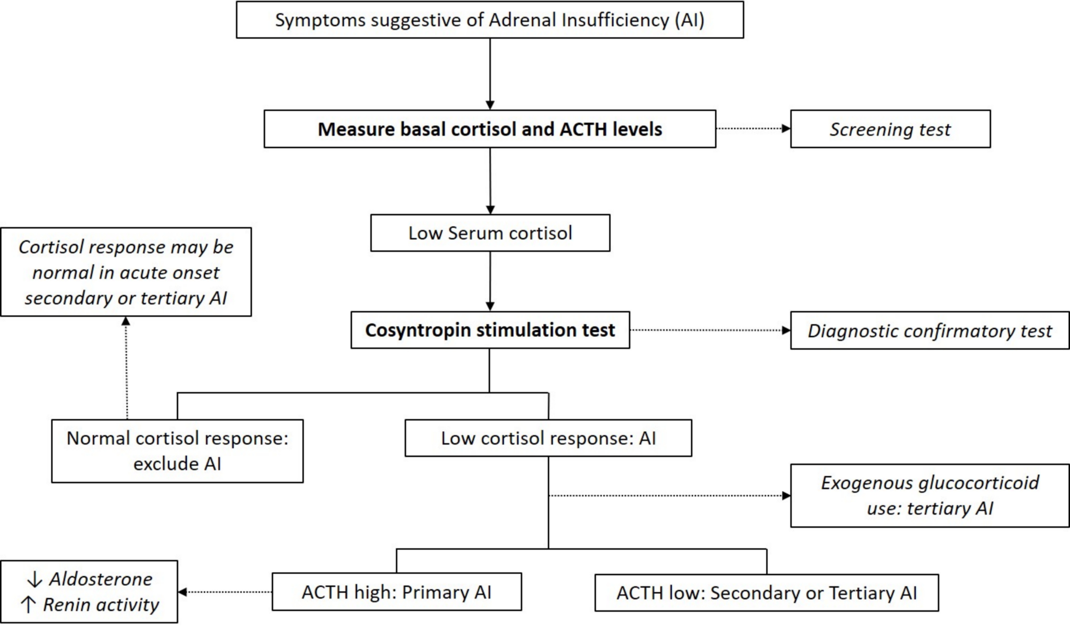
Adrenal insufficiency testing flowchart
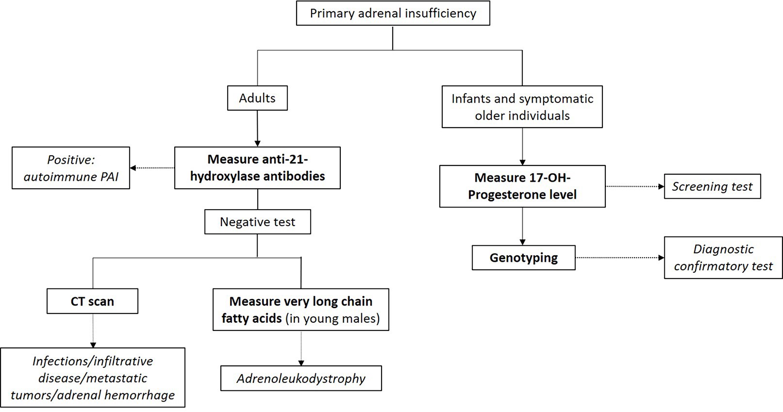
Primary adrenal insufficiency testing flowchart
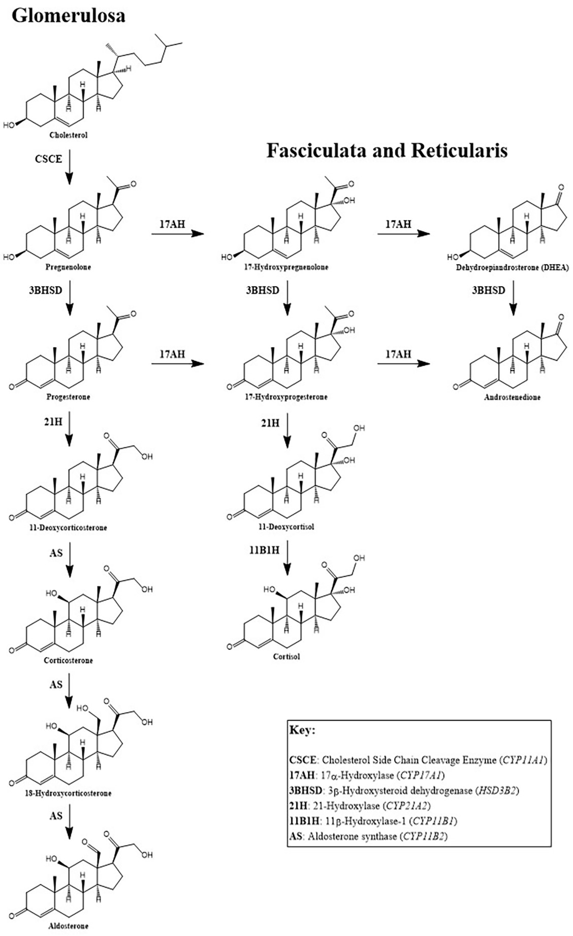
Adrenal steroid metabolism
Aldosterone
Diagrams / tables
Images hosted on other servers:

Aldosterone biosynthetic pathways
Anticardiolipin antibodies
Diagrams / tables
Images hosted on other servers:

2023 ACR / EULAR APS classification criteria
Calcitonin
Diagrams / tables
Images hosted on other servers:

Procalcitonin and calcitonin molecule

Calcitonin function

Interpretation of serum calcitonin measurement

Follow up of MTC

Calcium stimulation test
Videos
Regulation of blood calcium via PTH and calcitonin
Customizing imaging based on calcitonin levels in medullary thyroid cancer
Creatine kinase
Diagrams / tables
Images hosted on other servers:

Creatine kinase
Drugs of abuse
Diagrams / tables
Contributed by Vishnu Amaram Samara, Ph.D.
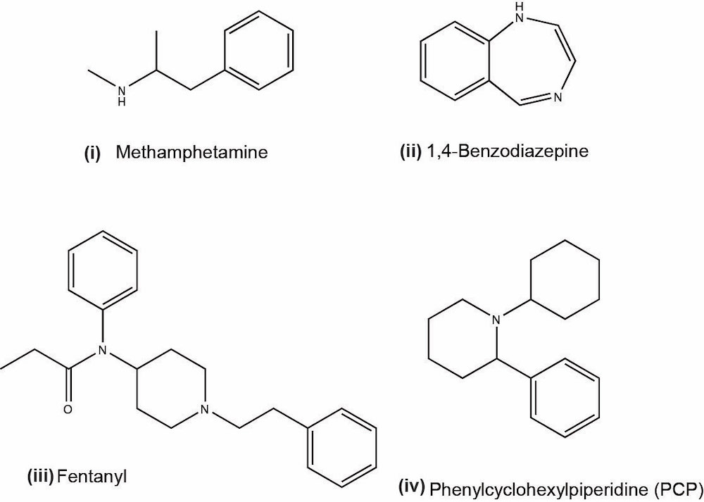
Chemical structures of a few common drugs of abuse
Images hosted on other servers:

Benzodiazepine metabolic pathways

Amphetamine metabolic pathway

Cannabinoid metabolic pathway

Opioid metabolic pathways
General cutoff limit concentrations of common drug classes by immunoassay screening and LC-MS / MS quantitative confirmation
| Drug class / metabolites
| Immunoassay screening
typical cutoff limits
(ng/mL)
| Quantitative cutoff
limits using LC-MS / MS
(ng/mL)
|
| Amphetamine
| 1,000
| 50
|
| Barbiturates
| 200
| 50
|
| Benzodiazepines
| 200
| 5
|
| Cannabinoids
| 50
| 15
|
| Cocaine
| 300
| 50
|
| Opiates
| 300
| 20
|
| Phencyclidine
| 25
| 10
|
Hepatitis B testing
Diagrams / tables
Images hosted on other servers:

Hepatitis B serologic course
Hyperaldosteronism
Diagrams / tables
Images hosted on other servers:

Synthesis pathway

Renin-angiotensinal-dosterone axis

Diagnostic pathways
Hypercortisolism
Clinical images
Images hosted on other servers:

Physical features

Patient description

Buffalo hump
Gross images
Images hosted on other servers:

Adrenal adenoma
Hyperlipidemia
Diagrams / tables
Images hosted on other servers:

Schematic of a chylomicron
Clinical images
Images hosted on other servers:

Xanthelasma palpebrarum
Interference in protein electrophoresis
Diagrams / tables
Contributed by Ahsan Farooq, M.D. and Yusheng Zhu, Ph.D.
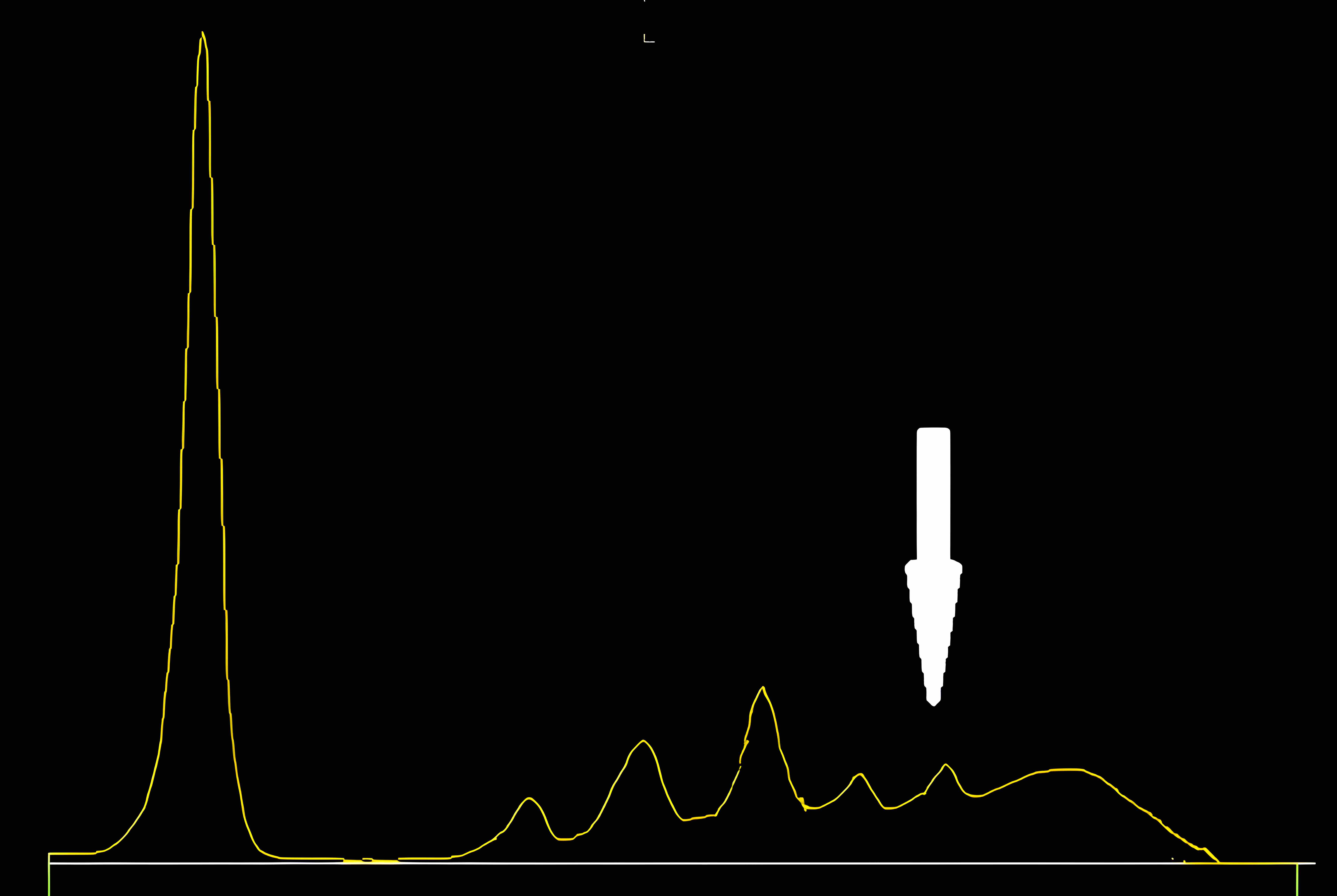
Fibrinogen
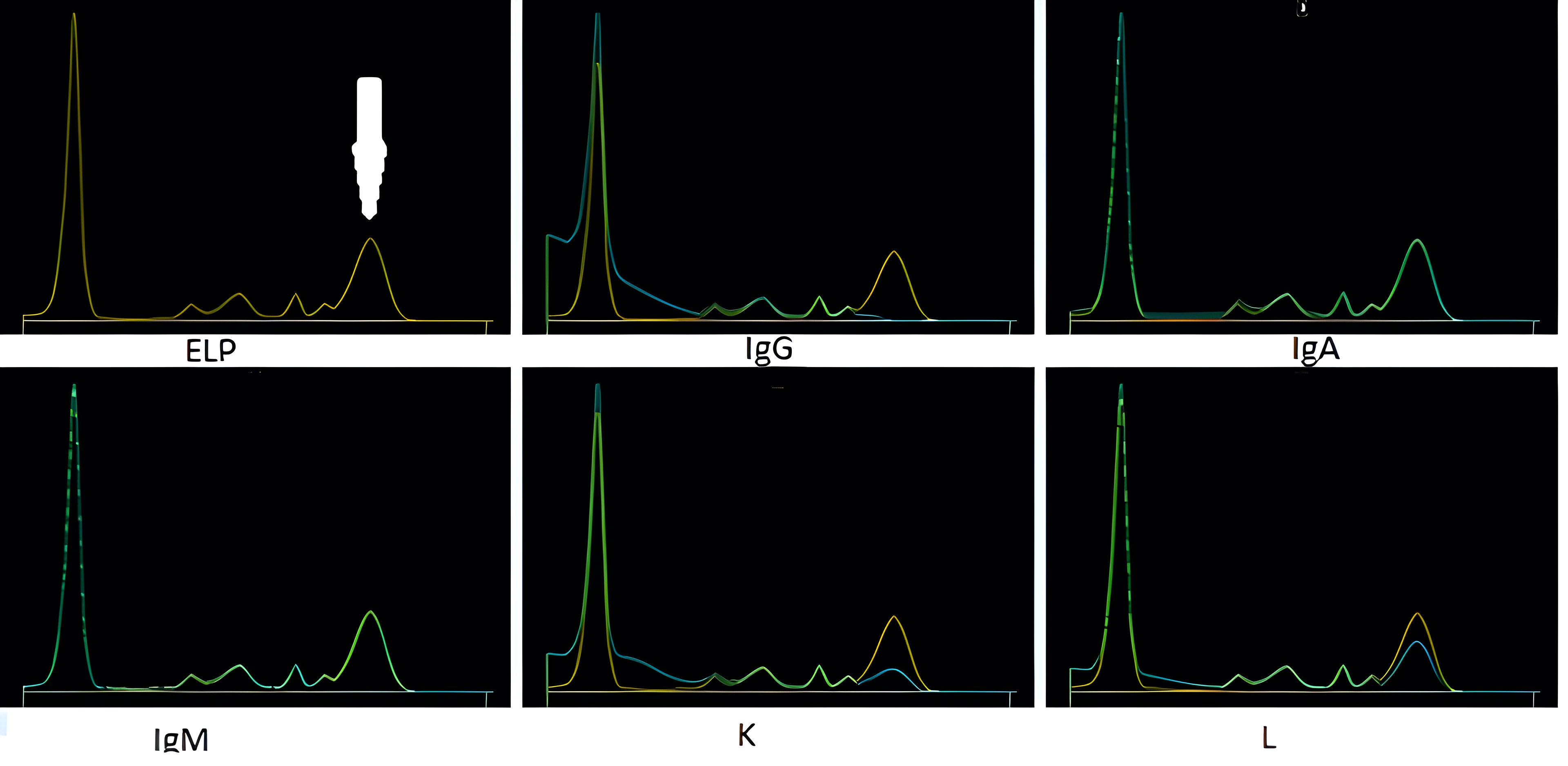
Patient with IgG4 RD
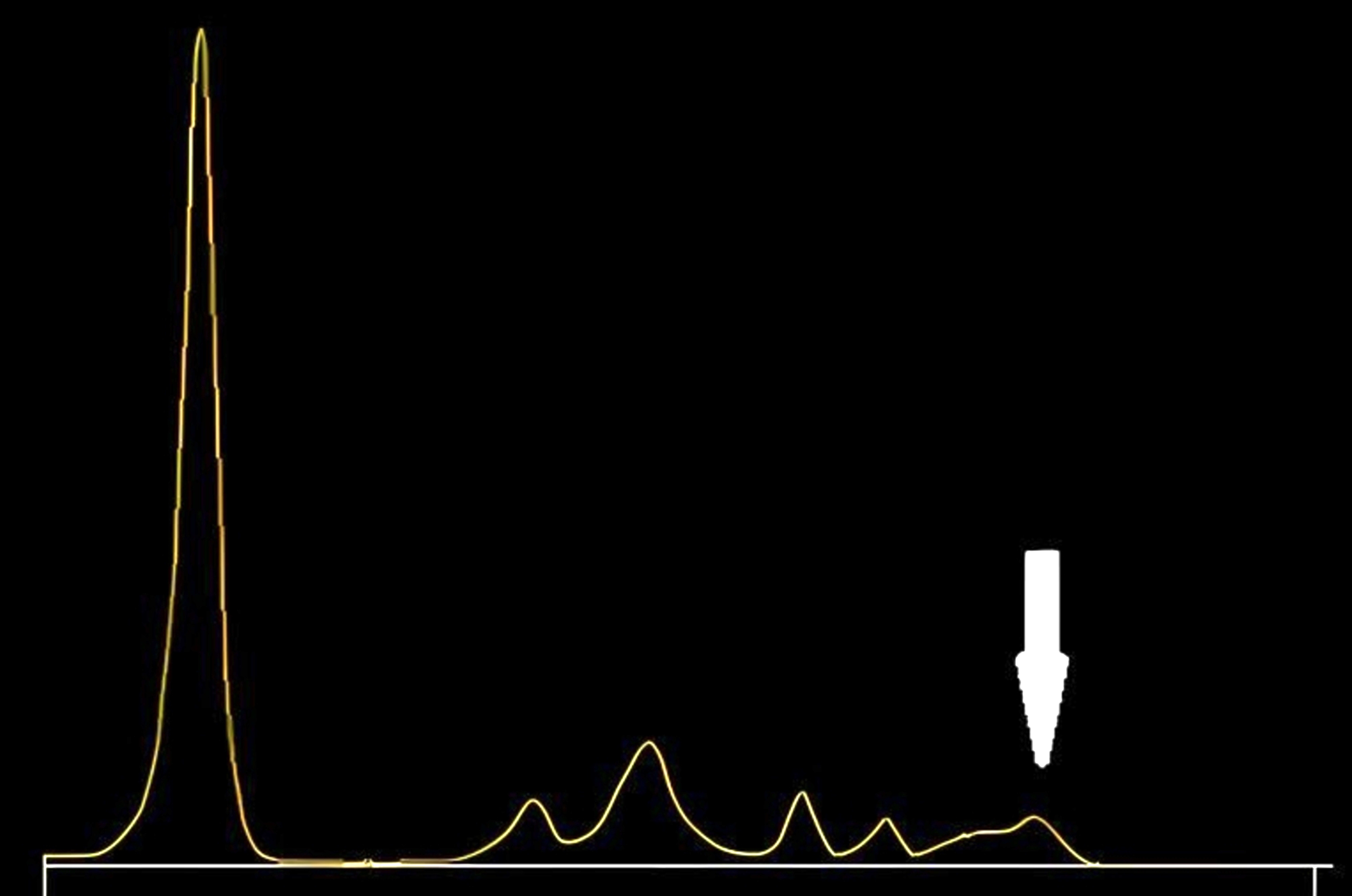
Patient on 5 fluorocytosine
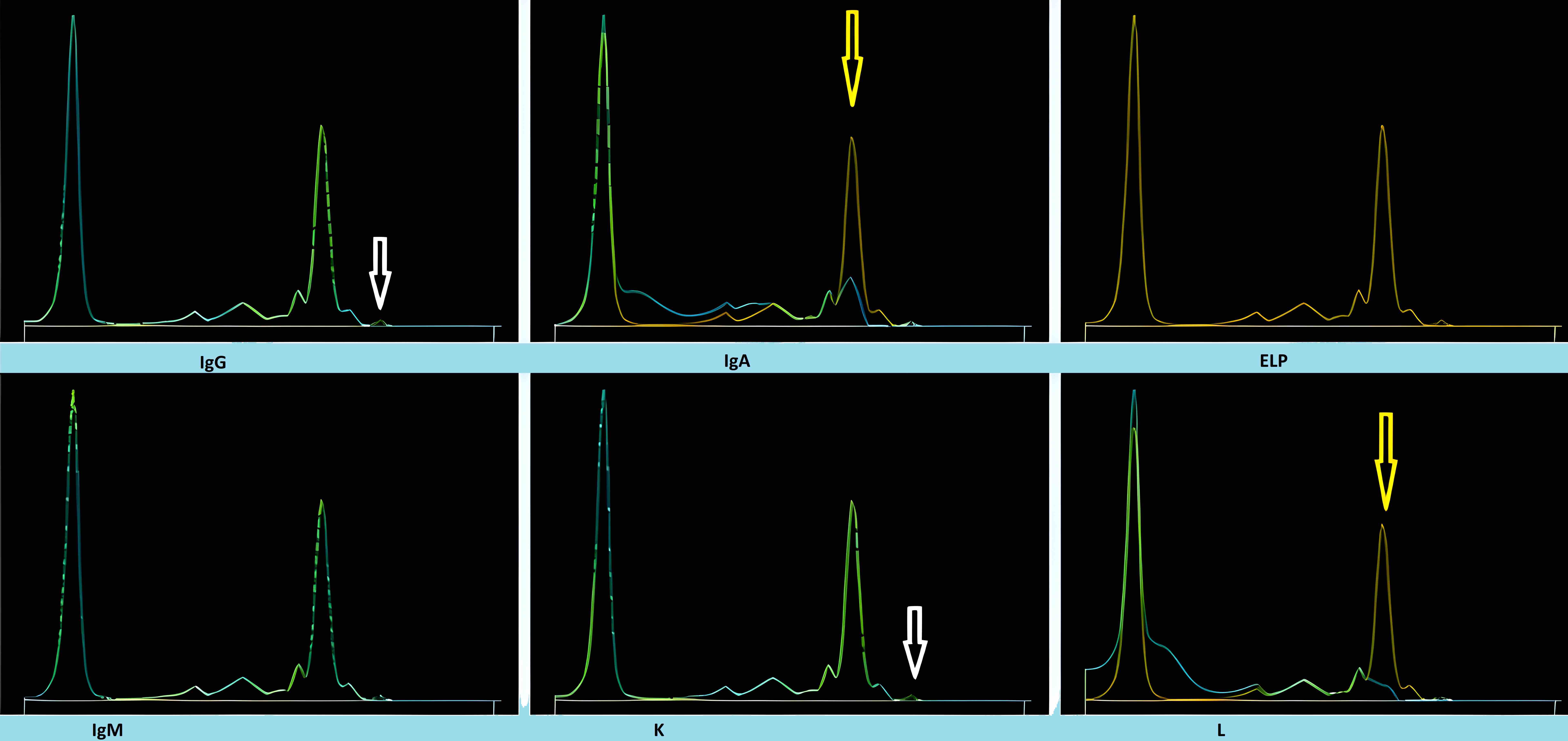
Multiple myeloma patient on daratumumab
Parathyroid hormone
Diagrams / tables
Images hosted on other servers:

Biologic effects of PTH and
vitamin D on calcium and
phosphate metabolism
Rheumatoid arthritis
Diagrams / tables
Images hosted on other servers:

Clinical manifestations of RA

2010 RA classification criteria

Classifying definite RA
Screening thyroid disorders
Diagrams / tables
Images hosted on other servers:

Diagnostic algorithm full

Diagnostic algorithm simplified

Classification of congenital hypothyroidism

Thyroid imaging and serum Tg in congenital hypothyroidism

Reference ranges

Genetic screening

Genetic diagnosis

Pregnancy and fetal hypothyroidism

Heel stick diagram
Syphilis
Diagrams / tables
Images hosted on other servers:

Serology response

Serologic testing algorithms
Systemic lupus erythematosus (SLE)
Diagrams / tables
Contributed by Jieli Shirley Li, M.D., Ph.D.
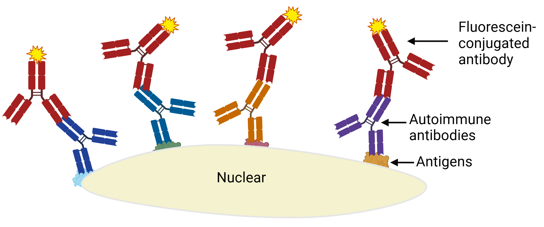
Fundamental principle of IFA ANA methods
Images hosted on other servers:

Comprehensive view of pathogenesis

IFA ANA testing classification tree

Classification criteria

Definitions of classification criteria
Therapeutic drug monitoring
Diagrams / tables
Contributed by Jingcai Wang, M.D., Ph.D.

Drug concentration versus half lives
Toxicology-general
Diagrams / tables
Contributed by Derek B. Laskar, M.D.
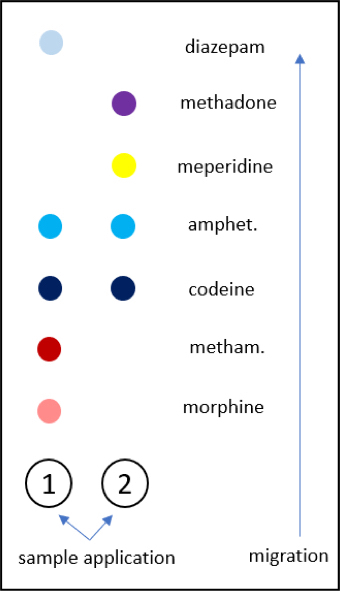
1: TLC for separation of major drugs of abuse
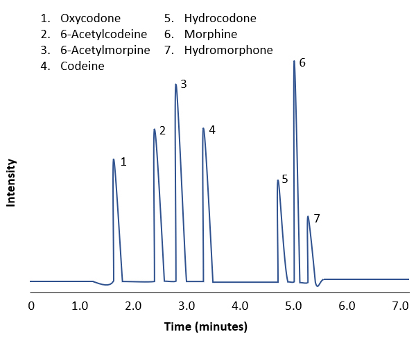
2: HPLC for separation of opiate analogs
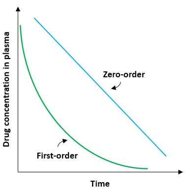
3: Zero order and first order elimination kinetics
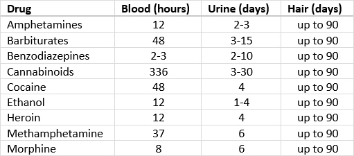
4: Window of detection for common drugs
Urine crystals & microscopy
Diagrams / tables
Contributed by Archana Shetty, M.B.B.S., M.D.
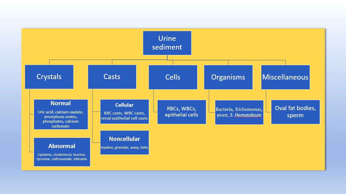
Urine sediment microscopy
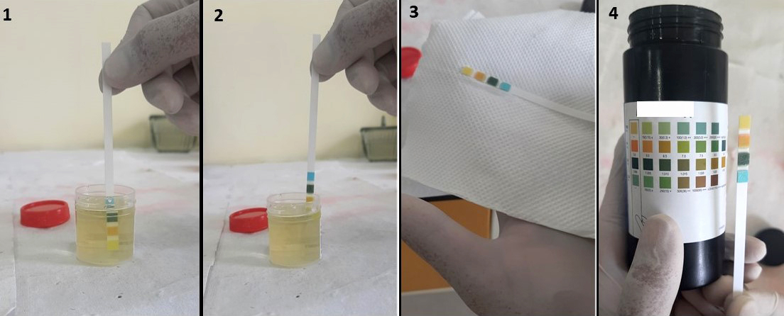
Steps in urine dipstick test
Microscopic (histologic) images
Contributed by Archana Shetty, M.B.B.S., M.D.
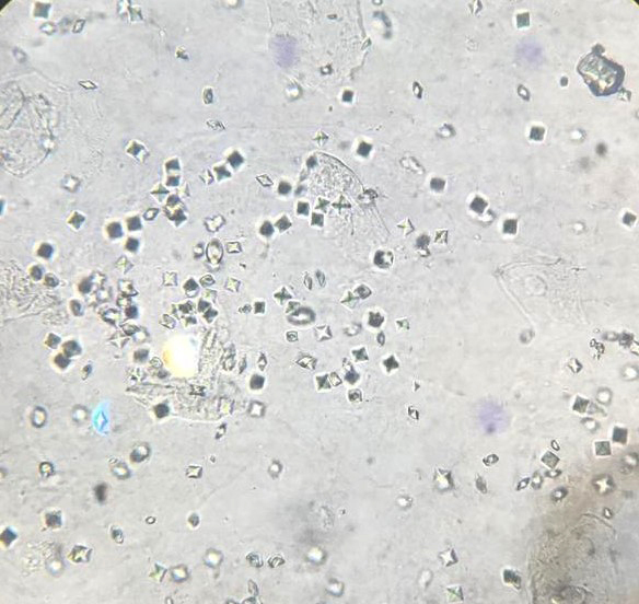
Calcium oxalate crystals
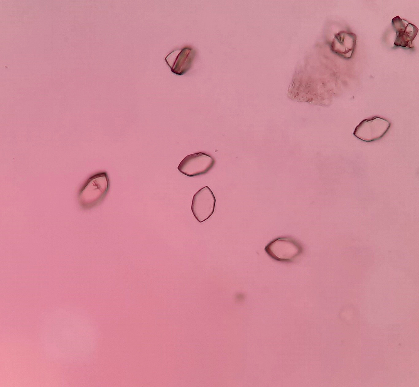
Uric acid crystals
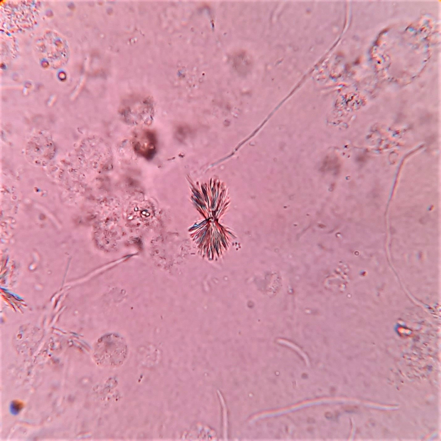
Sulfa drug crystals
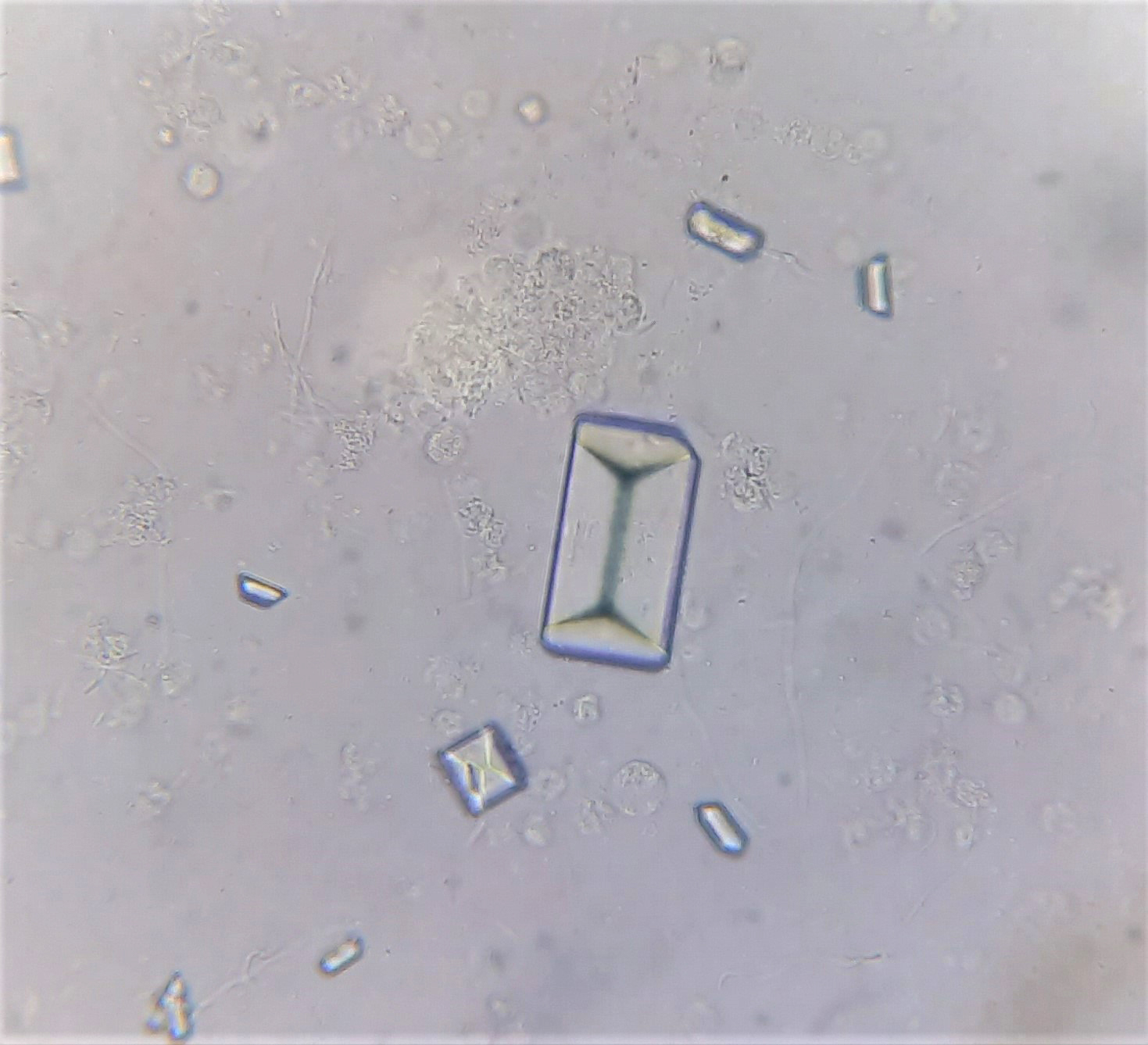
Triple phosphate crystals
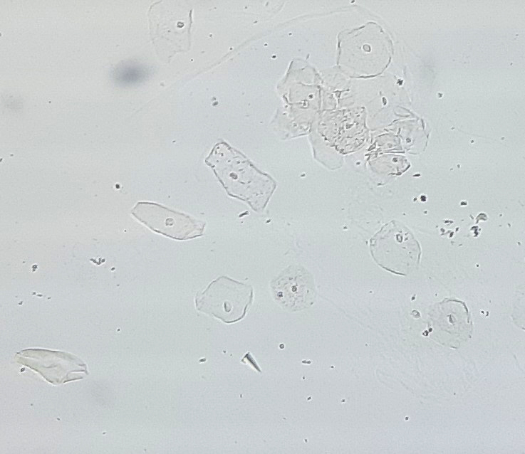
Squamous epithelial cells
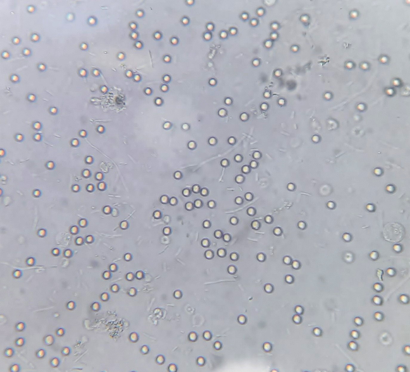
Red blood cells
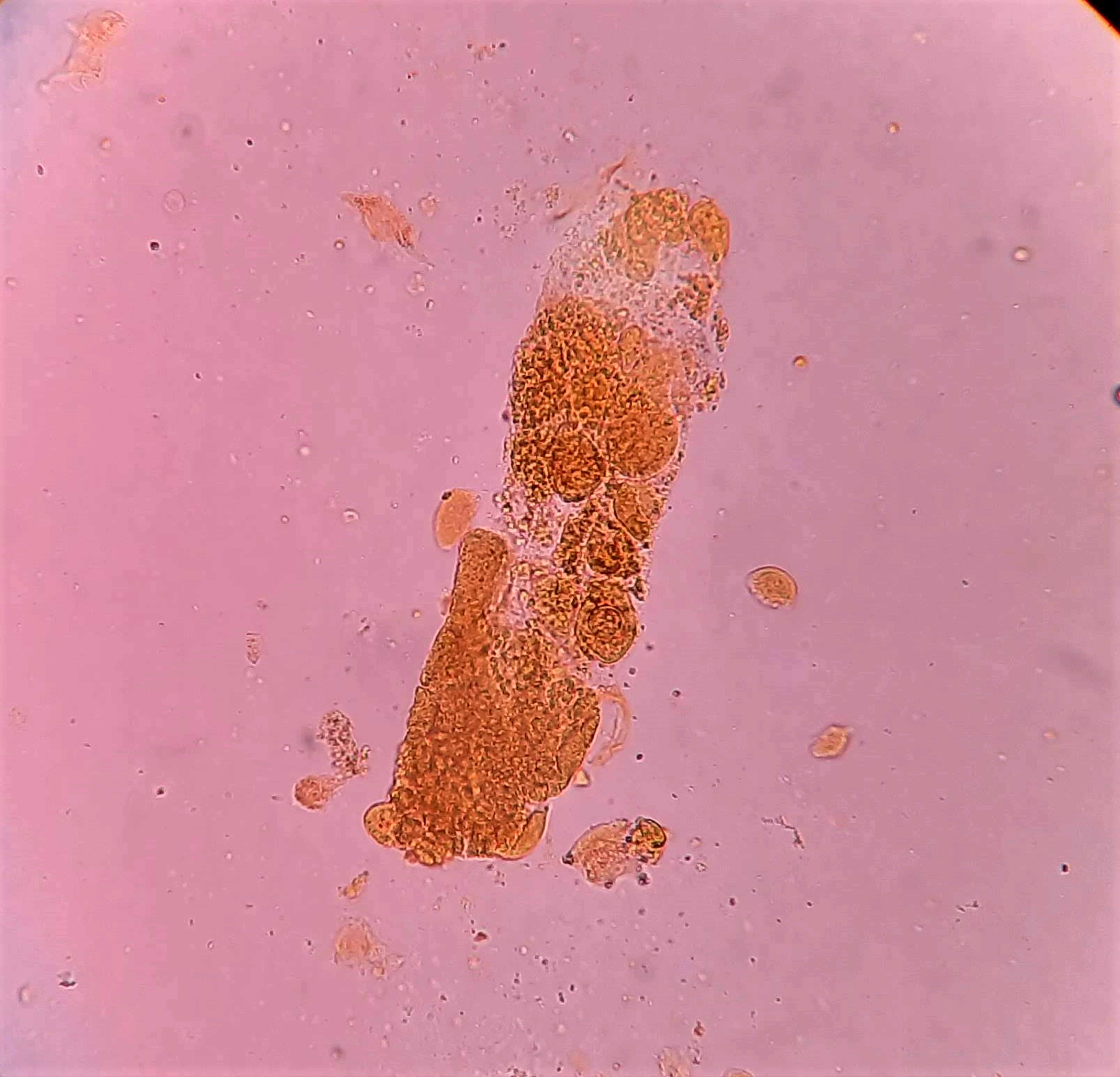
Red blood cell casts
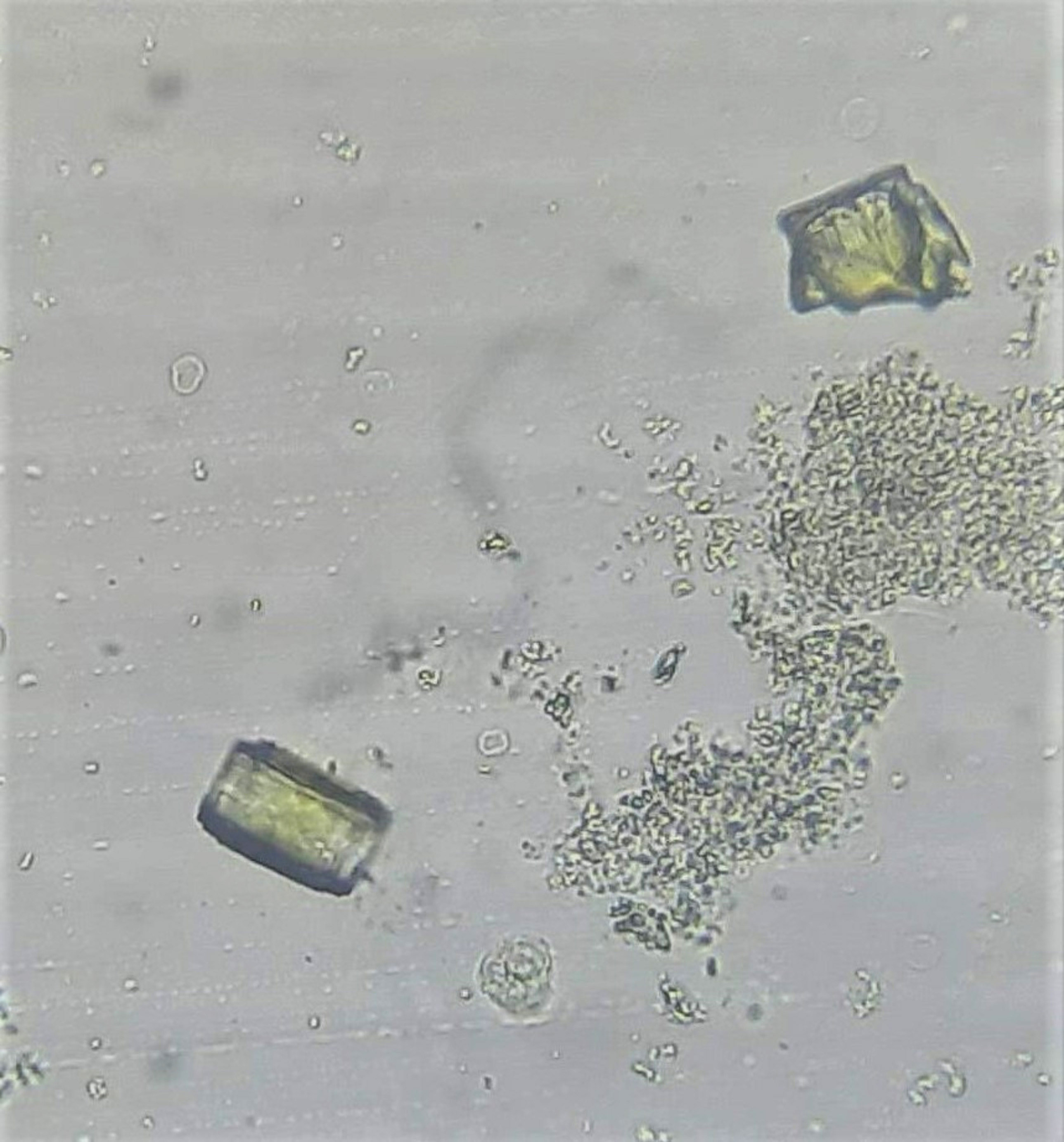
Amorphous urates in urine
Vitamin D
Diagrams / tables
Contributed by Zhicheng Jin, Ph.D.
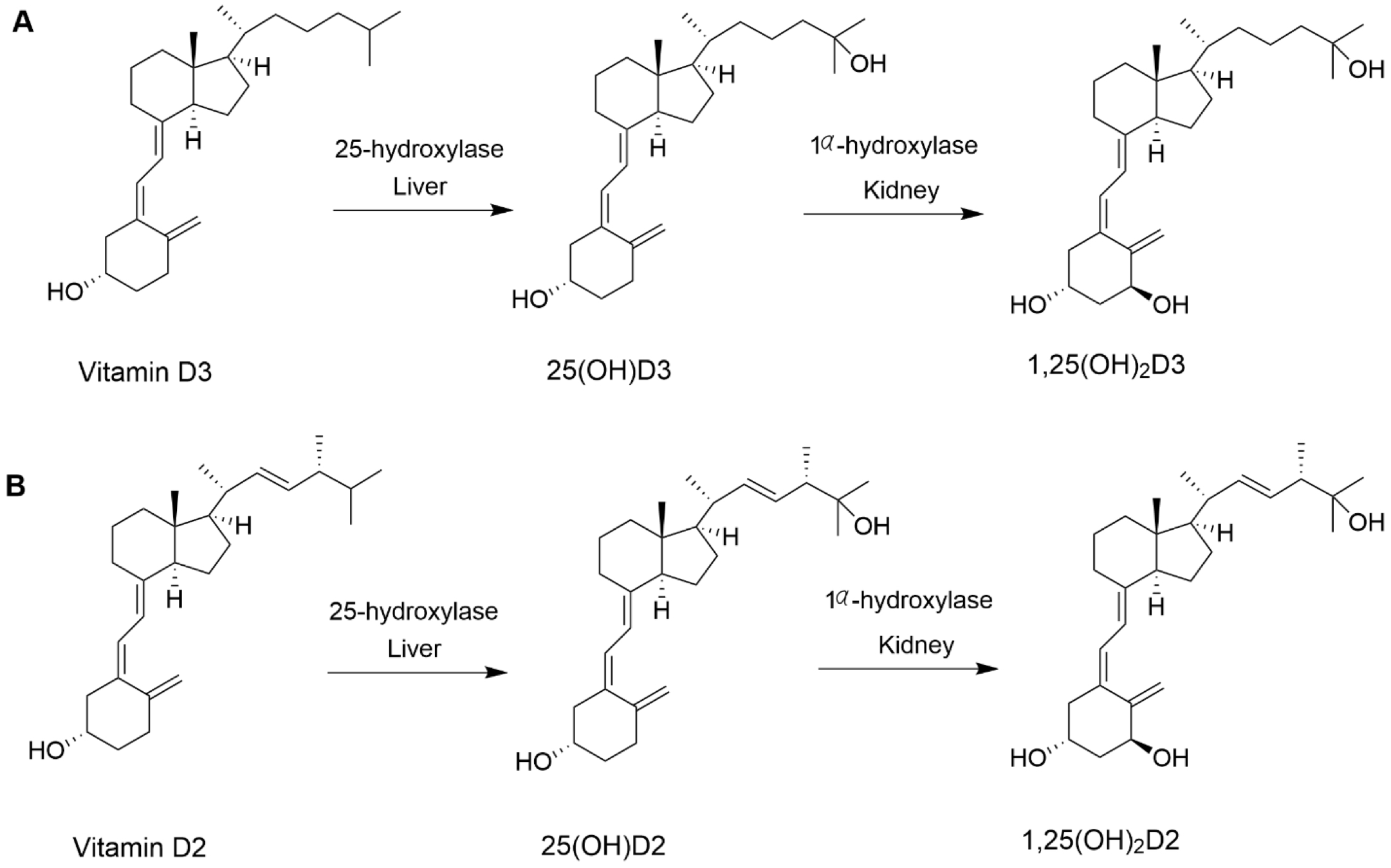
Structure and biological activation
lactate dehydrogenase
Diagrams / tables
Images hosted on other servers:


Pyruvate to Lactate conversion
Recent Chemistry, toxicology & urinalysis Pathology books
Haschek: 2022

Haschek-Hock: 2023

Magnani: 2020

Makowski: 2017

McPherson: 2021

Rifai: 2022

Snyder: 2020

Wagar: 2019
Find related Pathology books:
lab medicine,
management,
microbiology,
body fluid/urinalysis
Home > Chemistry, toxicology & urinalysis


