Table of Contents
Definition / general | Essential features | Epidemiology | Sites | Pathophysiology | Clinical features | Diagnosis | Radiology description | Prognostic factors | Case reports | Treatment | Gross images | Microscopic (histologic) description | Microscopic (histologic) images | Positive stains | Negative stains | Differential diagnosisCite this page: Chavez J, Zynger DL. OLD Male urethral carcinoma. PathologyOutlines.com website. https://www.pathologyoutlines.com/oldtopics/prostatemaleurethracarcinoma.html. Accessed December 26th, 2024.
Definition / general
- Rare primary neoplasm of epithelial origin
- Secondary involvement by urothelial carcinoma of the bladder is much more common than primary (Eur Urol 2013;64:823)
Essential features
- Urethral carcinoma is usually due to secondary involvement
- Primary urethral carcinoma is rare and the most frequent histologic types are urothelial carcinoma, squamous cell carcinoma and adenocarcinoma (not otherwise specified, clear cell)
Epidemiology
- Primary tumor accounts for < 1% of all genitourinary malignancies (Eur Urol 2013;64:823, BJU Int 2014;114:25)
- More common in women than men (Hematol Oncol Clin North Am 2012;26:1291)
- In men, more common in African Americans (Urology 2006;68:1164)
- Urothelial carcinoma is the predominant histologic type (54 - 65%), followed by squamous cell carcinoma (16 - 22%) and adenocarcinoma (10 - 16%) (Urology 2006;68:1164)
- Secondary involvement of urethra post-cystectomy is seen in 3 - 4% of patients (Eur Urol 2013;64:823, Int J Surg 2015;13:148)
Sites
- Type depends on sex and location:
- Male urethra divided into 4 anatomic regions: prostatic, membranous, bulbous and penile
- Prostatic and membranous portions: most commonly urothelial carcinoma and associated with bladder tumors (Semin Diagn Pathol 2015;32:238)
- Bulbous and membranous portions: most commonly squamous cell carcinoma and rarely associated with bladder tumors (Semin Diagn Pathol 2015;32:238)
- Adenocarcinoma presents in both sexes; may originate anywhere along the urethra
- Most commonly associated with diverticula and prostatic adenocarcinoma in men (Bostwick: Urologic Surgical Pathology, 3rd Edition, 2014)
- May arise from urothelial metaplastic mucosa or from periurethral glands in both sexes
- Clear cell adenocarcinoma is found predominantly in women and has a particular association with urethral diverticulum (J Urol 2008;180:2463, Int J Surg Oncol 2015;2015:790235)
- Indistinguishable from clear cell adenocarcinoma of the genital tract
- Male urethra divided into 4 anatomic regions: prostatic, membranous, bulbous and penile
Pathophysiology
- Predisposing factors include:
- Iatrogenic chronic irritation (chronic catheterization / urethroplasty) (Eur Urol 2013;64:823)
- Urethral strictures (BJU Int 2014;114:25)
- Radiation therapy (BJU Int 2008;101:964)
- Chronic urethritis secondary to sexually transmitted diseases
- Recurrent urinary tract infections
Clinical features
- Most patients present with symptoms associated with locally advanced disease (Eur Urol 2013;64:823)
- Gross hematuria or bloody urethral discharge, dysuria, extraurethral mass
- Bladder outlet obstruction, pelvic pain, urethrocutaneous fistula
- Abscess formation, dyspareunia
- Approximately 33% of men and women present with involved regional lymph nodes
Diagnosis
- Clinical examination with palpation of external genitalia for suspicious indurations and pelvic exam in women (Eur Urol 2013;64:823)
- Urinary cytology
- Diagnostic urethroscopy and biopsy
Radiology description
- Aims to assess local extent and detect lymphatic and distant metastatic spread
- Magnetic resonance imaging for evaluating extent of tumor and monitoring response to neoadjuvant chemotherapy (Eur Urol 2013;64:823)
Prognostic factors
- Both sexes (Hematol Oncol Clin North Am 2012;26:1291, Eur Urol 2013;64:823):
- Advanced age (≥ 65 years) and race (African American)
- Stage, grade, nodal involvement and metastasis
- Size and proximal tumor location
- Presence of concomitant bladder cancer
- Type and modality of treatment
- Men:
- Invasion of prostatic stroma signifies worse prognosis (Hematol Oncol Clin North Am 2012;26:1291)
- Lymphovascular, perineural invasion and histologic grade (squamous) (World J Urol 2009;27:169)
- Tumors arising in proximal urethra have worse prognosis than those arising in the distal (pendulous) portion
Case reports
- 39 year old man with a primary enteric type mucinous adenocarcinoma of the urethra (Int Surg 2014;99:669)
- 39 year old white man with metastatic urethral clear cell carcinoma (Clin Genitourin Cancer 2012;10:47)
- 49 year old man with squamous cell carcinoma of the bulbar urethra (J Clin Oncol 2011;29:e733)
Treatment
- Localized disease in men:
- Distal urethrectomy with negative surgical margins as an alternative to previously aggressive surgical excision with penile amputation (Eur Urol 2013;64:823)
- Advanced disease in both sexes:
- Chemotherapy with cisplatinum based agents
- Uncertain value of lymph node dissection (Curr Opin Urol 2015;25:129)
- References: Br J Urol 1998;82:835, Urology 1999;53:1126, Kufe: Cancer Medicine Review, 6th Edition, 2003
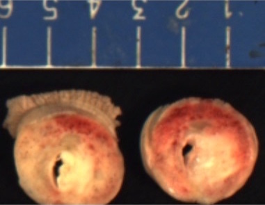
Gross images
Microscopic (histologic) description
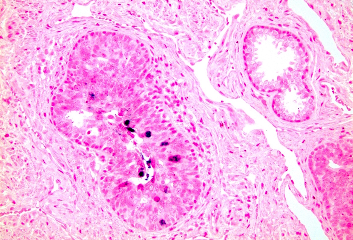
- Urothelial carcinoma
- Cytologically malignant urothelial cells with visible cell membranes, marked nucleomegaly, irregular nuclei, prominent nucleoli, dark chromatin and abundant mitosis (Bostwick: Urologic Surgical Pathology, 3rd Edition, 2014)
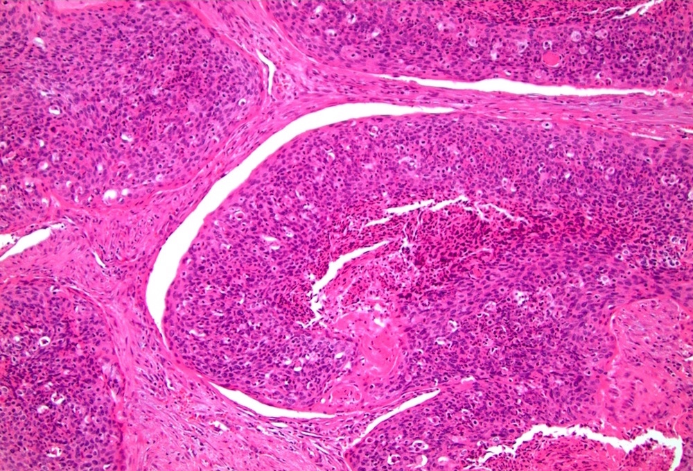
- Squamous cell carcinoma
- Sheets of large, pleomorphic tumor cells with focal or abundant keratinization (depending of grade of differentiation), ample cytoplasm, intercellular bridges, high mitotic activity, prominent nuclear atypia
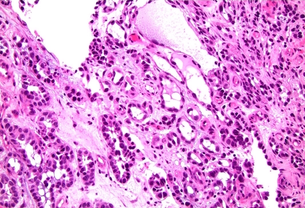
- Adenocarcinoma
- Composed of simple or pseudostratified columnar epithelium, apical cytoplasm and basally located hyperchromatic nuclei (Bostwick: Urologic Surgical Pathology, 3rd Edition, 2014)
- Occasional vacuolated cytoplasm with mucin or can be a true mucinous tumor with mucin pools
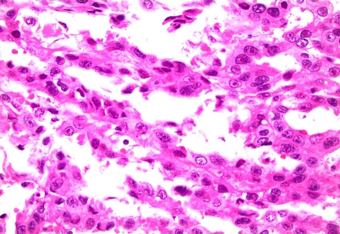
- Clear cell adenocarcinoma
- May have glandular, tubulocystic, solid / diffuse, papillary or micropapillary growth patterns
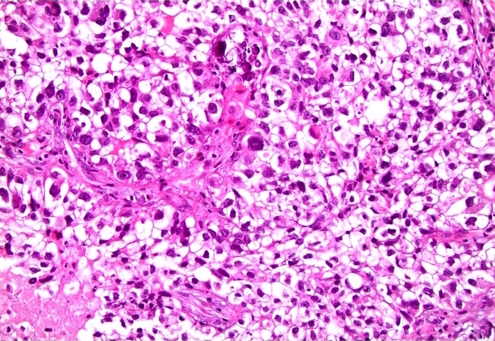
- Cuboidal, variably sized cells with abundant clear or eosinophilic cytoplasm and cytoplasmic vacuoles
- Nuclei that are hyperchromatic, pleomorphic and have prominent nucleoli
- Hobnail changes and extracellular mucoid material may be present
- Mitoses and necrosis are often seen
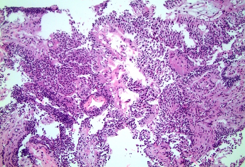
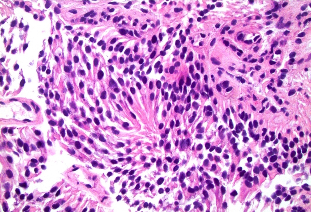
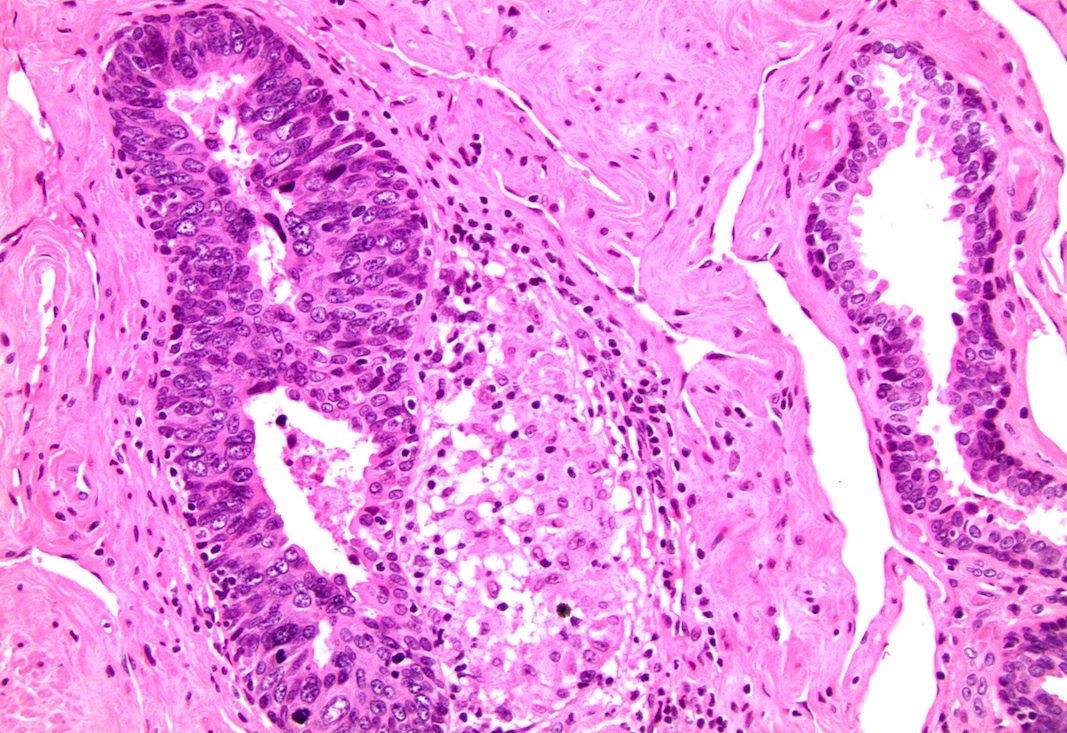
Microscopic (histologic) images
Contributed by Jesus Adrian Chavez, M.D. and Debra Zynger, M.D.
Clear cell adenocarcinoma:
Positive stains
- Adenocarcinoma
- CK20 (variable), CDX2 (variable), cytoplasmic beta catenin
- Clear cell adenocarcinoma
- AMACR (racemase) (75%), vimentin (75%), p53 (100%) (Virchows Arch 2013;462:193)
- PAX8 (50%), CK7 (50%) (Virchows Arch 2013;462:193)
- Squamous cell carcinoma
- High molecular weight cytokeratin (CK903, CK5/6), p63, p16 (HPV related)
- Urothelial carcinoma
- p53 (80%), CK20 (transurothelial in carcinoma in situ, variable in invasive urothelial carcinoma), CK7 , GATA3, high molecular weight cytokeratin (CK903, CK5/6), p63 (Hum Pathol 2013;44:2760)
Negative stains
- Urothelial carcinoma
- E-cadherin, CD44s, PAX8, PSA, PSAP
- Squamous cell carcinoma
- Adenocarcinoma
- Clear cell adenocarcinoma
Differential diagnosis
- Adenocarcinoma of adjacent anatomic sites (prostate and colon)
- Nephrogenic metaplasia (versus clear cell adenocarcinoma)
- Squamous cell carcinoma of penis with extension
- Urothelial carcinoma of bladder with extension (more frequent than primary)


