Table of Contents
Definition / general | Essential features | Epidemiology | Clinical features | Diagnosis | Radiology description | Radiology images | Prognostic factors | Case reports | Treatment | Gross description | Gross images | Microscopic (histologic) description | Microscopic (histologic) images | Positive stains | Negative stains | Molecular / cytogenetics description | Molecular / cytogenetics images | Videos | Sample pathology report | Differential diagnosis | Board review style question #1 | Board review style answer #1Cite this page: Anderson D, Tretiakova M. Pediatric cystic nephroma. PathologyOutlines.com website. https://www.pathologyoutlines.com/topic/kidneytumorpediatric.html. Accessed December 23rd, 2024.
Definition / general
- Uncommon, benign, exclusively cystic neoplasm of the kidney; lacks immature nephroblastic elements or solid growth (Hum Pathol 2016;48:81)
Essential features
- Multicystic neoplasm, often found in children under 4 years old, composed of fibrous septa and differentiated tubules
- Correlation to DICER1 mutations
- Excellent prognosis
Epidemiology
- Pediatric cystic nephroma (PCN) occurs most commonly in children younger than 4 years old, most of whom are boys (Am J Surg Pathol 2016;40:1591)
- Familial cases have been linked to DICER1 germline mutations and familial pleuropulmonary blastoma (PPB), whose main phenotypic spectrum includes PPB, PCN, ovarian Sertoli-Leydig tumors and multinodular goiter and less commonly, pineoblastoma, pituitary blastoma, nasal chondromesenchymal hamartoma and medulloepithelioma (Hum Pathol 2016;48:81)
Clinical features
- Palpable abdominal mass may be incidentally found by parents, caretakers or during routine physical examination
- Most often unilateral but familial forms may present bilaterally (J Med Genet 2010;47:863)
Diagnosis
- Imaging modalities (e.g., ultrasound, CT, MRI) show a multilocular cystic encapsulated mass
- Partial or radical nephrectomy will show diagnostic features
Radiology description
- On CT / MRI, appears as a cystic, multilocular mass, often with pseudocapsule defined as a thin rim of tissue demarcating the margin of the lesion from the adjacent renal parenchyma; may abut the renal pelvis or show protrusion / herniation into the renal pelvis (Hum Pathol 2016;48:81, Radiographics 1995;15:653)
- Ultrasonographic findings are multiple anechoic spaces separated by thin septa (Radiographics 1995;15:653)
Radiology images
Prognostic factors
- Prognosis is excellent: overall survival was 100% over a median followup of 2.4 years in one study (J Urol 2007;177:294)
- Presumed transformation to anaplastic sarcoma in rare cases; however, it is unknown whether all anaplastic sarcomas of the kidney arise from pre-existing cystic nephromas
- Anaplastic sarcomas show presence of cysts with solid areas that may be composed of undifferentiated spindle cells with anaplastic changes, benign or malignant chondroid differentiation; less common features include blastemal-like areas, foci of rhabdomyoblastic differentiation and small islands of osteoid
- Co-occurrence of TP53 mutation may be present with DICER1 mutation
- Longitudinal risks for transformation, responsiveness and prognosis are unknown
- References: Mod Pathol 2018;31:169, Mod Pathol 2014;27:1267
Case reports
- 5 month old girl with pleuropulmonary blastoma in association with cystic nephroma and DICER1 syndrome (Radiology 2014;273:622)
- 7 month old girl with histologic features predominately of cystic nephroma but with foci containing atypical mitotic figures and anaplastic nuclei thought to be nascent anaplastic sarcoma (Hum Pathol 2016;53:114)
- 16 month old girl with multilocular cystic renal tumor extending into the renal pelvis and ureter (J Urol Surg 2014;1:39)
- 2 year old girl with pediatric cystic nephroma and pleuropulmonary blastoma with mutation analysis and pedigree (BMC Cancer 2017;17:146)
- 8 year old girl with DICER1 mutation, anaplastic sarcoma and septated renal cysts, thought to be cystic nephroma (Pediatr Blood Cancer 2016;63:1272)
- 9 year old boy with bilateral and recurrent pediatric cystic nephroma (Can Urol Assoc J 2021;15:E290)
Treatment
- Adequately treated by resection with excellent prognosis if completely excised (J Urol 2007;177:294)
Gross description
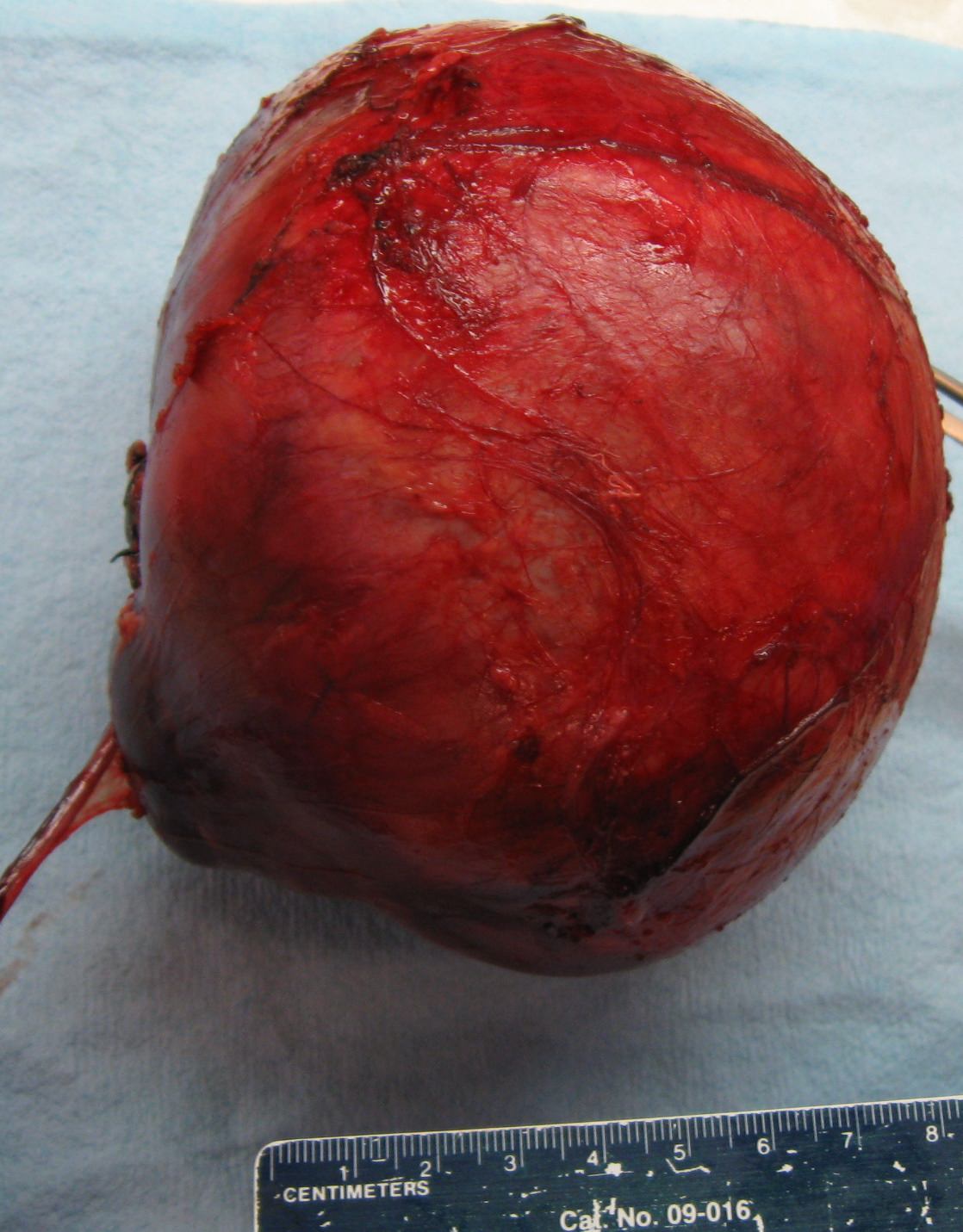
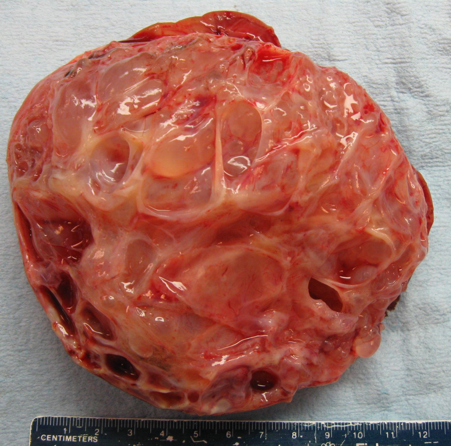
- Relatively large (9 cm average diameter) cystic lesion, well circumscribed from the surrounding normal kidney
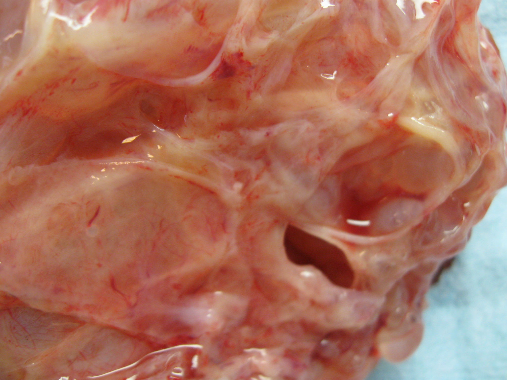
- Multiple smooth variably sized (mm to cm) cysts with clear fluid
- Thin septa with no expansile nodules altering cyst contours (Sebire: Diagnostic Pediatric Surgical Pathology, 1st Edition, 2009)
- Often adjacent, abutting, protruding or in direct contiguity to pelvicaliceal structures (Hum Pathol 2016;48:81)
Gross images
Microscopic (histologic) description
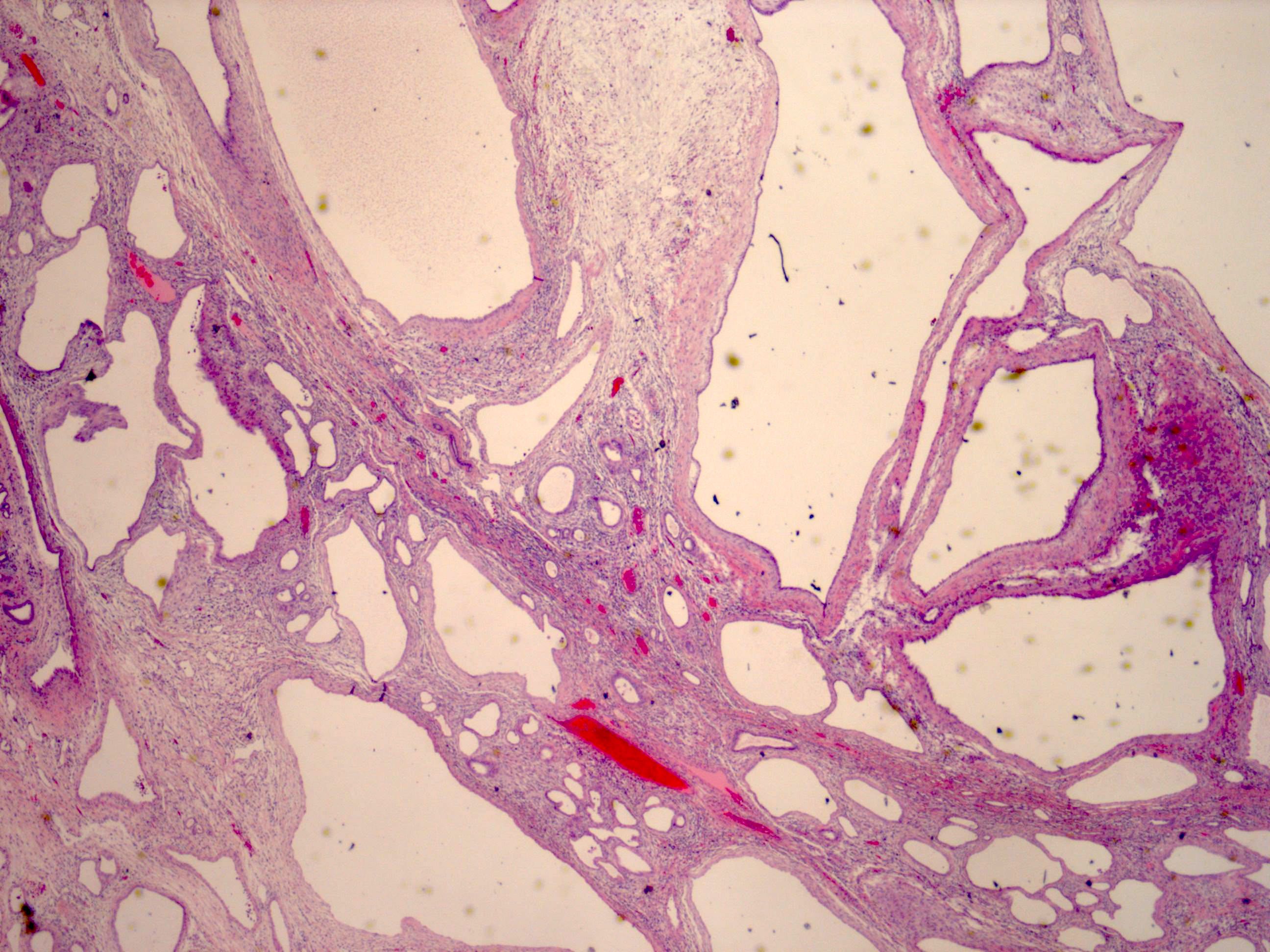
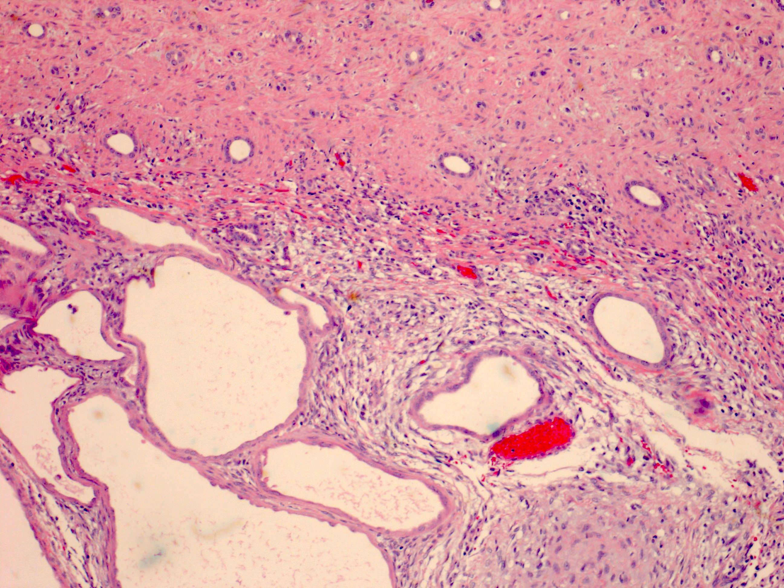
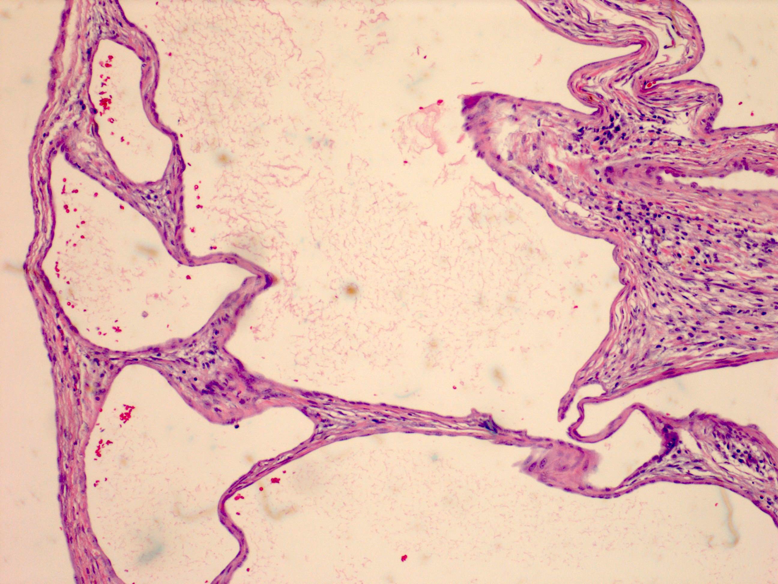
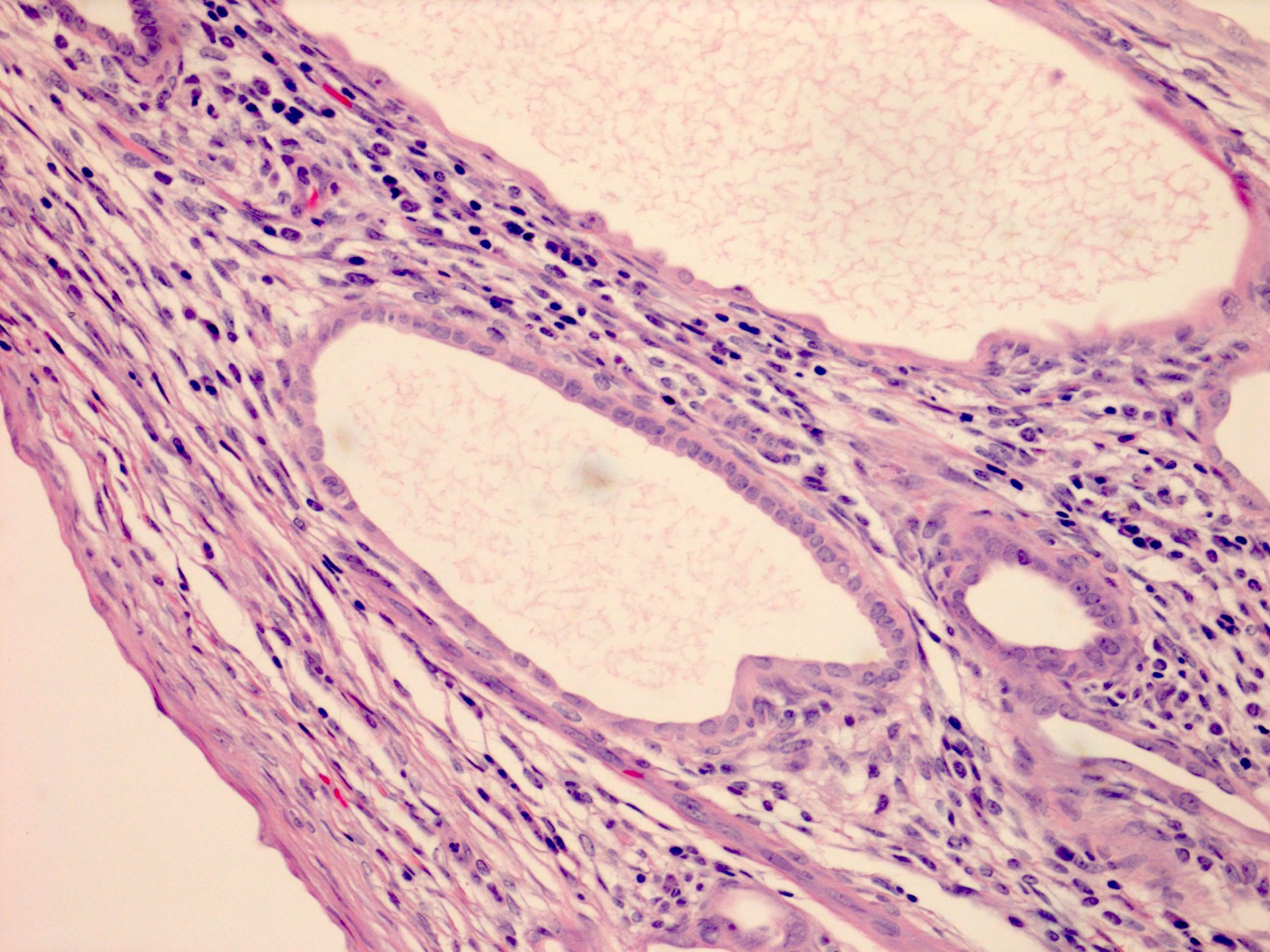
- Multicystic architecture with variably sized simple cysts lacking immature nephrogenic elements, solid areas and cytologic atypia
- Cystic septa are usually hypocellular and fibrous with variable amounts of mixed inflammation and rarely differentiated tubules
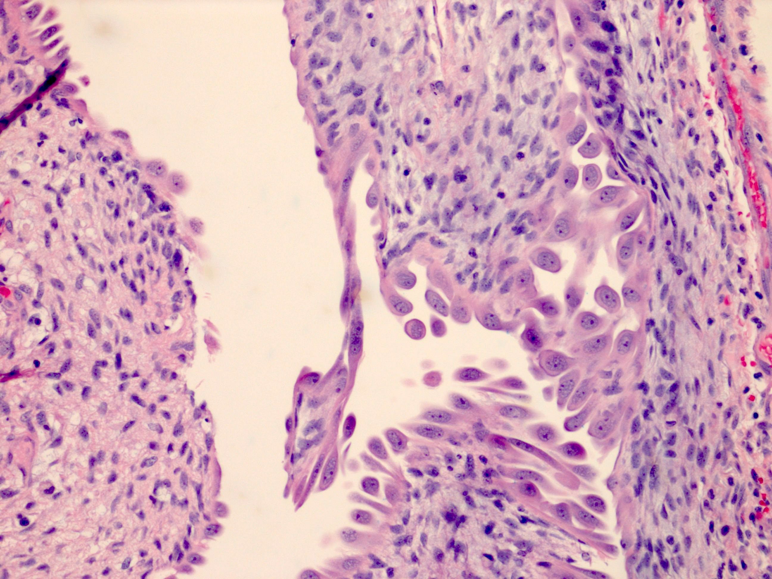
- Focally increased subepithelial cellularity may be present either due to spindle cells or inflammation
- Wavy / ropy collagen is absent
- Cyst lining epithelium is flattened to cuboidal, with frequent hobnailing
- No epithelial complexity (branching glands, papillary projection, cribriforming)
- Partial or complete fibrous pseudocapsule often containing entrapped tubules and glomeruli; some tumors intermingle with normal parenchyma (Hum Pathol 2016;48:81)
Microscopic (histologic) images
Positive stains
- ER positive in subepithelial stromal cells; PCN usually shows stronger and more diffuse positivity in contrast to adult cystic nephroma (Am J Surg Pathol 2017;41:472)
Negative stains
Molecular / cytogenetics description
- DICER1 is a multidomain protein with 2 type III endoribonucleases
- Involved in maturation of several classes of small noncoding RNAs such as miRNAs (Nat Rev Cancer 2014;14:662)
- PCNs usually carry DICER1 mutations, which may be somatic or germline
- Majority of mutations are nonsense and frameshift (truncating) substitutions and missense substitutions
- Somatic mutations often involve RNase IIIb and germline mutations may involve multiple DICER1 domains (Nat Rev Cancer 2014;14:662, Hum Pathol 2016;48:81, Mod Pathol 2014;27:1267)
Videos
Pediatric renal tumors: molecular diagnostics and COG updates
Sample pathology report
- Left kidney, partial nephrectomy:
- Pediatric cystic nephroma; margins are negative (see comment)
- Comment: Pediatric cystic nephroma is characterized by mutations in the DICER1 gene and may occur as a part of DICER1 syndrome. Clinical correlation and correlation with germline DICER1 mutation analysis is recommended.
Differential diagnosis
- Adult cystic nephroma:
- Separate pathologic entity mainly in women who are > 30 years and lack the DICER1 mutation (Hum Pathol 2016;48:81, Am J Surg Pathol 2016;40:1591)
- Usually have thicker septa composed of more cellular ovarian type stroma and ropy / wavy collagen
- Stromal cells are often inhibin positive at least focally while PCN is inhibin negative
- ER positivity is seen in both but typically more diffuse and intense in PCN (Am J Surg Pathol 2017;41:472)
- Segmental polycystic kidney disease:
- Clinical, radiologic correlation and family history may be helpful
- Lack of well defined fibrous wall
- Extension of cysts into the adjacent renal parenchyma with intermingling of nephrons; in contrast, PCN usually pushes aside native nephrons
- Lack of DICER1 mutation
- Cystic partially differentiated nephroblastomas and cystic Wilms tumor:
- Cystic Wilms tumor has immature nephrogenic elements plus solid nodules distorting the septal contours (Hum Pathol 2016;48:81)
- Both lack DICER1 mutations (Mod Pathol 2014;27:1267)
- Cystic renal dysplasia:
- Usually bilateral and diffuse
- Disorganized architecture with cysts, primitive tubules and undifferentiated mesenchyme
- Other cystic lesions of the kidney
Board review style question #1
A 2 year old boy with metastatic pineoblastoma was noted to have a palpable kidney mass. CT imaging showed a low attenuating cystic mass arising from the left kidney. Partial nephrectomy was performed and showed the above morphology on H&E. Given the findings, what syndromic condition needs to be considered?
- Denys-Drash Syndrome
- DICER1 syndrome
- Hereditary retinoblastoma
- Von Hippel-Lindau (VHL) syndrome
Board review style answer #1
B. DICER1 syndrome. The image shows features of pediatric cystic nephroma. DICER1 syndrome is characterized by an increased risk of developing pleuropulmonary blastoma, multinodular goiter, pediatric cystic nephroma, Sertoli-Lydig cell tumors and embryonal rhabdomyosarcoma. Less commonly, DICER1 mutations have been documented in pineoblastoma, pituitary blastoma, nasal chondromesenchymal hamartoma and medulloepithelioma, among others (Acta Neuropathol 2014;128:583, Acta Neuropathol 2014;128:111, Hum Genet 2014;133:1443, Eye (Lond) 2013;27:896).
Hereditary retinoblastoma is associated with pineablastoma and retinoblastoma. VHL is associated with hemangioblastomas, pheochromocytomas, renal cell carcinomas and endolymphatic sac tumors. Denys-Drash syndrome is a WT1 related Wilms tumor syndrome and associated with disorders of sexual development and Wilms tumor.
Comment Here
Reference: Pediatric cystic nephroma


