16 September 2021 - Case of the Month #507
All cases are archived on our website. To view them sorted by case number, diagnosis or category, visit our main Case of the Month page. To subscribe or unsubscribe to Case of the Month or our other email lists, click here.
Thanks to Dr. Catherine E. Hagen, Mayo Clinic, Rochester, Minnesota (USA) for contributing this case and the discussion and to Dr. Aaron R. Huber, University of Rochester, Rochester, New York (USA), for reviewing the discussion.

Advertisement
Case of the Month #507
Clinical history:
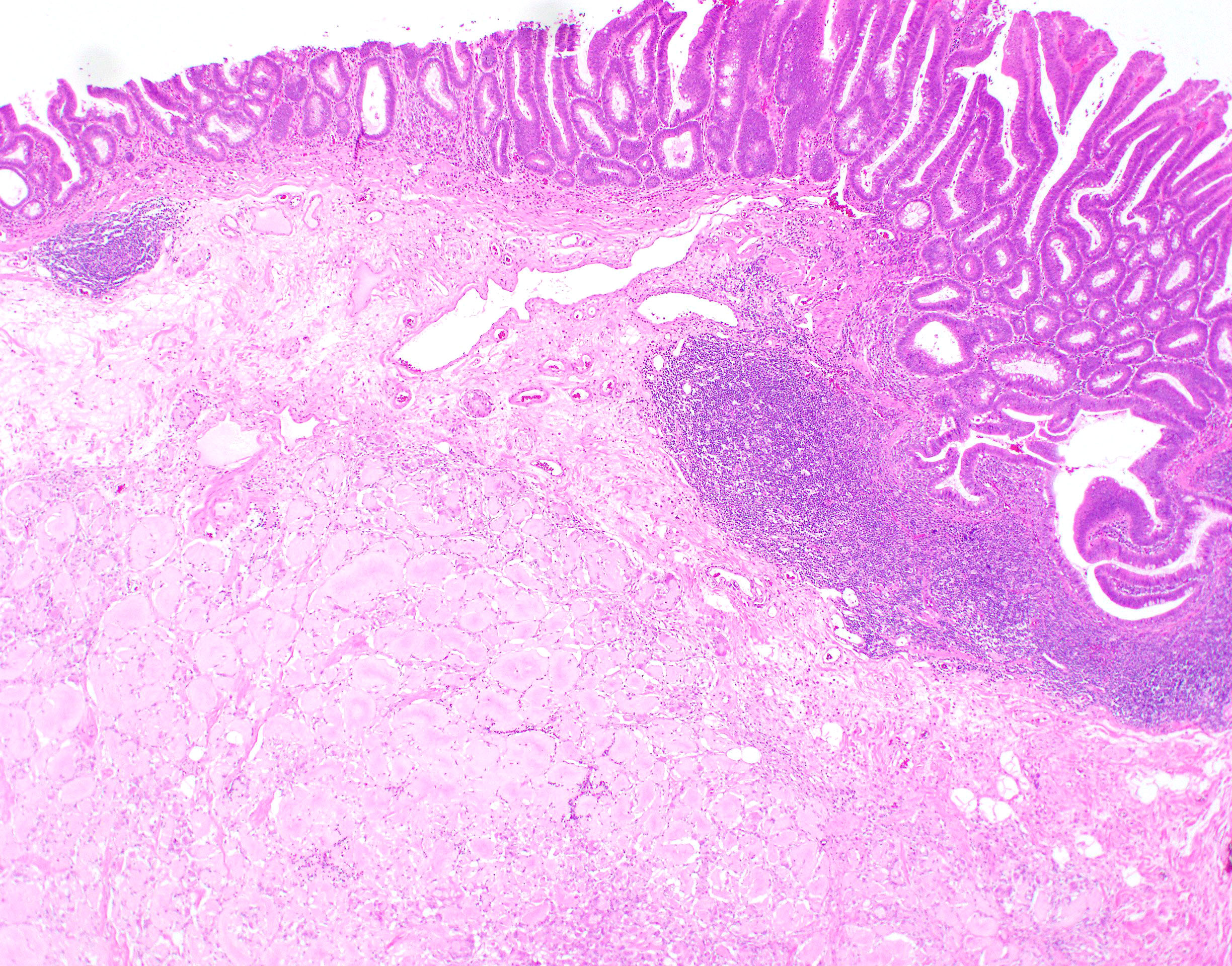
54 year old man had a partial colonic resection for an endoscopically unresectable tubular adenoma of the right colon.
Histopathology images:
What is your diagnosis?
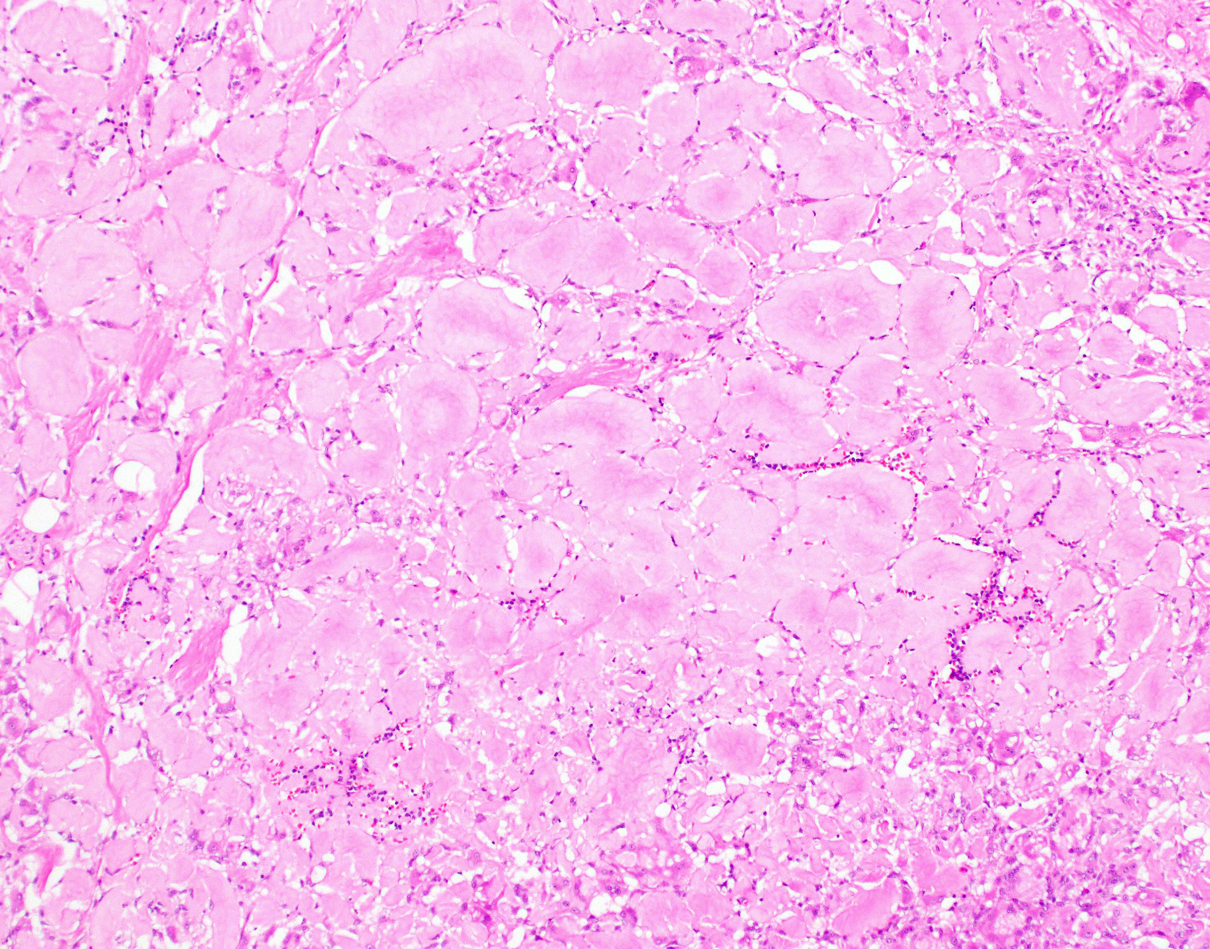
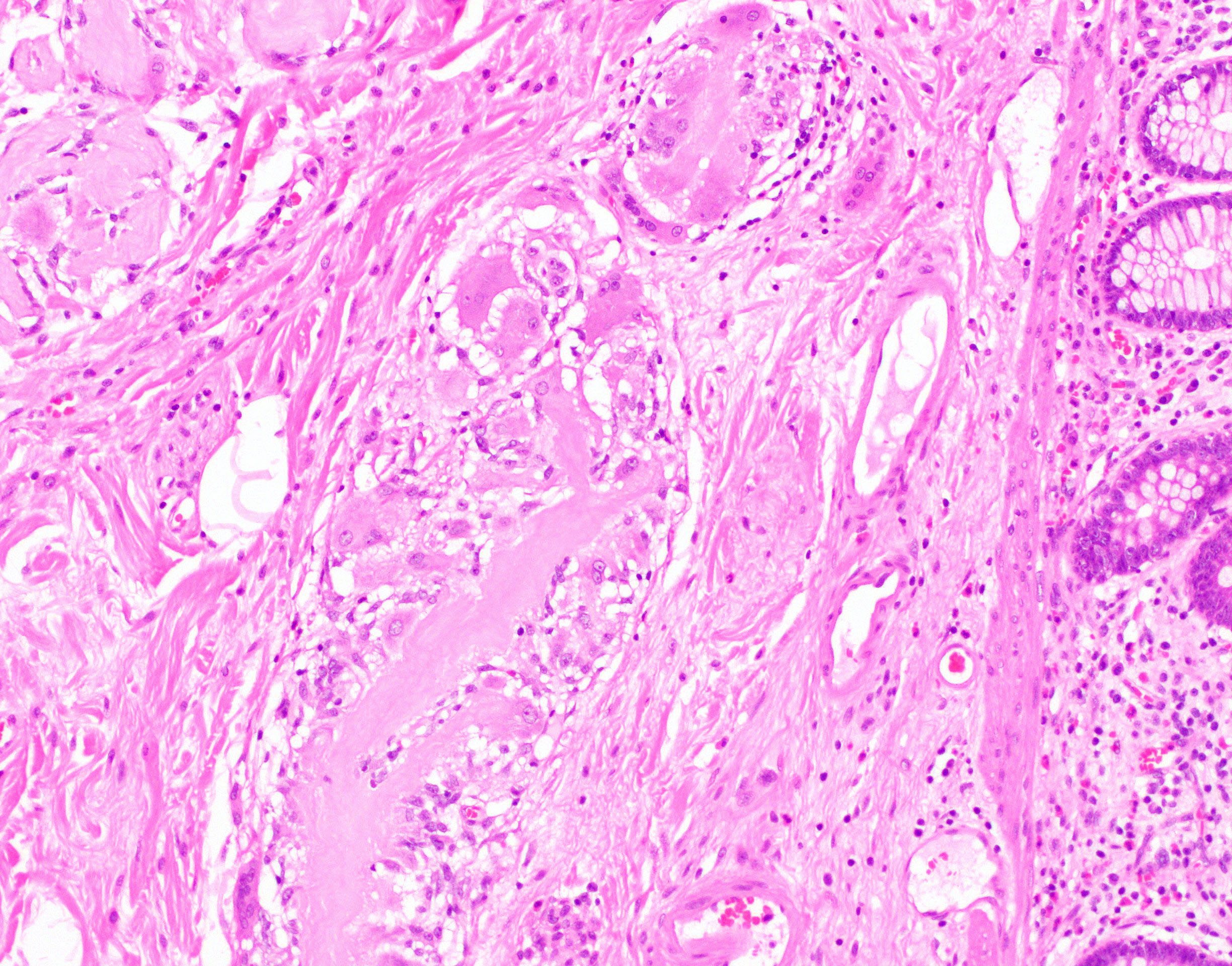
Diagnosis: Lifting agent granuloma
Test question (answer at the end):
Which of the following features is characteristic of submucosal lifting agents (Eleview or ORISE)?
A. Association with ruptured diverticula
B. Involvement of vascular walls
C. Positive for Congo red stain
D. Surrounding giant cell reaction
Discussion:
Eleview and ORISE are newly approved submucosal lifting agents utilized to visualize and resect sessile lesions of the gastrointestinal tract during endoscopic procedures. Compared with traditional lifting agents, these newer agents have the advantage of immediate availability without preparation and the need for fewer repeat injections, leading to decreased procedure time (Arch Pathol Lab Med 2021 Feb 11 [Epub ahead of print]).
Both agents appear histologically similar but their appearance varies with time. Immediately after injection, the agents have a basophilic, bubbly appearance, similar to acellular mucin. With time, the agents take on a pink, hyalinized appearance and often show surrounding foreign body giant cell reaction and fibrosis. At the hyalinized stage, the material may mimic amyloid or a pulse granuloma. Distinction from amyloid is usually straightforward, as amyloidosis frequently involves vessels, lacks a giant cell reaction and is positive for Congo red stain. Pulse granulomas are giant cell reactions to vegetable material and are frequently seen in association with diverticulosis with perforation or mucosal ulceration (Am J Surg Pathol 2020;44:793, Gastrointest Endosc 2021;93:470, Am J Clin Pathol 2020;153:630).
In the early stages these agents can show variable positivity for mucicarmine but do not stain for PAS. In the late stages, the material has a light blue appearance on trichrome or Alcian blue and will stain blue-purple on AFB and pink with PAS. Congo red, Movat, mucicarmine, GMS, colloidal iron and von Kossa stains are negative at this stage (Arch Pathol Lab Med 2021 Feb 11 [Epub ahead of print]).
These lifting agents are safe and cause no adverse effects to the patient but awareness of the histologic appearance and correlation with a history of submucosal lifting agent use can help avoid misclassification.
Test question answer:
D. Surrounding giant cell reaction. Submucosal lifting agents frequently show a surrounding giant cell reaction. Amyloid will stain positive for Congo red and frequently is seen within vessels walls. Pulse granulomas are frequently seen in association with ruptured diverticula.
All cases are archived on our website. To view them sorted by case number, diagnosis or category, visit our main Case of the Month page. To subscribe or unsubscribe to Case of the Month or our other email lists, click here.
Thanks to Dr. Catherine E. Hagen, Mayo Clinic, Rochester, Minnesota (USA) for contributing this case and the discussion and to Dr. Aaron R. Huber, University of Rochester, Rochester, New York (USA), for reviewing the discussion.

Advertisement
Case of the Month #507
Clinical history:
54 year old man had a partial colonic resection for an endoscopically unresectable tubular adenoma of the right colon.
Histopathology images:
What is your diagnosis?
Click here for diagnosis, test question and discussion:
Diagnosis: Lifting agent granuloma
Test question (answer at the end):
Which of the following features is characteristic of submucosal lifting agents (Eleview or ORISE)?
A. Association with ruptured diverticula
B. Involvement of vascular walls
C. Positive for Congo red stain
D. Surrounding giant cell reaction
Discussion:
Eleview and ORISE are newly approved submucosal lifting agents utilized to visualize and resect sessile lesions of the gastrointestinal tract during endoscopic procedures. Compared with traditional lifting agents, these newer agents have the advantage of immediate availability without preparation and the need for fewer repeat injections, leading to decreased procedure time (Arch Pathol Lab Med 2021 Feb 11 [Epub ahead of print]).
Both agents appear histologically similar but their appearance varies with time. Immediately after injection, the agents have a basophilic, bubbly appearance, similar to acellular mucin. With time, the agents take on a pink, hyalinized appearance and often show surrounding foreign body giant cell reaction and fibrosis. At the hyalinized stage, the material may mimic amyloid or a pulse granuloma. Distinction from amyloid is usually straightforward, as amyloidosis frequently involves vessels, lacks a giant cell reaction and is positive for Congo red stain. Pulse granulomas are giant cell reactions to vegetable material and are frequently seen in association with diverticulosis with perforation or mucosal ulceration (Am J Surg Pathol 2020;44:793, Gastrointest Endosc 2021;93:470, Am J Clin Pathol 2020;153:630).
In the early stages these agents can show variable positivity for mucicarmine but do not stain for PAS. In the late stages, the material has a light blue appearance on trichrome or Alcian blue and will stain blue-purple on AFB and pink with PAS. Congo red, Movat, mucicarmine, GMS, colloidal iron and von Kossa stains are negative at this stage (Arch Pathol Lab Med 2021 Feb 11 [Epub ahead of print]).
These lifting agents are safe and cause no adverse effects to the patient but awareness of the histologic appearance and correlation with a history of submucosal lifting agent use can help avoid misclassification.
Test question answer:
D. Surrounding giant cell reaction. Submucosal lifting agents frequently show a surrounding giant cell reaction. Amyloid will stain positive for Congo red and frequently is seen within vessels walls. Pulse granulomas are frequently seen in association with ruptured diverticula.


