16 October 2019 - Case of the Month #484
All cases are archived on our website. To view them sorted by case number, diagnosis or category, visit our main Case of the Month page. To subscribe or unsubscribe to Case of the Month or our other email lists, click here.
Thanks to Dr. Debra Zynger, The Ohio State University Wexner Medical Center, Columbus, Ohio (USA), for contributing this case and Dr. Maria Tretiakova, University of Washington, Seattle, Washington (USA), for writing the discussion.

Advertisement
Case of the Month #484
Clinical history:
A 34 year old man with a 1.4 cm mass underwent a partial nephrectomy.
Gross image:
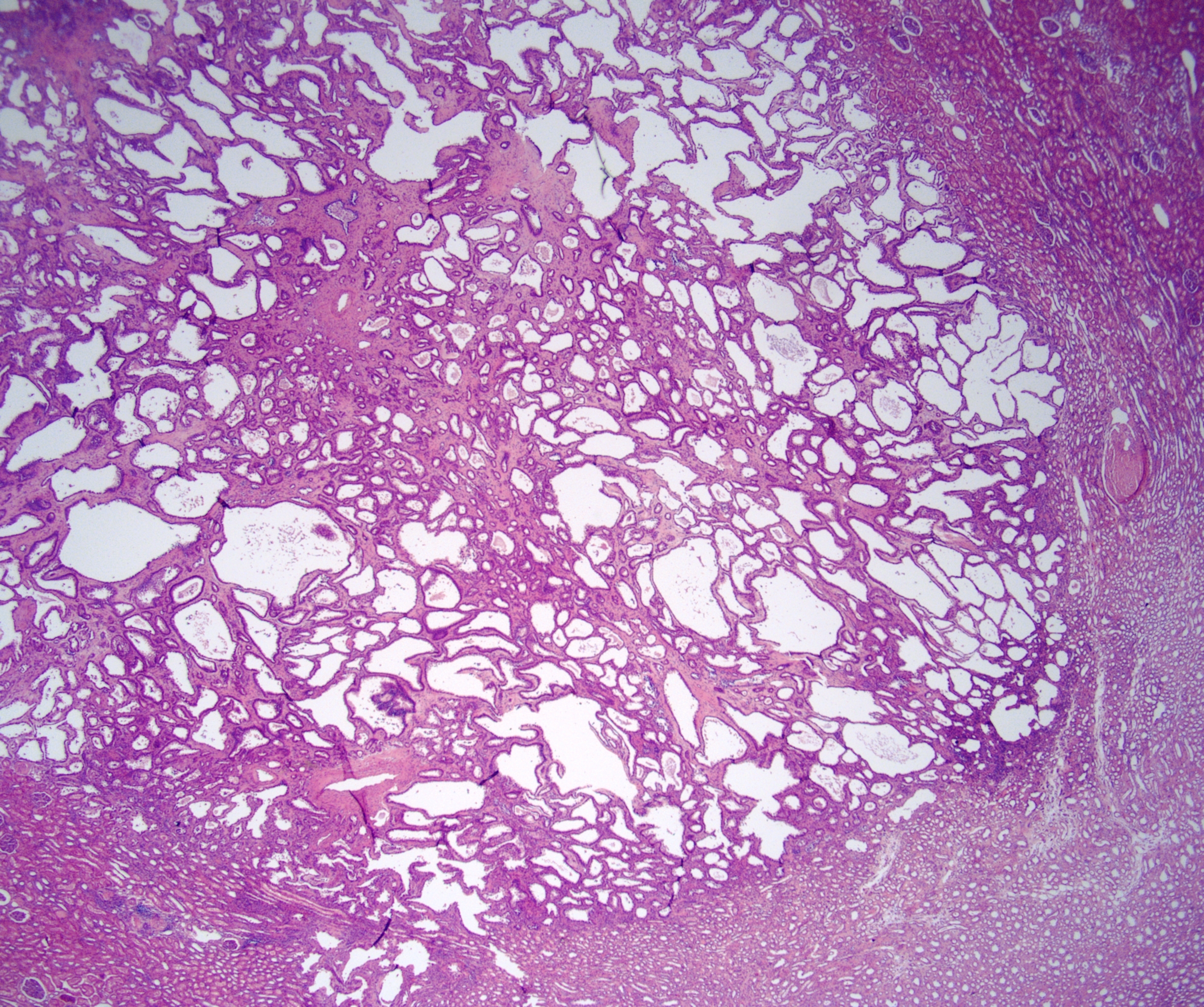
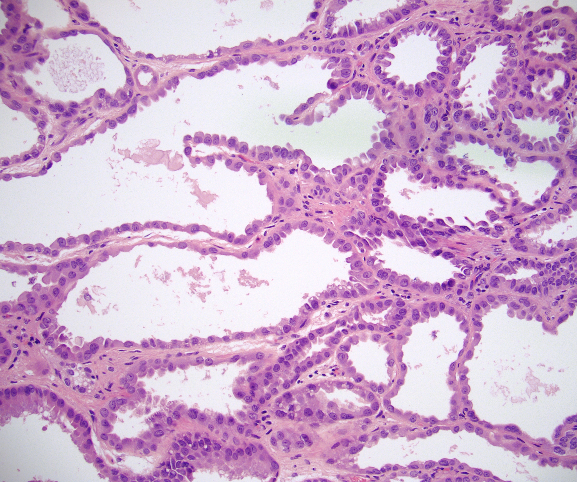
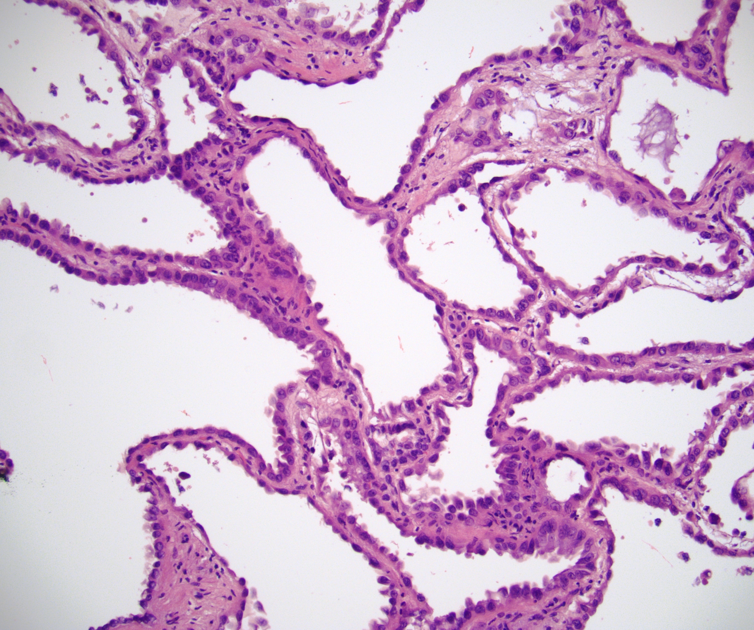
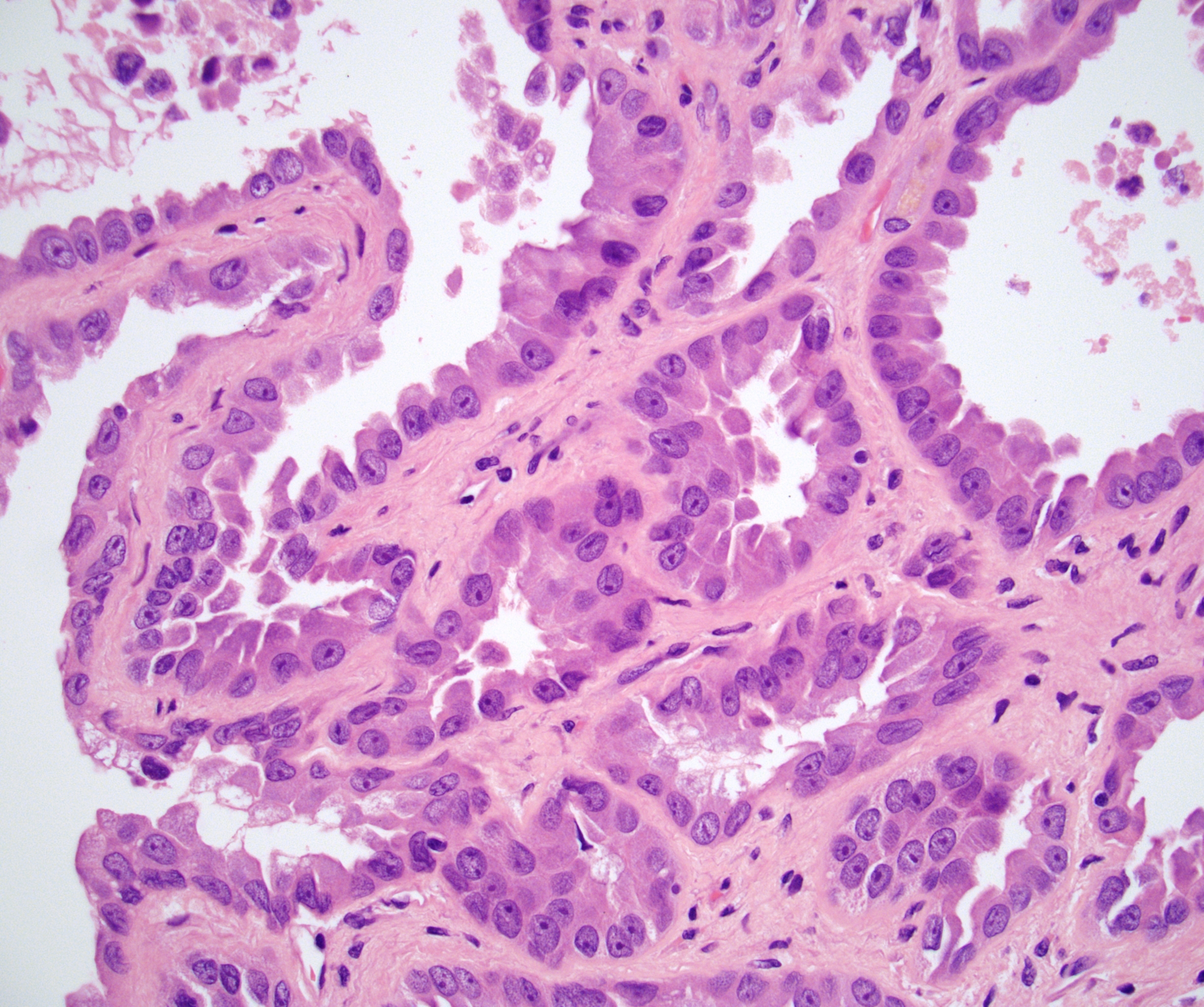
Histopathology images:
What is your diagnosis?
Diagnosis: Tubulocystic renal cell carcinoma
Test question (answer at the end):
What is the most accurate way to grossly describe tubulocystic renal cell carcinoma?
A. Large hemorrhagic tumor of renal medulla
B. Encapsulated multiloculated cystic and solid mass
C. Honeycomb-like yellow tumor
D. Well circumscribed mass with spongy or bubblewrap appearance
Stains:
Discussion:
Tubulocystic renal cell carcinoma was recognized as a distinct entity in the 2016 WHO classification of genitourinary tumors. These tumors are rare, predominantly affect males (M:F >7:1) and have a wide age range at presentation. They generally have indolent behavior (Eur Urol 2016;70:93, J Mol Diagn 2018;20:34).
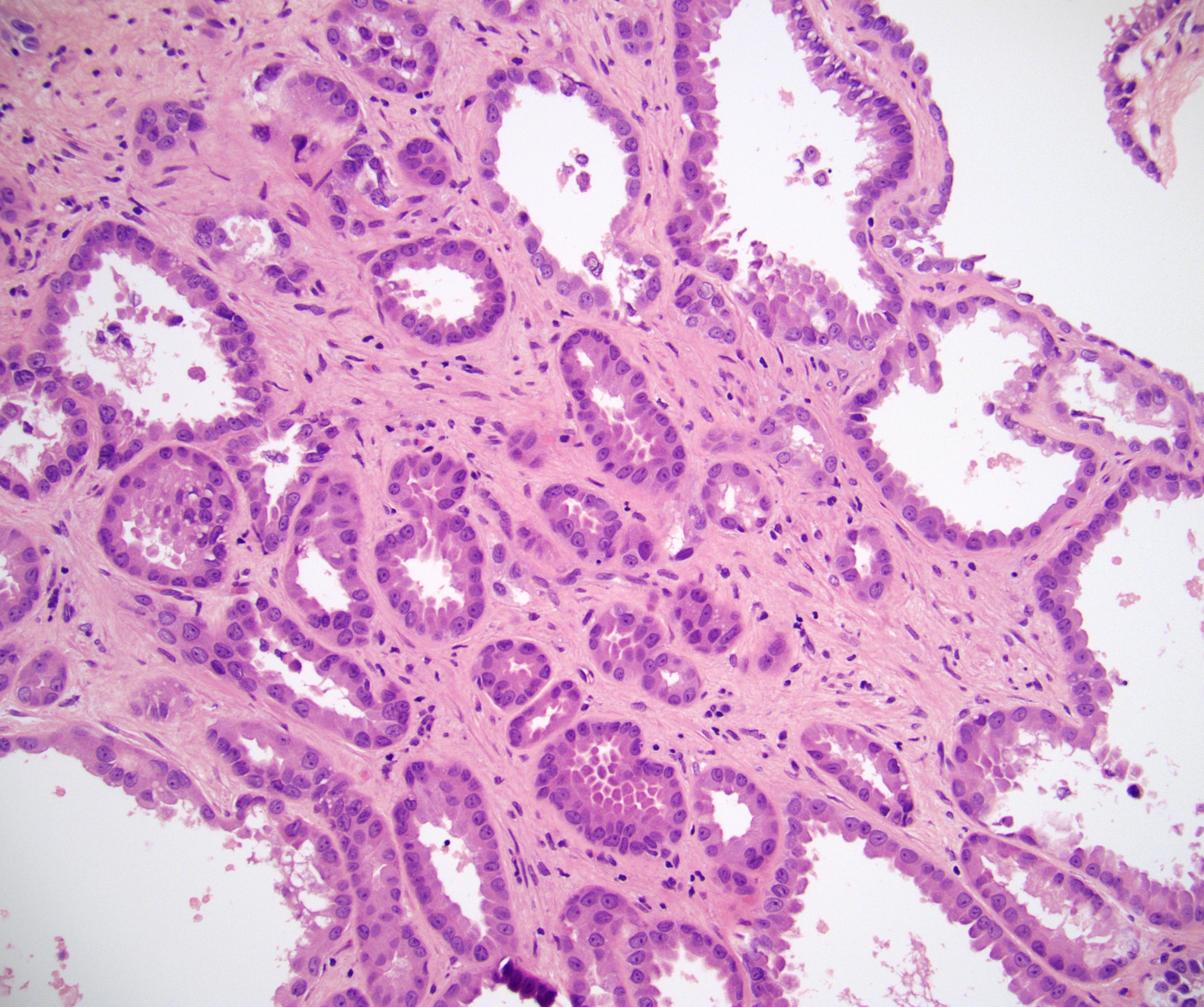
Tubulocystic renal cell carcinoma has a very peculiar gross morphology and microscopic appearance. Macroscopically, these tumors are well circumscribed, spongy and devoid of solid areas. They are often compared to bubble wrap, a sponge or Swiss cheese. Microscopically, they are composed of small to intermediate sized tubules and cysts with scant intervening stroma. Tubules are lined by a single layer of oncocytic hobnail cells with large nuclei and prominent nucleoli (Am J Surg Pathol 2008;32:177, Am J Surg Pathol 2009;33:384).
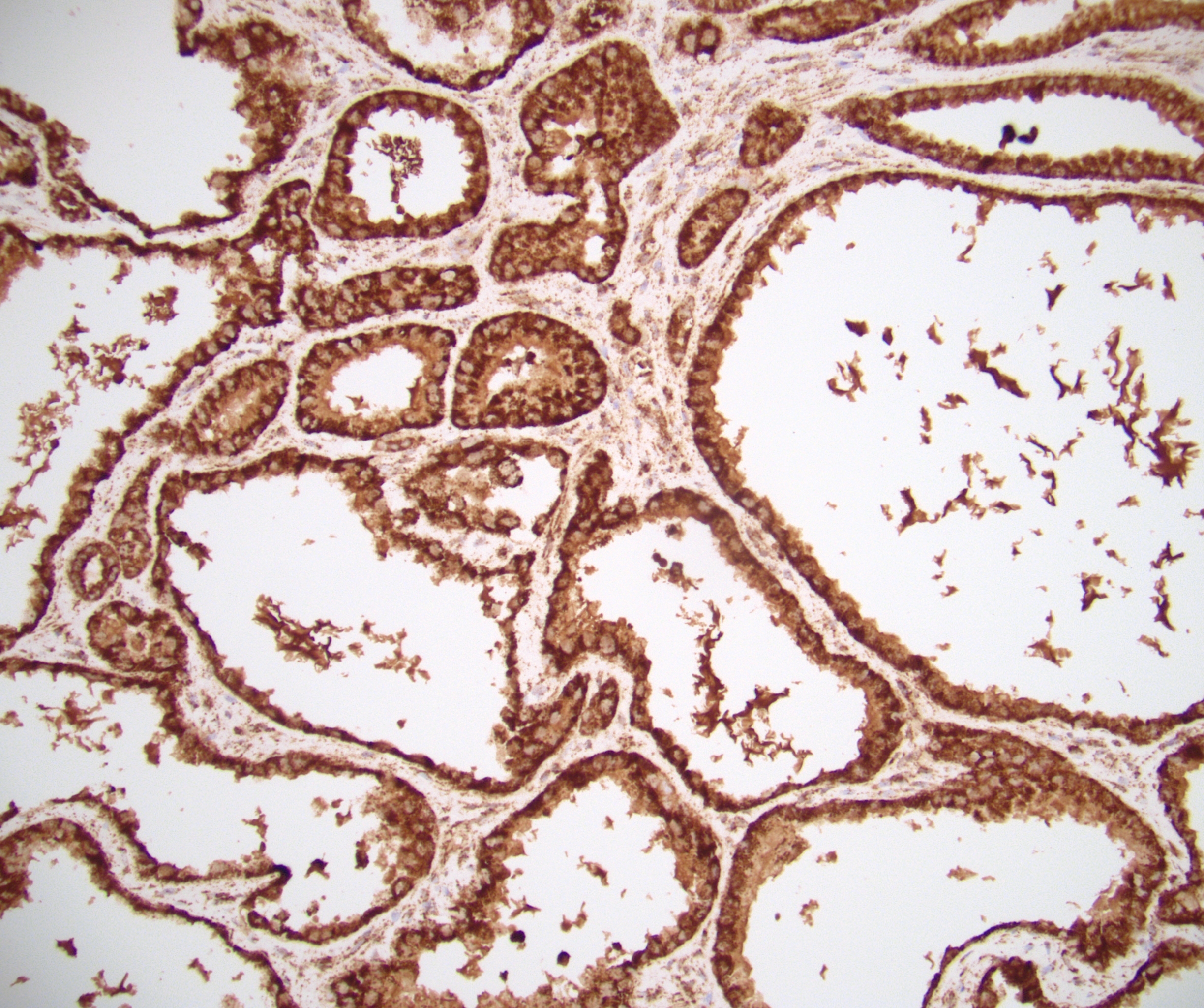
Most cases are diffusely positive for PAX8, CK7, CD10, P504S / AMACR, fumarate hydratase (FH) and focally for high molecular weight cytokeratin. Although this immunoprofile overlaps papillary renal cell carcinoma and collecting duct carcinoma, tubulocystic renal cell carcinoma has a small tumor size, is located in the renal cortex and lacks desmoplastic stroma and papillary architecture.
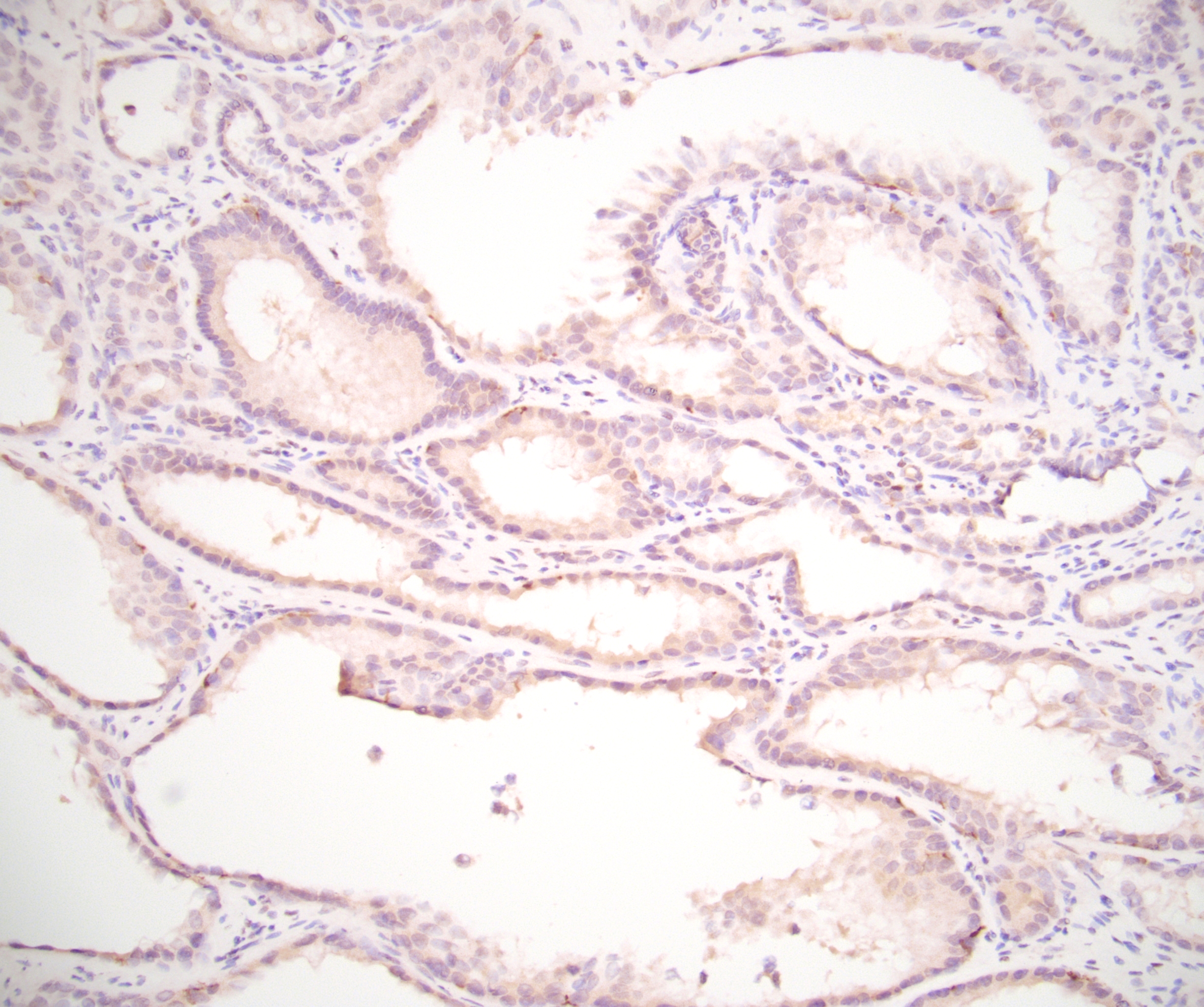
The differential diagnosis also includes adult cystic nephroma and mixed epithelial and stromal tumor. However, both are immunoreactive for ER and PR (unlike this case), typically affect women, contain ovarian-type stroma and usually lack hobnail cells or abundant tubules.
Test question answer:
D. Tubulocystic renal cell carcinoma has a very peculiar gross morphology, often compared to bubble wrap, sponge and Swiss cheese.
All cases are archived on our website. To view them sorted by case number, diagnosis or category, visit our main Case of the Month page. To subscribe or unsubscribe to Case of the Month or our other email lists, click here.
Thanks to Dr. Debra Zynger, The Ohio State University Wexner Medical Center, Columbus, Ohio (USA), for contributing this case and Dr. Maria Tretiakova, University of Washington, Seattle, Washington (USA), for writing the discussion.

Advertisement
Case of the Month #484
Clinical history:
A 34 year old man with a 1.4 cm mass underwent a partial nephrectomy.
Gross image:
Histopathology images:
What is your diagnosis?
Click here for diagnosis, test question and discussion:
Diagnosis: Tubulocystic renal cell carcinoma
Test question (answer at the end):
What is the most accurate way to grossly describe tubulocystic renal cell carcinoma?
A. Large hemorrhagic tumor of renal medulla
B. Encapsulated multiloculated cystic and solid mass
C. Honeycomb-like yellow tumor
D. Well circumscribed mass with spongy or bubblewrap appearance
Stains:
Discussion:
Tubulocystic renal cell carcinoma was recognized as a distinct entity in the 2016 WHO classification of genitourinary tumors. These tumors are rare, predominantly affect males (M:F >7:1) and have a wide age range at presentation. They generally have indolent behavior (Eur Urol 2016;70:93, J Mol Diagn 2018;20:34).
Tubulocystic renal cell carcinoma has a very peculiar gross morphology and microscopic appearance. Macroscopically, these tumors are well circumscribed, spongy and devoid of solid areas. They are often compared to bubble wrap, a sponge or Swiss cheese. Microscopically, they are composed of small to intermediate sized tubules and cysts with scant intervening stroma. Tubules are lined by a single layer of oncocytic hobnail cells with large nuclei and prominent nucleoli (Am J Surg Pathol 2008;32:177, Am J Surg Pathol 2009;33:384).
Most cases are diffusely positive for PAX8, CK7, CD10, P504S / AMACR, fumarate hydratase (FH) and focally for high molecular weight cytokeratin. Although this immunoprofile overlaps papillary renal cell carcinoma and collecting duct carcinoma, tubulocystic renal cell carcinoma has a small tumor size, is located in the renal cortex and lacks desmoplastic stroma and papillary architecture.
The differential diagnosis also includes adult cystic nephroma and mixed epithelial and stromal tumor. However, both are immunoreactive for ER and PR (unlike this case), typically affect women, contain ovarian-type stroma and usually lack hobnail cells or abundant tubules.
Test question answer:
D. Tubulocystic renal cell carcinoma has a very peculiar gross morphology, often compared to bubble wrap, sponge and Swiss cheese.


