11 April 2018 - Case of the Week #453 (revised 15 June 2020)
All cases are archived on our website. To view them sorted by case number, diagnosis or category, visit our main Case of the Week page. To subscribe or unsubscribe to Case of the Week or our other email lists, click here.
Thanks to Dr. Sajna V.M. Kutty, Aster MIMS, Kerala (India) for contributing this case and Dr. Belinda Lategan, St. Boniface Hospital, Winnipeg, Manitoba (Canada) for writing the discussion. This case was reviewed in May 2020 by Dr. Jennifer Bennett, University of Chicago and Dr. Carlos Parra-Herran, University of Toronto.

Advertisement
Website news:
(1) We are changing the name of the Website News email blast to Monthly updates, to avoid confusion with the quarterly What's New newsletter. To subscribe to any of our newsletters, click on the Newsletters button in the Header or Footer.
(2) March saw a record number of sessions (905,045) and page views (2,162,466). Traffic is up 23% from March 2017. We are nearing completion of our topic conversions to our new format and continue to add authors to keep the textbook up to date. Thanks again for making PathologyOutlines the #1 website for pathologists.
(3) Please add CommentsPathout@gmail.com to your SafeList / Address book, so your subscriptions to our emails go directly into your inbox.
Visit and follow our Blog to see recent updates to the website.
Case of the Week #453
Clinical history:
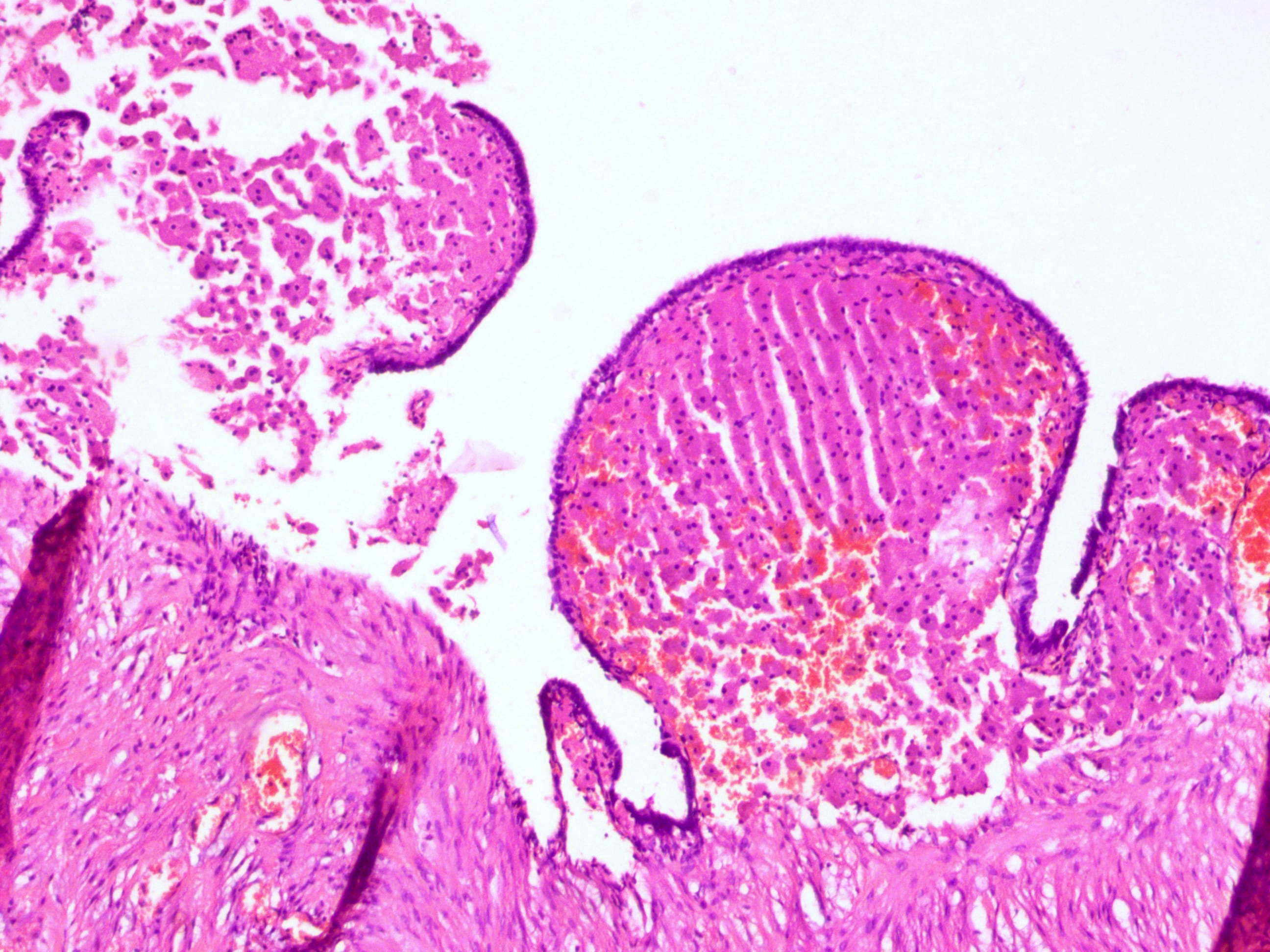
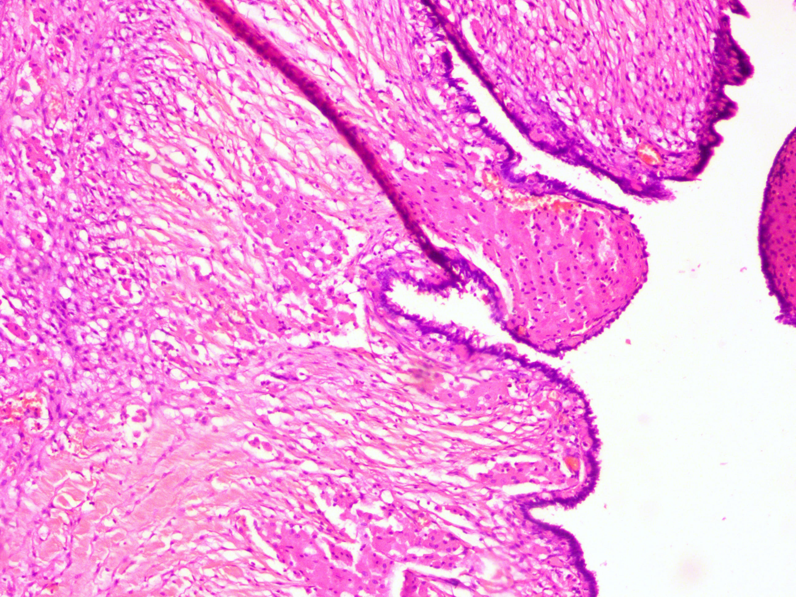
A 43 year old woman with a history of right ovarian cystectomy underwent interval sterilization. Intraoperatively, bilateral hydrosalpinges and dense adhesions were noted. On macroscopy, one fallopian tube also had a thickened wall.
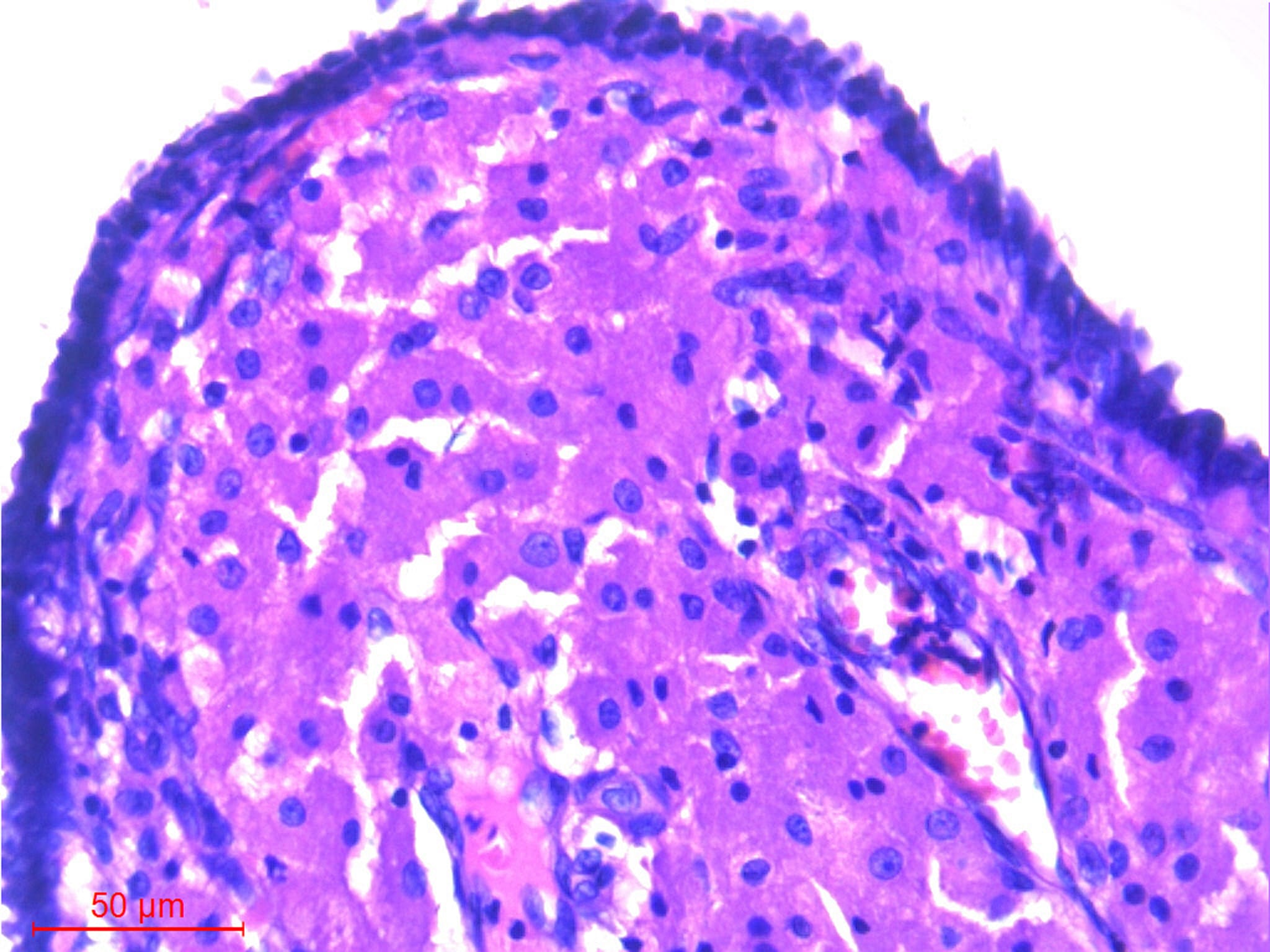
Histopathology images (fallopian tube with thickened wall):
What is your diagnosis?
Diagnosis:
Xanthelasma, fallopian tube
Test question (answer at the end):
Which of the following statement(s) are true in relation to xanthelasma of the fallopian tube?
A. It is associated with chronic endometriosis of the fallopian tube.
B. The accumulation of lipofuscin-laden macrophages and a chronic inflammatory infiltrate is a hallmark of this diagnosis.
C. It is a localized collection of tissue histiocytes containing lipid and lacks a significant associated inflammatory reaction.
D. The macrophages contain bacilli highlighted by acid fast stains (ZN and modified Fite stain).
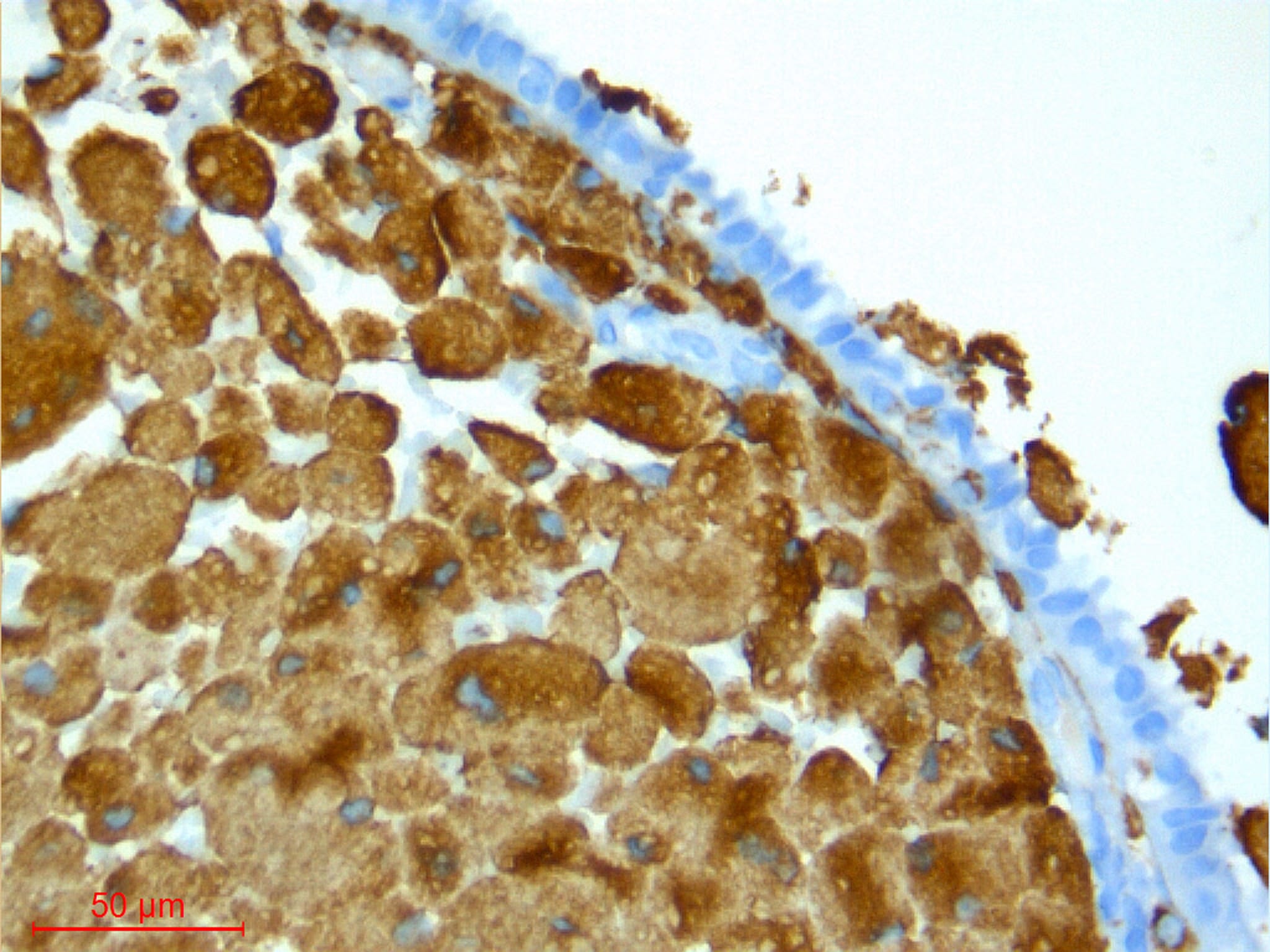
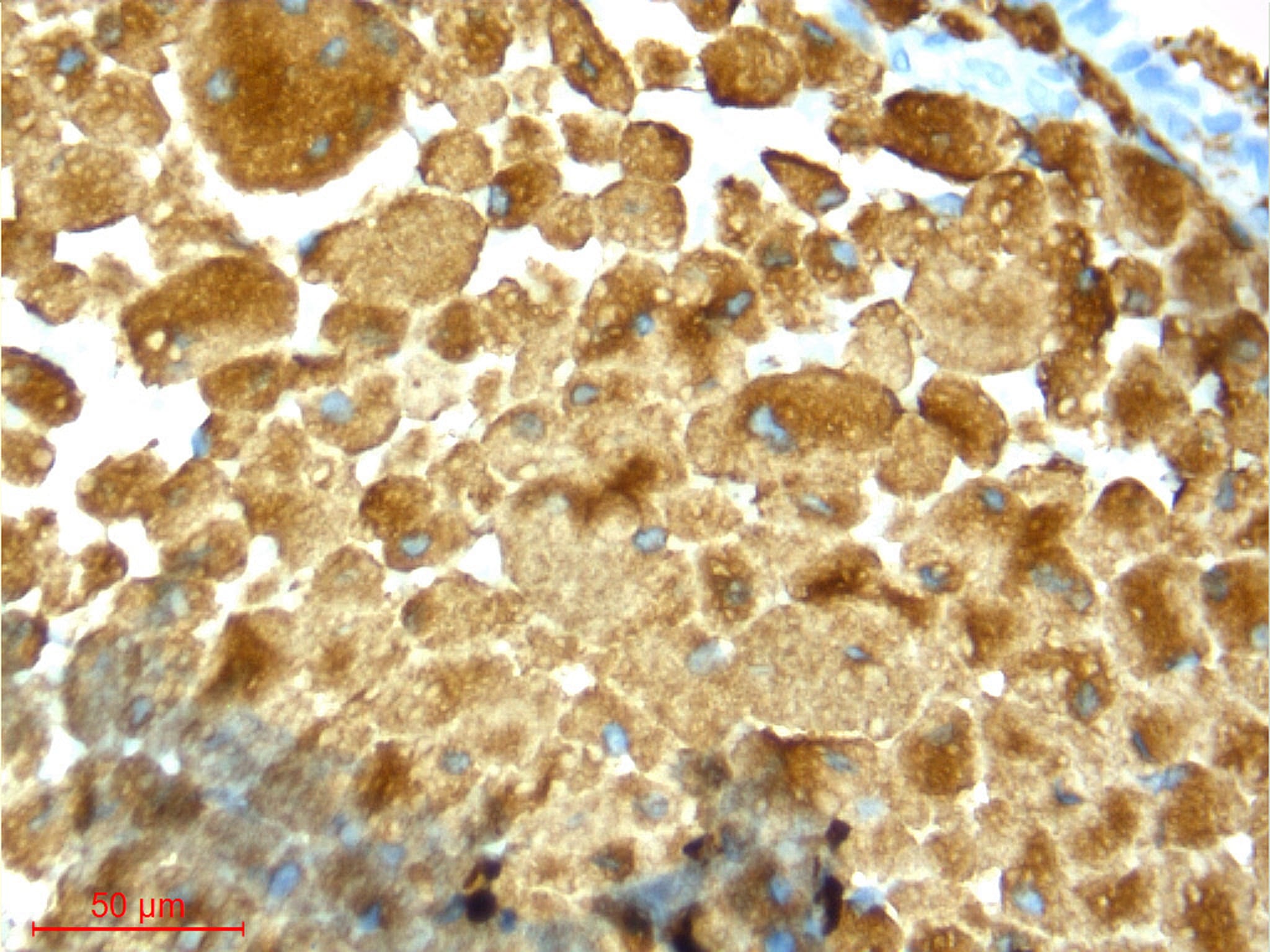
Special stains:
Discussion:
Xanthelasma or xanthoma, defined as localized aggregates of lipid laden macrophages, is described in many organ sites, most commonly the GI tract (gastric xanthoma, colonic xanthomatous polyp) and skin. Although cutaneous xanthelasma is associated with systemic abnormalities of lipid metabolism, especially in younger patients, xanthelasma at other sites is not.
This case illustrates xanthelasma/xanthoma of the fallopian tube, incidentally found at tubal ligation. The thickened wall of one of the fallopian tubes contains aggregates of foamy histiocytes without any associated granulomatous inflammation and no significant neutrophilic or lymphoplasmacytic inflammation. The lipid laden cells are highlighted by CD68 but negative for keratins and inhibin (Arch Pathol Lab Med 2003;127:e417).
The differential diagnosis for xanthelasma/xanthoma of the fallopian tube includes benign inflammatory entities in addition to malignancy. Xanthogranulomatous salpingitis is a destructive process of uncertain etiology in which foamy macrophages and lymphoplasmacytic inflammation accumulate in either or both fallopian tubes. It may present with symptoms of a mass lesion. Xanthogranulomatous inflammation may occur throughout the female genital tract (BMJ Case Rep 2015;2015:bcr2015210642 , J Cancer 2012;3:100 , Arch Pathol Lab Med 2001;125:260). Specific infectious organisms including mycobacteria may present with aggregates of foamy histiocytes. Special stains for organisms and correlation with the clinical picture aids in diagnosis. Malignancy, either primary from the female genital tract or metastatic from other sites, should also be considered. Metastatic lobular carcinoma of the breast (histiocytoid variant), may also present as aggregates of foamy or “histiocytic” cells. The clinical history and use of a keratin stain is helpful in ruling out this diagnosis.
Test Question Answer:
C. It is a localized collection of tissue histiocytes containing lipid and lacks significant associated inflammation.
All cases are archived on our website. To view them sorted by case number, diagnosis or category, visit our main Case of the Week page. To subscribe or unsubscribe to Case of the Week or our other email lists, click here.
Thanks to Dr. Sajna V.M. Kutty, Aster MIMS, Kerala (India) for contributing this case and Dr. Belinda Lategan, St. Boniface Hospital, Winnipeg, Manitoba (Canada) for writing the discussion. This case was reviewed in May 2020 by Dr. Jennifer Bennett, University of Chicago and Dr. Carlos Parra-Herran, University of Toronto.

Advertisement
Website news:
(1) We are changing the name of the Website News email blast to Monthly updates, to avoid confusion with the quarterly What's New newsletter. To subscribe to any of our newsletters, click on the Newsletters button in the Header or Footer.
(2) March saw a record number of sessions (905,045) and page views (2,162,466). Traffic is up 23% from March 2017. We are nearing completion of our topic conversions to our new format and continue to add authors to keep the textbook up to date. Thanks again for making PathologyOutlines the #1 website for pathologists.
(3) Please add CommentsPathout@gmail.com to your SafeList / Address book, so your subscriptions to our emails go directly into your inbox.
Visit and follow our Blog to see recent updates to the website.
Case of the Week #453
Clinical history:
A 43 year old woman with a history of right ovarian cystectomy underwent interval sterilization. Intraoperatively, bilateral hydrosalpinges and dense adhesions were noted. On macroscopy, one fallopian tube also had a thickened wall.
Histopathology images (fallopian tube with thickened wall):
What is your diagnosis?
Diagnosis:
Xanthelasma, fallopian tube
Test question (answer at the end):
Which of the following statement(s) are true in relation to xanthelasma of the fallopian tube?
A. It is associated with chronic endometriosis of the fallopian tube.
B. The accumulation of lipofuscin-laden macrophages and a chronic inflammatory infiltrate is a hallmark of this diagnosis.
C. It is a localized collection of tissue histiocytes containing lipid and lacks a significant associated inflammatory reaction.
D. The macrophages contain bacilli highlighted by acid fast stains (ZN and modified Fite stain).
Special stains:
Discussion:
Xanthelasma or xanthoma, defined as localized aggregates of lipid laden macrophages, is described in many organ sites, most commonly the GI tract (gastric xanthoma, colonic xanthomatous polyp) and skin. Although cutaneous xanthelasma is associated with systemic abnormalities of lipid metabolism, especially in younger patients, xanthelasma at other sites is not.
This case illustrates xanthelasma/xanthoma of the fallopian tube, incidentally found at tubal ligation. The thickened wall of one of the fallopian tubes contains aggregates of foamy histiocytes without any associated granulomatous inflammation and no significant neutrophilic or lymphoplasmacytic inflammation. The lipid laden cells are highlighted by CD68 but negative for keratins and inhibin (Arch Pathol Lab Med 2003;127:e417).
The differential diagnosis for xanthelasma/xanthoma of the fallopian tube includes benign inflammatory entities in addition to malignancy. Xanthogranulomatous salpingitis is a destructive process of uncertain etiology in which foamy macrophages and lymphoplasmacytic inflammation accumulate in either or both fallopian tubes. It may present with symptoms of a mass lesion. Xanthogranulomatous inflammation may occur throughout the female genital tract (BMJ Case Rep 2015;2015:bcr2015210642 , J Cancer 2012;3:100 , Arch Pathol Lab Med 2001;125:260). Specific infectious organisms including mycobacteria may present with aggregates of foamy histiocytes. Special stains for organisms and correlation with the clinical picture aids in diagnosis. Malignancy, either primary from the female genital tract or metastatic from other sites, should also be considered. Metastatic lobular carcinoma of the breast (histiocytoid variant), may also present as aggregates of foamy or “histiocytic” cells. The clinical history and use of a keratin stain is helpful in ruling out this diagnosis.
Test Question Answer:
C. It is a localized collection of tissue histiocytes containing lipid and lacks significant associated inflammation.


