10 June 2015 - Case #354
All cases are archived on our website. To view them sorted by case number, diagnosis or category, visit our main Case of the Month page. To subscribe or unsubscribe to Case of the Month or our other email lists, click here.
Thanks to Dr. Rajib Gupta, University of Tennessee Health Science Center and Dr. Jesse Jenkins, St. Jude Children's Research Hospital, Tennessee (USA), for contributing this case.
Advertisement
Case #354
Clinical history:
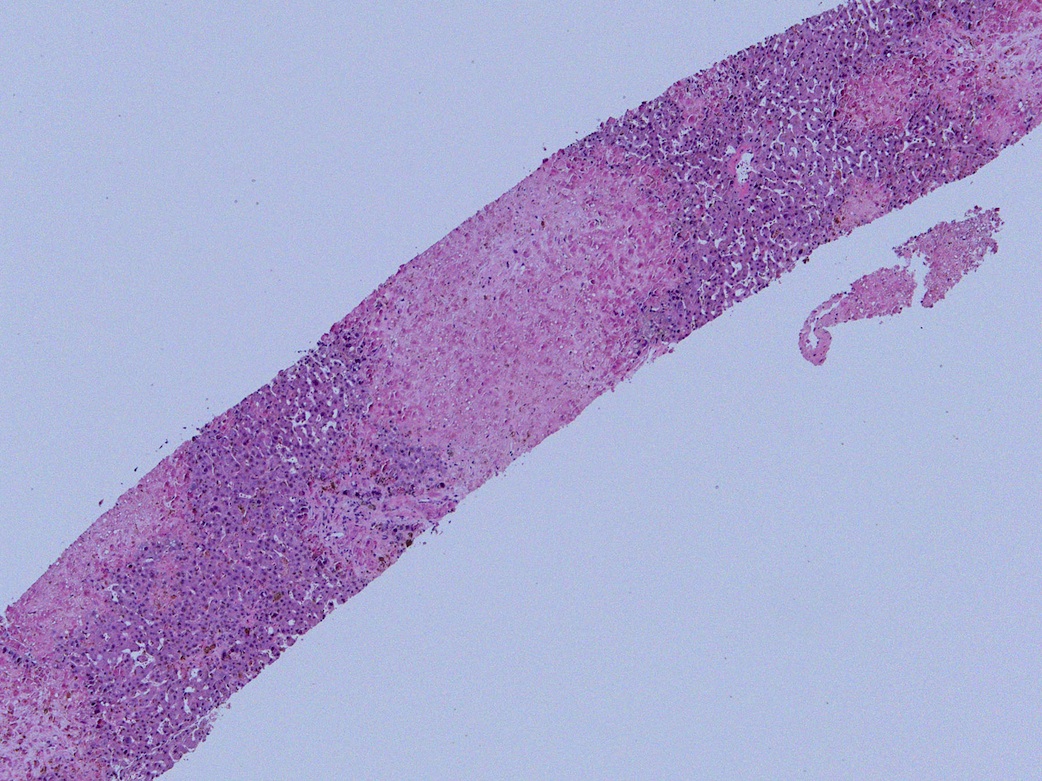
A 20 month old girl, post-stem cell transplant for MLL rearranged acute myeloid leukemia (AML), presented with features of AML relapse, including bone marrow blasts, multifocal liver lesions and worsening kidney function. CT guided liver biopsy was performed.
Microscopic images:
What is your diagnosis?
Diagnosis: Adenovirus hepatitis with necrosis, post-stem cell transplant
Immunostains:
Discussion:
The adenovirus immunostains above confirm the diagnosis.
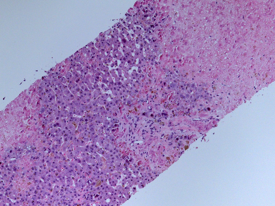
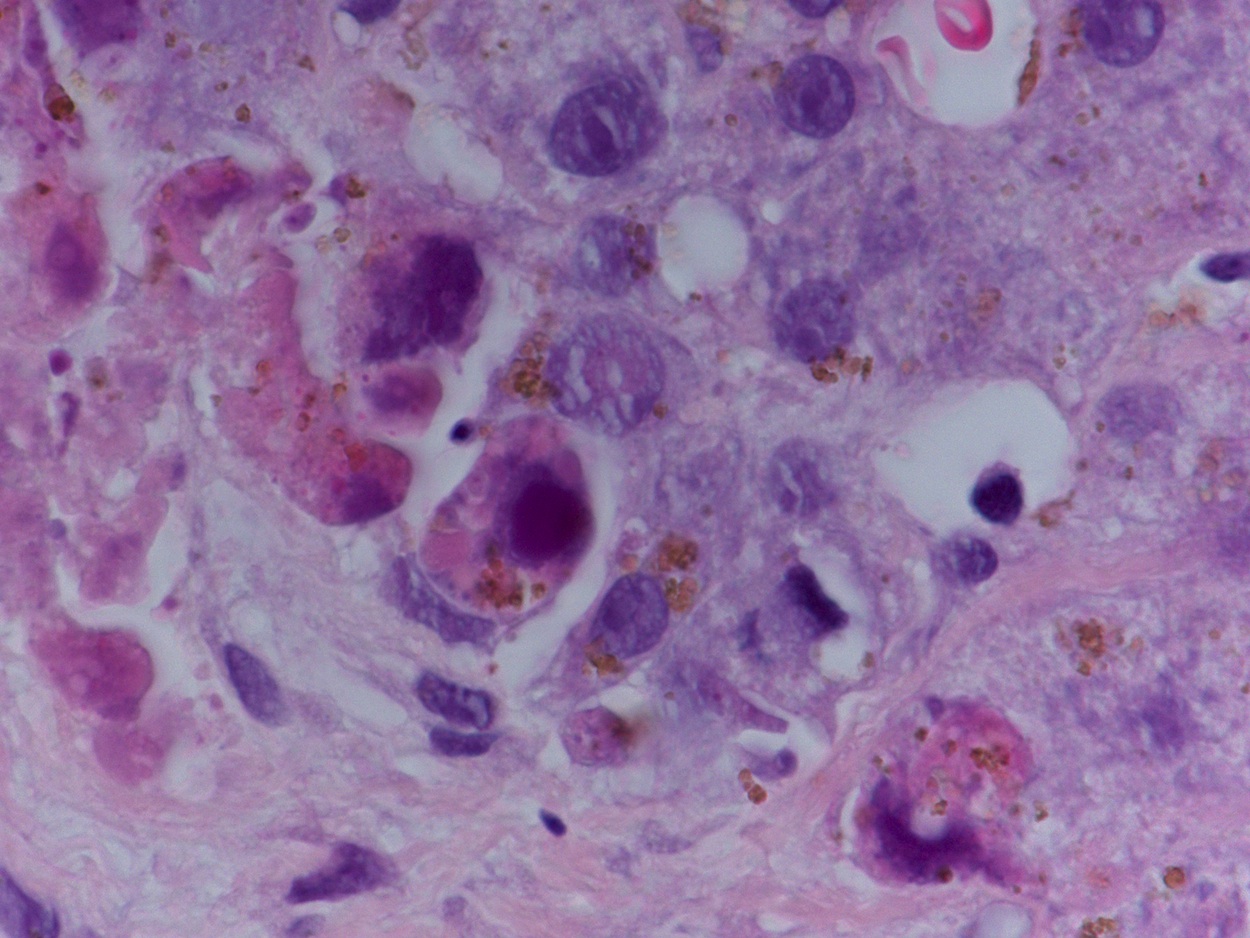
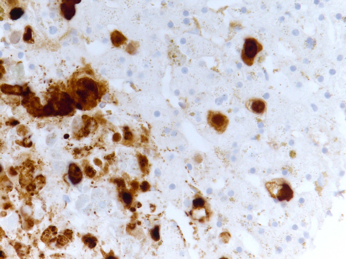
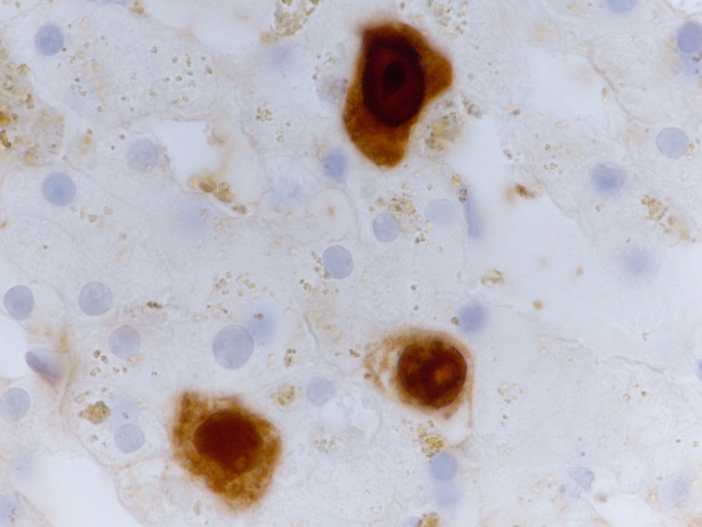
Figure 1 is a panoramic view of the liver biopsy core, showing remnant hepatic tissue on either side of a large necrotic focus. Figure 2 shows a low power view of a few hepatocytes with dark purple-red nuclear inclusion bodies. Figure 3 shows a high power view of hepatocytes showing characteristic nuclear and cytoplasmic viral inclusion bodies. Figure 4 shows a high power view of nuclear and cytoplasmic positivity of adenovirus immunohistochemistry in the hepatocytes. The positive nuclear immunostain on the right side of the image corresponds to hepatocytes in the viable area while the immunostain on the left corresponds to dead hepatocytes within the necrotic area. Figure 5 is an oil immersion view of adenovirus immunostain in 3 affected hepatocytes.
Adenovirus is a major cause of non-relapse morbidity and mortality after allogeneic hematopoietic stem cell transplantation for hematological malignancies and may be associated with fulminant hepatic failure (Intern Med 2006;45:975, Case Rep Infect Dis 2012;2012:463569). The histology described in this case is classic.
Treatment with anti-virals, including cidofovir, may be effective (Ann Hepatol 2014;13:827, Transplant Proc 2013;45:293, J Pediatr Hematol Oncol 2012;34:e298, Medscape: Cidofovir [Accessed 10 October 2023]).
All cases are archived on our website. To view them sorted by case number, diagnosis or category, visit our main Case of the Month page. To subscribe or unsubscribe to Case of the Month or our other email lists, click here.
Thanks to Dr. Rajib Gupta, University of Tennessee Health Science Center and Dr. Jesse Jenkins, St. Jude Children's Research Hospital, Tennessee (USA), for contributing this case.
- Silverberg's Principles and Practice of Surgical Pathology and Cytopathology (2015) by Mark R. Wick, Virginia A. LiVolsi, John D. Pfeifer, Edward B. Stelow and Paul E. Wakely Jr. Silverberg's Principles and Practice of Surgical Pathology and Cytopathology is one of the most durable reference texts in pathology. Thoroughly revised and updated, this state-of-the-art new edition encompasses the entire fields of surgical pathology and cytopathology in a single source. Its practice-oriented format uniquely integrates these disciplines to present all the relevant features of a particular lesion, side by side. Over 3500 color images depict clinical features, morphological attributes, histochemical and immunohistochemical findings and molecular characteristics of all lesions included. This edition features new highly experienced and academically accomplished editors, while chapters are written by the leading experts in the field (several new to this edition, bringing a fresh approach).
For more information, visit our New Books page.
Website news:
(1) We have posted four new lectures from ARUP Laboratories on our CME / Apps / Board Review for Pathology or Laboratory Medicine page.
(2) We have added 3 new articles to our Management of Pathology Practices chapter in May, written by Mick Raich, President, Vachette Pathology.
(3) We are thinking of creating a new Pathology books app for Smart Phones / Tablets, but need your feedback. The app would list new books recently added to the website, the most popular books year to date and possibly other pathology related book information. We anticipate it would be updated monthly. Is this is something that you would use to purchase books? Let Dr. Pernick know (helpful or not helpful or other comments) at NatPernick@gmail.com.
Visit and follow our Blog to see recent updates to the website.
Case #354
Clinical history:
A 20 month old girl, post-stem cell transplant for MLL rearranged acute myeloid leukemia (AML), presented with features of AML relapse, including bone marrow blasts, multifocal liver lesions and worsening kidney function. CT guided liver biopsy was performed.
Microscopic images:
What is your diagnosis?
Click here for diagnosis and discussion:
Diagnosis: Adenovirus hepatitis with necrosis, post-stem cell transplant
Immunostains:
Discussion:
The adenovirus immunostains above confirm the diagnosis.
Figure 1 is a panoramic view of the liver biopsy core, showing remnant hepatic tissue on either side of a large necrotic focus. Figure 2 shows a low power view of a few hepatocytes with dark purple-red nuclear inclusion bodies. Figure 3 shows a high power view of hepatocytes showing characteristic nuclear and cytoplasmic viral inclusion bodies. Figure 4 shows a high power view of nuclear and cytoplasmic positivity of adenovirus immunohistochemistry in the hepatocytes. The positive nuclear immunostain on the right side of the image corresponds to hepatocytes in the viable area while the immunostain on the left corresponds to dead hepatocytes within the necrotic area. Figure 5 is an oil immersion view of adenovirus immunostain in 3 affected hepatocytes.
Adenovirus is a major cause of non-relapse morbidity and mortality after allogeneic hematopoietic stem cell transplantation for hematological malignancies and may be associated with fulminant hepatic failure (Intern Med 2006;45:975, Case Rep Infect Dis 2012;2012:463569). The histology described in this case is classic.
Treatment with anti-virals, including cidofovir, may be effective (Ann Hepatol 2014;13:827, Transplant Proc 2013;45:293, J Pediatr Hematol Oncol 2012;34:e298, Medscape: Cidofovir [Accessed 10 October 2023]).


