3 September 2014 - Case #325
All cases are archived on our website. To view them sorted by case number, diagnosis or category, visit our main Case of the Month page. To subscribe or unsubscribe to Case of the Month or our other email lists, click here.
Thanks to Dr. Nasir Uddin, Aga Khan University Hospital (Pakistan), for contributing this case.
Advertisement
Case #325
Clinical history:
A 66 year old woman had chronic headaches. CT scan showed an isodense area and calcification in the left frontoparietal region of the brain. A biopsy was performed.
Microscopic images:
What is your diagnosis?
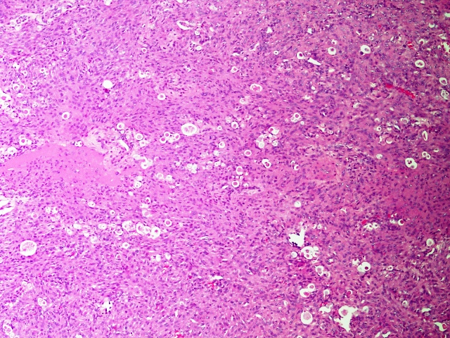
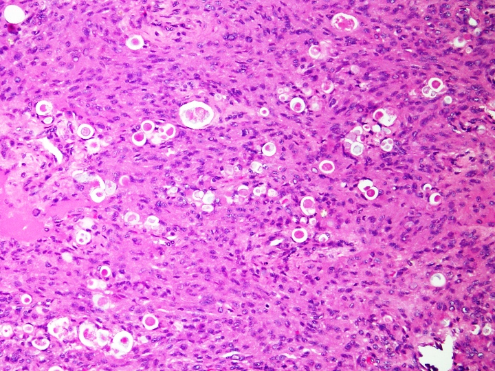
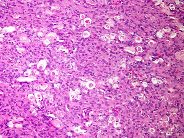
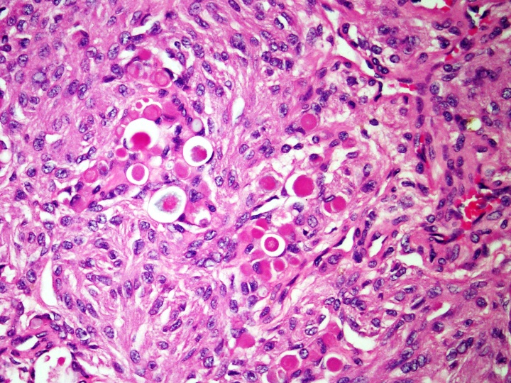
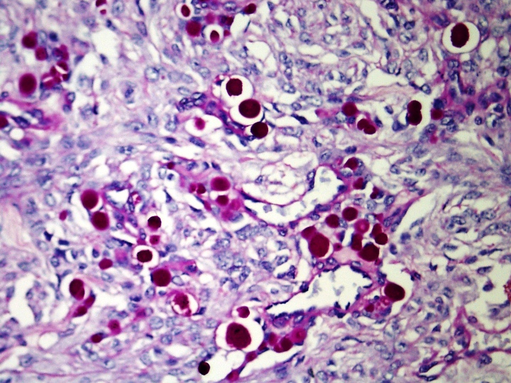
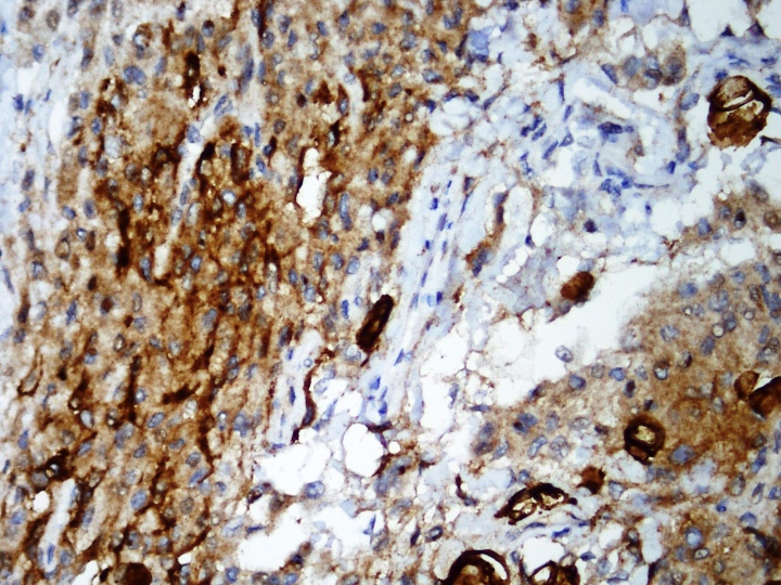
Diagnosis: Secretory meningioma, WHO grade I
Special stain and immunostain images:
Discussion:
Secretory meningioma is a rare, WHO grade I tumor with benign behavior, representing 1 - 3% of all meningiomas (Int J Clin Exp Pathol 2013;6:358). Clinically, it is more common in women and may have an elevated serum CEA. On imaging, its preferred location is at the cranial base. Whole exome sequencing analysis of DNA indicates that these tumors are defined by combined KLF4 K409Q and TRAF7 mutations (Acta Neuropathol 2013;125:351).
Histology shows cells with classic meningioma histology, as well as foci with intracellular lumina with eosinophilic, granular or hyalinized secretions referred to as pseudopsammoma bodies. These secretions are PASD+ and CEA+. Abundant mast cells may also be present (Neurosurg Rev 2006;29:41).
Differential diagnosis includes:
Prognosis is related to completeness of surgical excision and surgical risk factors. Postoperatively, these tumors are frequently associated with severe peritumoral edema (Neuro Oncol 2009;11:819). Residual tumor grows slowly and reacts well to gamma knife therapy.
All cases are archived on our website. To view them sorted by case number, diagnosis or category, visit our main Case of the Month page. To subscribe or unsubscribe to Case of the Month or our other email lists, click here.
Thanks to Dr. Nasir Uddin, Aga Khan University Hospital (Pakistan), for contributing this case.

Advertisement
Website news:
(1) Don't forget to check out our Promotions page / email to see the special products and services offered by our advertisers!
(2) We are now adding thumbnails and links of new books to the chapter pages each month, to help visitors learn about new offers. Remember, if you click on any of these links and then buy that book or anything else from Amazon.com, you are helping to support our website.
(3) We hope you will stop by our booth this year at CAP '14 in Chicago! We are booth #623. Please let us know how we can make the website more useful to you. We can also take your picture for our Facebook page.
Visit and follow our Blog to see recent updates to the website.
Case #325
Clinical history:
A 66 year old woman had chronic headaches. CT scan showed an isodense area and calcification in the left frontoparietal region of the brain. A biopsy was performed.
Microscopic images:
What is your diagnosis?
Click here for diagnosis and discussion:
Diagnosis: Secretory meningioma, WHO grade I
Special stain and immunostain images:
Discussion:
Secretory meningioma is a rare, WHO grade I tumor with benign behavior, representing 1 - 3% of all meningiomas (Int J Clin Exp Pathol 2013;6:358). Clinically, it is more common in women and may have an elevated serum CEA. On imaging, its preferred location is at the cranial base. Whole exome sequencing analysis of DNA indicates that these tumors are defined by combined KLF4 K409Q and TRAF7 mutations (Acta Neuropathol 2013;125:351).
Histology shows cells with classic meningioma histology, as well as foci with intracellular lumina with eosinophilic, granular or hyalinized secretions referred to as pseudopsammoma bodies. These secretions are PASD+ and CEA+. Abundant mast cells may also be present (Neurosurg Rev 2006;29:41).
Differential diagnosis includes:
- Metastatic carcinoma: usually CK7-, CK20+ (Adv Clin Path 1999;3:47)
- Microcystic, clear cell or chordoid meningioma
Prognosis is related to completeness of surgical excision and surgical risk factors. Postoperatively, these tumors are frequently associated with severe peritumoral edema (Neuro Oncol 2009;11:819). Residual tumor grows slowly and reacts well to gamma knife therapy.

