15 June 2012 - Case #245
All cases are archived on our website. To view them sorted by case number, diagnosis or category, visit our main Case of the Month page. To subscribe or unsubscribe to Case of the Month or our other email lists, click here.
Thanks to Dr. Sean Williamson, Indiana University School of Medicine, for contributing this case.
Advertisement
Case #245
Clinical history:
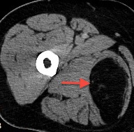
A 39 year old man had surgery for an intracranial tumor. PET / CT imaging incidentally revealed an 8.9 cm fat density soft tissue lesion within the right adductor magnus muscle (Wikipedia: Adductor Magnus Muscle [Accessed 15 April 2024]). PET demonstrated evidence of increased metabolism within the mass (SUV 3.1), raising a differential diagnosis of benign intramuscular lipoma and liposarcoma (Wikipedia: Standardized Uptake Value [Accessed 15 April 2024]).
Radiology image:
Gross image:
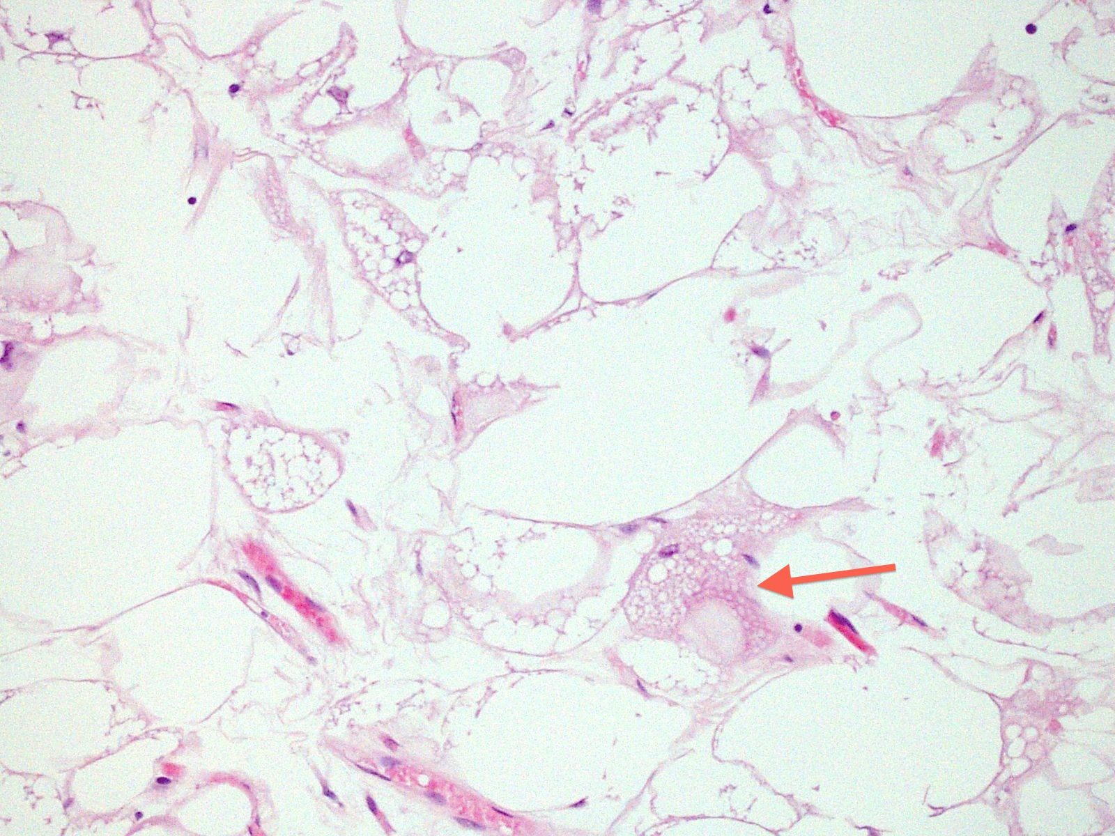
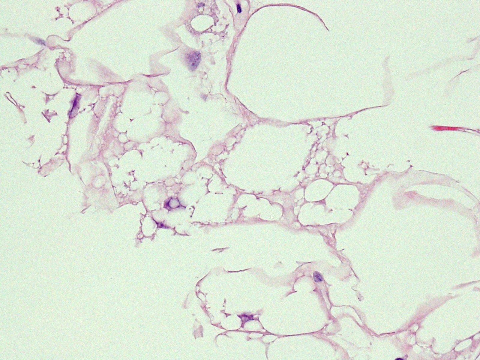
Microscopic images:
What is your diagnosis?
Diagnosis: Hibernoma
Discussion:
Hibernoma is a lipoma subtype containing prominent brown adipocytes that resembles normal brown fat, as a classic lipoma resembles white fat. It is rare, representing 2% of lipomas; 60% occur in males, and the mean age is 26 - 38 years. Hibernomas occur most commonly in the thigh (as in this case), shoulder, back, neck, axilla and mediastinum (Am J Surg Pathol 2001;25:809). Thigh lesions may mimic liposarcoma by CT / MRI (Med Sci Monit 2009;15:CS117).
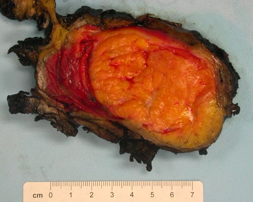
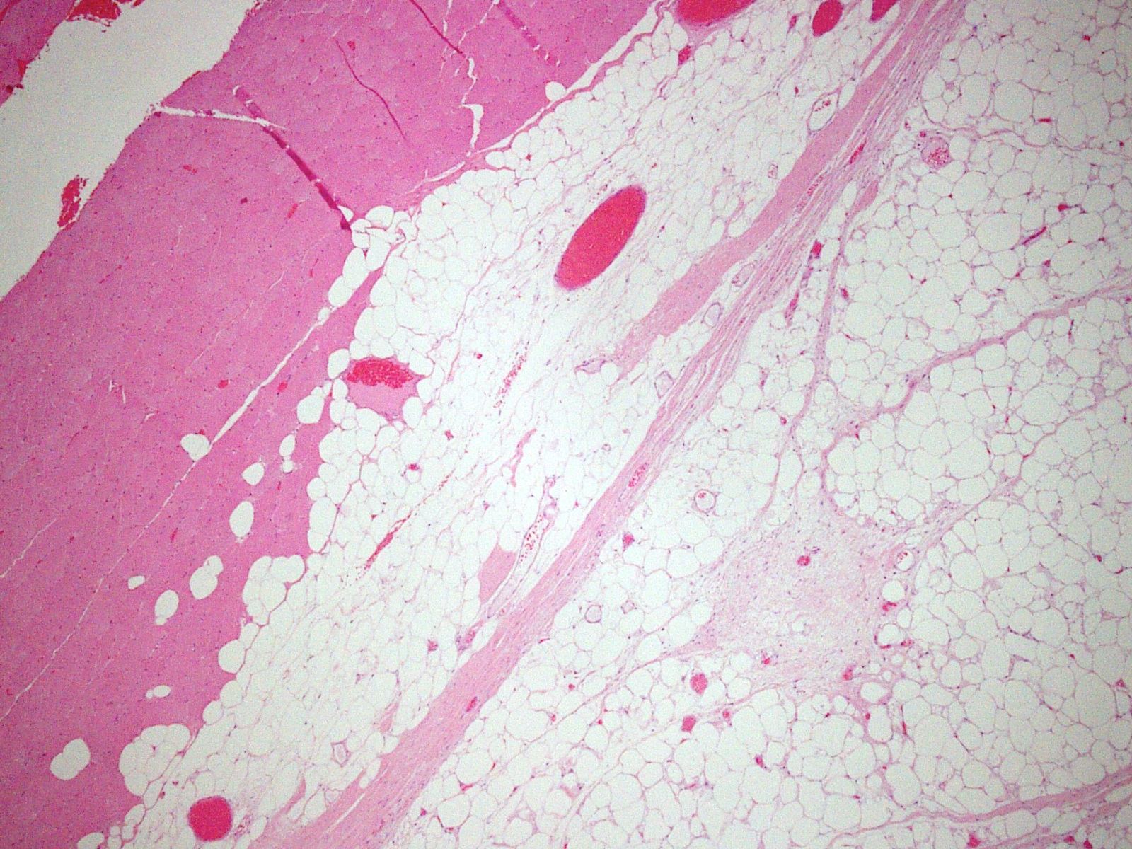
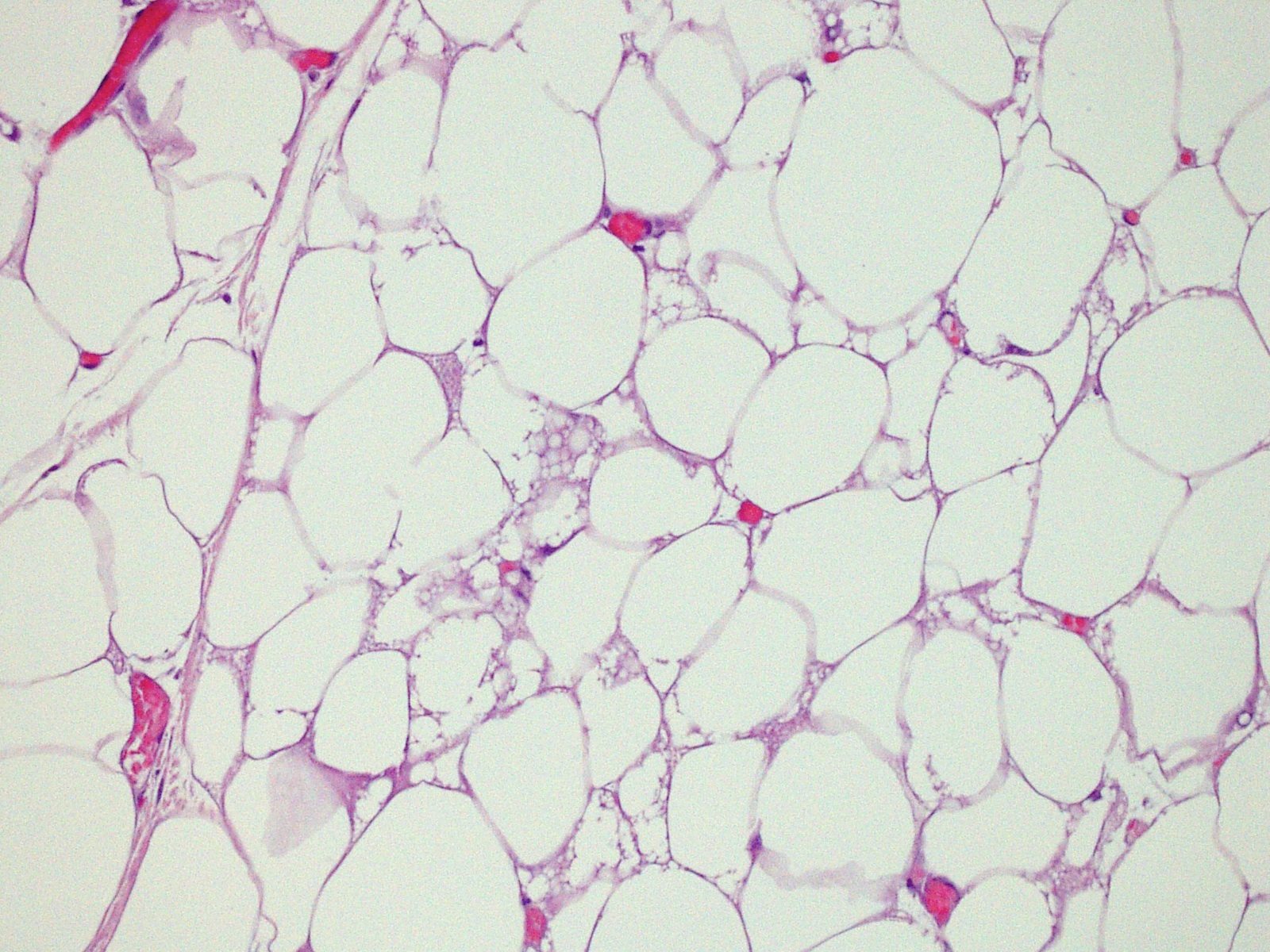
Grossly, hibernomas have a red/brown cut surface resembling the brown fat in some hibernating animals. The brown color is due to vasculature or mitochondria. These tumors are soft, lobulated, well delineated or encapsulated. In 10% of cases, they infiltrate adjacent striated muscle. Histologically, there is an organoid arrangement of uniform large cells resembling brown fat with coarsely granular to multivacuolated cytoplasm that is eosinophilic or pale. Vacuoles are small and stain for neutral fat. The nuclei are small and central, with no / rare atypia. They often contain mixtures of white fat, as well as loose basophilic matrix. Subtypes include lipoma-like, myxoid and spindle cell.
The differential diagnosis includes atypical lipomatous tumor, lipoma and normal brown fat. Atypical lipomatous tumor / well differentiated liposarcoma is a deep tumor with atypical cells that are positive for MDM2 and CDK4 by FISH or real time PCR. In classic lipoma, the lipocytes are not multivacuolated. Children may have residual brown fat around cervical or axillary lymph nodes but it does not form a distinct mass.
In this case, the tumor was predominantly lipoma-like, with only rare multivacuolated cells and rare cells with a more eosinophilic appearance. No large hyperchromatic cells specific for atypical lipomatous tumor / well differentiated liposarcoma were identified.
Complete surgical excision is associated with an excellent long term clinical outcome.
All cases are archived on our website. To view them sorted by case number, diagnosis or category, visit our main Case of the Month page. To subscribe or unsubscribe to Case of the Month or our other email lists, click here.
Thanks to Dr. Sean Williamson, Indiana University School of Medicine, for contributing this case.
Best Practices for Anatomic Pathology Quality
Pathology Leadership: Free 45 minute webinar
Wednesday June 27th at 12:00pm Eastern
Register (link invalid)
Sponsored by: AccuPathology - Pathology Quality Management Software
● Integrate with your LIS to automate tasks, identifying potential QA to be performed
● Identify pre-analytic and post-analytic variables that contribute to diagnostic errors
● Automate mandated OPPE, FPPE processes with quantitative data
● Dashboards and pro-forma reports for improved operations, CAP and The Joint Commission audits
Spend less time, getting better information to make REAL changes while always being compliant.
Website
Phone: (267) 564-5015
Website news:
(1) The Ovary-nontumor chapter has now been updated, based on reviews by Mohiedean Ghofrani, M.D. and Shahidul Islam, M.D.
(2) Our Feature Page for the month highlights Diagnostic testing / reagents, and includes Advanced Cell Diagnostics, Inc. (ACD), bioTheranostics, Covance, Epitomics, Horizon Diagnostics, Leica Microsystems and Ventana Medical.
(3) The Colon tumor chapter has now been updated, based on reviews by Jela Bandovic, M.D., Shilpa Jain, M.D. and Charanjeet Singh, M.D.
Visit and follow our Blog to see recent updates to the website.
Case #245
Clinical history:
A 39 year old man had surgery for an intracranial tumor. PET / CT imaging incidentally revealed an 8.9 cm fat density soft tissue lesion within the right adductor magnus muscle (Wikipedia: Adductor Magnus Muscle [Accessed 15 April 2024]). PET demonstrated evidence of increased metabolism within the mass (SUV 3.1), raising a differential diagnosis of benign intramuscular lipoma and liposarcoma (Wikipedia: Standardized Uptake Value [Accessed 15 April 2024]).
Radiology image:
Gross image:
Microscopic images:
What is your diagnosis?
Click here for diagnosis and discussion:
Diagnosis: Hibernoma
Discussion:
Hibernoma is a lipoma subtype containing prominent brown adipocytes that resembles normal brown fat, as a classic lipoma resembles white fat. It is rare, representing 2% of lipomas; 60% occur in males, and the mean age is 26 - 38 years. Hibernomas occur most commonly in the thigh (as in this case), shoulder, back, neck, axilla and mediastinum (Am J Surg Pathol 2001;25:809). Thigh lesions may mimic liposarcoma by CT / MRI (Med Sci Monit 2009;15:CS117).
Grossly, hibernomas have a red/brown cut surface resembling the brown fat in some hibernating animals. The brown color is due to vasculature or mitochondria. These tumors are soft, lobulated, well delineated or encapsulated. In 10% of cases, they infiltrate adjacent striated muscle. Histologically, there is an organoid arrangement of uniform large cells resembling brown fat with coarsely granular to multivacuolated cytoplasm that is eosinophilic or pale. Vacuoles are small and stain for neutral fat. The nuclei are small and central, with no / rare atypia. They often contain mixtures of white fat, as well as loose basophilic matrix. Subtypes include lipoma-like, myxoid and spindle cell.
The differential diagnosis includes atypical lipomatous tumor, lipoma and normal brown fat. Atypical lipomatous tumor / well differentiated liposarcoma is a deep tumor with atypical cells that are positive for MDM2 and CDK4 by FISH or real time PCR. In classic lipoma, the lipocytes are not multivacuolated. Children may have residual brown fat around cervical or axillary lymph nodes but it does not form a distinct mass.
In this case, the tumor was predominantly lipoma-like, with only rare multivacuolated cells and rare cells with a more eosinophilic appearance. No large hyperchromatic cells specific for atypical lipomatous tumor / well differentiated liposarcoma were identified.
Complete surgical excision is associated with an excellent long term clinical outcome.


