12 October 2011 - Case #220
All cases are archived on our website. To view them sorted by case number, diagnosis or category, visit our main Case of the Month page. To subscribe or unsubscribe to Case of the Month or our other email lists, click here.
This case was contributed by Dr. Eddie Fridman, The Chaim Sheba Medical Center, Israel.
CancerTYPE ID is a standardized, objective molecular test based on the differential expression of 92 genes, that classifies tumors by matching the gene expression pattern of a patients tumor tissue to a database of known tumor types and histological subtypes.
CancerTYPE IDs database includes 2,206 tumors from multiple tumor banks, selected to provide broader and deeper representation of the heterogeneity of tumors. The 92-gene assay does not overlap with IHC markers, providing complementary data to standard tumor diagnosis.
CancerTYPE ID uses real-time reverse transcription polymerase chain reaction (RT-PCR). A very low copy number of RNA molecules can be detected, thus reducing the sample tissue required for testing. Testing is conducted and results are generated at bioTheranostics' CAP-accredited, CLIA-certified laboratory.
Advertisement
Case #220
Clinical history:
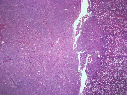
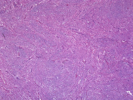
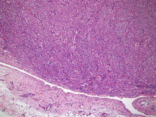
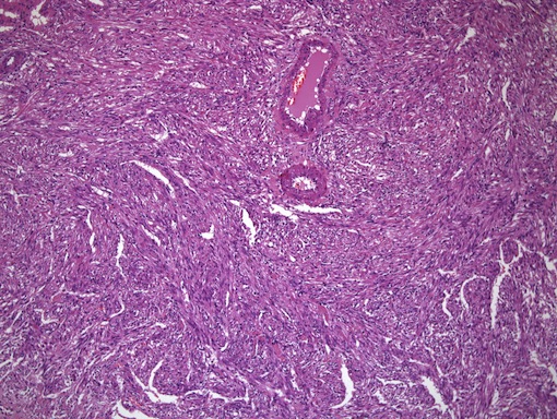
A 37 year old woman had a right renal mass, and underwent a partial nephrectomy.
Microscopic images:
What is your diagnosis?
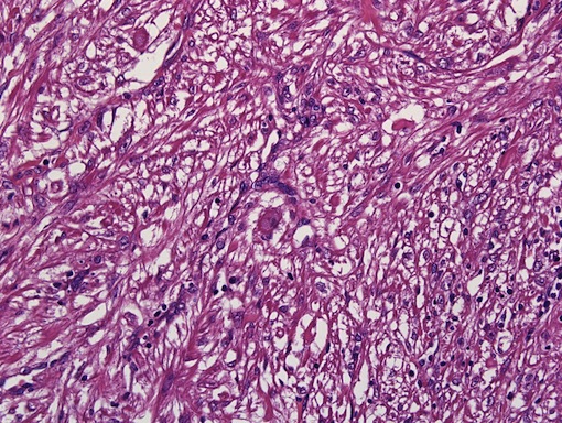
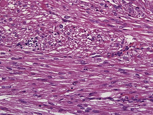
Diagnosis: Renal angiomyolipoma with predominant smooth muscle component
Immunostains:

Discussion:
Angiomyolipoma (AML) is a neoplasm composed of blood vessels, smooth muscle and adipose tissue in varying proportions. It derives from the perivascular epithelioid cells, first described in 1991 but with no known normal cellular counterpart (Arch Pathol Lab Med 2009;133:648, Am J Surg Pathol 1991;15:644). No precursor lesion for PEComas has been described.
PEC related tumors include angiomyolipoma, clear cell sugar tumor, lymphangioleiomyomatosis and other rare tumors labeled PEC, NOS. These tumors are often perivascular, and have varied histology. The tumor cells have clear to granular, lightly eosinophilic cytoplasm, with small, central nuclei and indistinct nucleoli. There is typically no / mild atypia and no / rare mitotic activity, although exceptions occur. In angiomyolipoma, adipose tissue typically predominates, although cases with mostly smooth muscle, as with this patient, do occur, often in the renal capsule (capsuloma). Smooth muscle cells are intimately associated with the vessel walls and may appear to spin off the walls in a radial fashion. The blood vessels are thick walled and tortuous, resembling arteriovenous malformations.
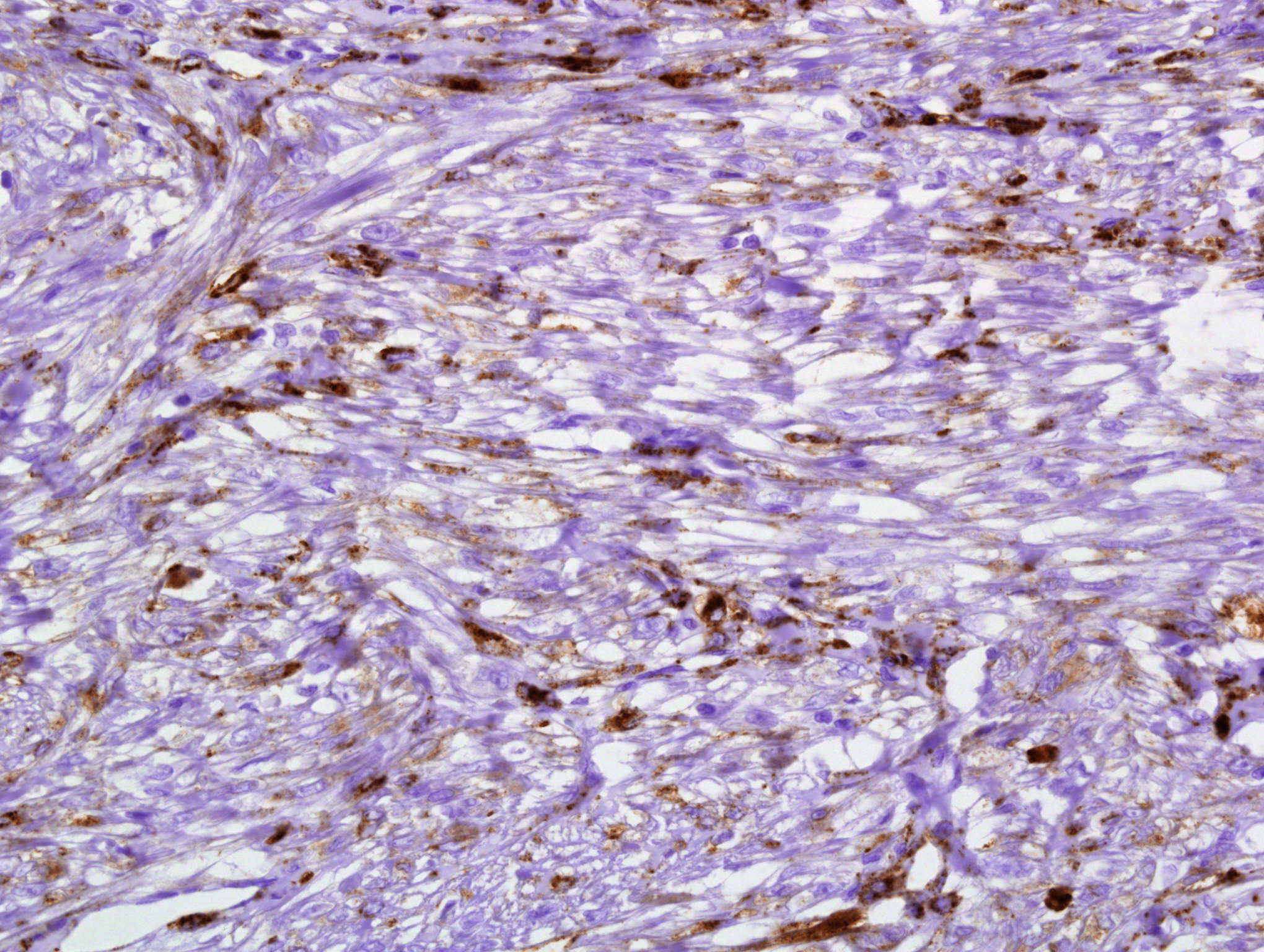
PEComas have a distinctive immunohistochemistry profile, with reactivity for myoid (desmin, smooth muscle actin, calponin) and melanocytic (HMB45, MelanA) markers. Keratins are typically negative, as in this case.
Angiomyolipomas are circumscibed but not encapsulated. They often become quite large, and can cause life threatening hemorrhage. Although 80% of patients with tuberous sclerosis have angiomyolipomas, most patients with angiomyolipoma do not have tuberous sclerosis (OMIM: 191100 [Accessed 13 May 2024]).
The differential diagnosis of smooth muscle predominant angiomyolipoma includes leiomyoma and leiomyosarcoma. Leiomyoma lacks the characteristic blood vessels and adipose tissue of AML. Leiomyosarcoma typically has prominent nuclear atypia, tumor cell necrosis and mitotic figures.
AML typically has benign behavior, although epithelioid tumors are considered to be malignant with the ability to recur and metastasize. Treatment consists of selective arterial embolization or excision (Eur Urol 2009;55:1155).
References: Murphy: Tumors of the Kidney Bladder and Related Urinary Structures, 4th Edition, 2004
All cases are archived on our website. To view them sorted by case number, diagnosis or category, visit our main Case of the Month page. To subscribe or unsubscribe to Case of the Month or our other email lists, click here.
This case was contributed by Dr. Eddie Fridman, The Chaim Sheba Medical Center, Israel.
CancerTYPE ID is a standardized, objective molecular test based on the differential expression of 92 genes, that classifies tumors by matching the gene expression pattern of a patients tumor tissue to a database of known tumor types and histological subtypes.
CancerTYPE IDs database includes 2,206 tumors from multiple tumor banks, selected to provide broader and deeper representation of the heterogeneity of tumors. The 92-gene assay does not overlap with IHC markers, providing complementary data to standard tumor diagnosis.
CancerTYPE ID uses real-time reverse transcription polymerase chain reaction (RT-PCR). A very low copy number of RNA molecules can be detected, thus reducing the sample tissue required for testing. Testing is conducted and results are generated at bioTheranostics' CAP-accredited, CLIA-certified laboratory.
Website news:
(1) We are looking for more cases of the week (we are down to a two month supply). We are also looking for a reviewer(s) for the Salivary Gland chapter. For more information, click here.
(2) We are looking for pathologists with expertise in the topic to write a 250 word article on "What's New" for these future Feature Pages: Consumable Lab Products (Nov11), Grossing workstations / lab furniture (Dec11), Billing / consulting / management (Jan12), Microscopes / slide imaging (Feb12), for $50 per article.
Visit and follow our Blog to see recent updates to the website.
Case #220
Clinical history:
A 37 year old woman had a right renal mass, and underwent a partial nephrectomy.
Microscopic images:
What is your diagnosis?
Click here for diagnosis and discussion:
Diagnosis: Renal angiomyolipoma with predominant smooth muscle component
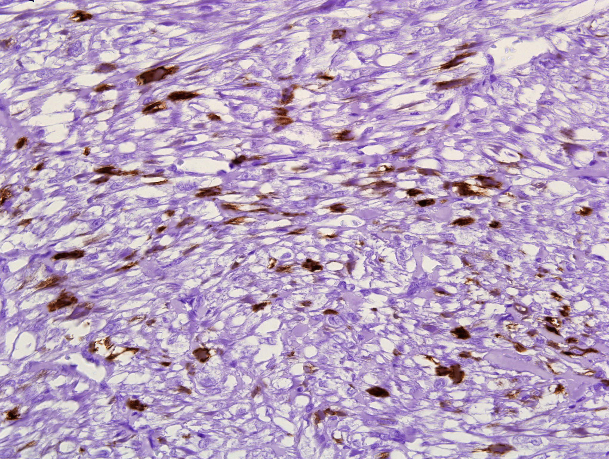
Immunostains:
MNF116 (pankeratin)
HMB45
Desmin
Discussion:
Angiomyolipoma (AML) is a neoplasm composed of blood vessels, smooth muscle and adipose tissue in varying proportions. It derives from the perivascular epithelioid cells, first described in 1991 but with no known normal cellular counterpart (Arch Pathol Lab Med 2009;133:648, Am J Surg Pathol 1991;15:644). No precursor lesion for PEComas has been described.
PEC related tumors include angiomyolipoma, clear cell sugar tumor, lymphangioleiomyomatosis and other rare tumors labeled PEC, NOS. These tumors are often perivascular, and have varied histology. The tumor cells have clear to granular, lightly eosinophilic cytoplasm, with small, central nuclei and indistinct nucleoli. There is typically no / mild atypia and no / rare mitotic activity, although exceptions occur. In angiomyolipoma, adipose tissue typically predominates, although cases with mostly smooth muscle, as with this patient, do occur, often in the renal capsule (capsuloma). Smooth muscle cells are intimately associated with the vessel walls and may appear to spin off the walls in a radial fashion. The blood vessels are thick walled and tortuous, resembling arteriovenous malformations.
PEComas have a distinctive immunohistochemistry profile, with reactivity for myoid (desmin, smooth muscle actin, calponin) and melanocytic (HMB45, MelanA) markers. Keratins are typically negative, as in this case.
Angiomyolipomas are circumscibed but not encapsulated. They often become quite large, and can cause life threatening hemorrhage. Although 80% of patients with tuberous sclerosis have angiomyolipomas, most patients with angiomyolipoma do not have tuberous sclerosis (OMIM: 191100 [Accessed 13 May 2024]).
The differential diagnosis of smooth muscle predominant angiomyolipoma includes leiomyoma and leiomyosarcoma. Leiomyoma lacks the characteristic blood vessels and adipose tissue of AML. Leiomyosarcoma typically has prominent nuclear atypia, tumor cell necrosis and mitotic figures.
AML typically has benign behavior, although epithelioid tumors are considered to be malignant with the ability to recur and metastasize. Treatment consists of selective arterial embolization or excision (Eur Urol 2009;55:1155).
References: Murphy: Tumors of the Kidney Bladder and Related Urinary Structures, 4th Edition, 2004